Method Article
嗅觉系统作为模型来研究轴突生长的模式和形态
摘要
We describe a protocol for in vivo labeling of olfactory sensory neurons by electroporation and subsequent confocal laser-scanning or multiphoton microscopy to visualize neuronal morphology and its development over time.
摘要
The olfactory system has the unusual capacity to generate new neurons throughout the lifetime of an organism. Olfactory stem cells in the basal portion of the olfactory epithelium continuously give rise to new sensory neurons that extend their axons into the olfactory bulb, where they face the challenge to integrate into existing circuitry. Because of this particular feature, the olfactory system represents a unique opportunity to monitor axonal wiring and guidance, and to investigate synapse formation. Here we describe a procedure for in vivo labeling of sensory neurons and subsequent visualization of axons in the olfactory system of larvae of the amphibian Xenopus laevis. To stain sensory neurons in the olfactory organ we adopt the electroporation technique. In vivo electroporation is an established technique for delivering fluorophore-coupled dextrans or other macromolecules into living cells. Stained sensory neurons and their axonal processes can then be monitored in the living animal either using confocal laser-scanning or multiphoton microscopy. By reducing the number of labeled cells to few or single cells per animal, single axons can be tracked into the olfactory bulb and their morphological changes can be monitored over weeks by conducting series of in vivo time lapse imaging experiments. While the described protocol exemplifies the labeling and monitoring of olfactory sensory neurons, it can also be adopted to other cell types within the olfactory and other systems.
引言
The lifelong turnover of sensory neurons distinguishes the olfactory system from many other sensory and neuronal systems1,2. Newly formed sensory neurons are continuously generated in the basal portion of the olfactory epithelium3 and extend their axons into the olfactory bulb, the first relay station of the olfactory system4. However, the cellular and molecular mechanisms controlling the formation and maintenance of the olfactory map are far from being fully understood4,5.
Here, we describe a protocol for labeling sensory neurons of the olfactory organ of larval X. laevis by in vivo electroporation of fluorophore-coupled dextrans. The presented protocol allows visualization of axonal morphology and connectivity, track axonal development over time and study mechanisms regulating axonal wiring and guidance.
Electroporation is a well established method to introduce charged macromolecules, like dextran-coupled dyes and DNA, into cells6,7. The cell membrane is permeabilized by application of short voltage pulses and the molecules are electrophoretically delivered into the cytosol8. Spatially restricted electroporation using a micropipette permits selective labeling of cells including neurons and has been applied in various neuronal systems including the visual system of X. laevis9,10.
We show how the electroporated animals can be used to study axonal growth patterns and morphology in living animals using confocal laser-scanning or multiphoton microscopy. The described procedure allows identifying the coarse topology of axonal projections of sensory neurons of the main and accessory olfactory system11,12. Using in vivo time lapse imaging, it is also suitable to supervise the glomerular connections of single mature sensory neurons, and to monitor the evolution of the axonal projection patterns of immature sensory neurons12. The described protocol can be applied to investigate the structure and formation of olfactory circuits in the intact animal and can be adapted to other cell types within the olfactory and other neuronal systems.
研究方案
注:动物的处理和实验进行批准的哥廷根大学委员会,伦理动物实验。
1.准备的仪器和吸管制作
- 确保电穿孔装置由具有大的工作距离在立体显微镜的,并配备了荧光照明和过滤套用于所使用的染料。
- 电穿孔使用一个专用的单个细胞电穿孔仪或附连到示波器的通用方波脉冲发生器。连接所述电穿孔仪输出到移液管保持器和一个浴电极。脉冲发生器的正极端子连接到微管保持器和所述负极端子向浴电极。确保两个吸管保持器和浴电极含有涂有一层薄薄的氯化银的银导线。
- 安装在显微吸管持有人允许精确定位。
- 从制造硼硅玻璃毛细管内的灯丝电微量。
- 使用水平微量拉马和所描述的贝斯曼等人 13施加改性协议移液器用于膜片钳实验的制造。
- 适应参数,以产生一个较长的刀柄和一个较小的喷嘴开口导致约15-20兆欧姆的单细胞电穿孔较高的吸液管电阻。移液管电阻要低, 例如 ,3-4兆欧,对细胞群体的标签。
- 测量可以直接在吸液管电阻与一个专用的单细胞电穿孔仪或测定用应用程序定义的电压脉冲的后示波器电流流动后用欧姆定律计算出来。
2.制备电穿孔的解决方案
- 溶于青蛙铃声(98毫米氯化钠,2毫米氯化钾荧光基团偶联葡聚糖,1毫米氯化钙2,2毫米MGC升2,5mM葡萄糖,5mM的丙酮酸钠,10mM的HEPES,pH调至7.8,克分子渗透压浓度为230 mOsmol /升)以3毫摩尔的浓度存在。这些细胞可能不那么明亮,如果较低的染料浓度使用标记。准备大量库存解决方案,并把它分成小等份。冻结,储存(稳定数月)。
注:葡聚糖是在不同尺寸和各种发射/激发光谱可用的( 例如 ,页面488葡聚糖10KD,Alexa的546葡聚糖10KD,Alexa的568葡聚糖10KD,Alexa的594葡聚糖10KD,TMR-葡聚糖3KD。)。 - 回填具有细长枪头用小体积的葡聚糖溶液(1-5微升)微量。仔细轻弹微量用手指从枪头去除残留的气泡。
- 装在吸管架微量。确保吸液管内的银线是在与染料溶液接触。
3.选择幼虫X的蟾</ EM>
- 使用X的白化幼虫蟾用于实验。野生型动物中具有该显示期间共焦/多光子成像的自发荧光发射,并且因此不适合用于所描述的实验,颜料填充的黑素细胞。
- 舞台新科普和法贝尔14后premetamorphotic蝌蚪。选择阶段45-53的实验蝌蚪。
4.电穿孔荧光基团偶联葡聚糖
- 放置一小块组织培养皿中并覆盖它具有小体积的含0.02%三卡因水(3-氨基苯甲酸乙酯甲磺酸盐,调节至pH 7)。
- 麻醉在水中含0.02%三卡因的蝌蚪。几分钟后,这些动物停止移动。通过触摸蝌蚪确定适当的麻醉。它们应是无响应。
- 小心地从麻醉转移蝌蚪的组织覆盖的培养皿。
- 确保洗浴ELECTR颂关闭电电路。确保电极与湿纸巾接触;直接接触到蝌蚪是没有必要的。
- 定位枪头接近使用显微嗅觉器官。
- 穿透皮肤覆盖的嗅觉器官与枪头和尖小心翼翼地提前进入主嗅上皮,或犁鼻上皮的位于市中心的感觉神经元层。
- 引发正极的方形电压脉冲,以染料转移到的感觉神经元。应用单一电压脉冲( 如 ,25 V,脉冲长度为25毫秒)或多个脉冲序列( 例如 ,50 V,脉冲宽度300微秒,400毫秒的火车时间在200赫兹)。
注:确定最佳的电压脉冲参数所需的扩展标签。减小电压脉冲的幅度,持续时间和重复次数,以降低标记的细胞的数目。适用于高电压脉冲的幅度,对期限和数量ulses为更广泛的标签。 - 可视化成功的染料挤压和电利用体视显微镜的荧光照明触发脉冲。成功的电穿孔后的染料迅速扩散到胞体和树突。
- 重复步骤4.5-4.9的蝌蚪的第二个嗅觉器官。
- 转移蝌蚪放入盛有淡水回收的烧杯中。经过约5分钟蝌蚪从麻醉中醒来,并开始正常的游泳动作。
- 后24小时,将电穿孔的染料扩散,在感觉神经元中,并最终到达经由轴突运输嗅球。
5.安装动物体内可视化细胞及轴突的过程
- 麻醉在水中含0.02%三卡因的蝌蚪。
- 仔细蝌蚪转移到成像室, 比如一个小的硅橡胶盖培养皿蝌蚪大小的凹陷。
- 切一个小矩形变成封口膜的条纹。盖上封口膜的蝌蚪,离开前端脑通过切口窗口暴露出来。修正了上盘针的封口膜,而不伤及蝌蚪。
- 确保蝌蚪被淹没在含有0.02%三卡因足够的水。
- 安装成像室上直立多光子显微镜或共聚焦显微镜的阶段。
- 获得嗅球的图像的三维堆叠。确保该成像程序是尽可能地短,并且不超过10-15分钟。
- 返回蝌蚪变成正常的水在一个单独的油箱,并防止暴露在光线下。 5分钟后,蝌蚪从麻醉中醒来。
- 每天指定的时间间隔后,重复步骤5.1-5.7, 如:,。
6.安装动物的细胞和轴突的进程离体可视化
- 另外第5该协议的,使用切除大脑准备可视化标记的感觉神经元。
- 麻醉,杀死蝌蚪由大脑的横断处过渡到脊髓。切除组织含有嗅觉器官,嗅神经和端脑前一个块。
- 转移的组织块以蛙振铃溶液和除去结缔组织与细剪至脑的腹侧边露出。
- 转移组织块的成像室,并与铂框架弦用尼龙线固定。
- 装入成像室上的共焦/多光子显微镜的阶段。
- 获得嗅球的图像的三维堆叠。
7.图像处理和数据评估
- 优化,并应用滤波以提高图像质量和神经元结构的可视化的非本地装置所描述的双门轿跑车,P。等人 15。
注:数据来自于活标本更深的组织层成像实验往往是嘈杂。 - 创建图像栈概述的最大强度的预测。
- 时间推移成像实验筛选数据集的感觉神经元的单明确标识轴突。
- 所描述的鹏等人通过神经元突起的软件辅助重建跟踪细胞形态。16。
结果
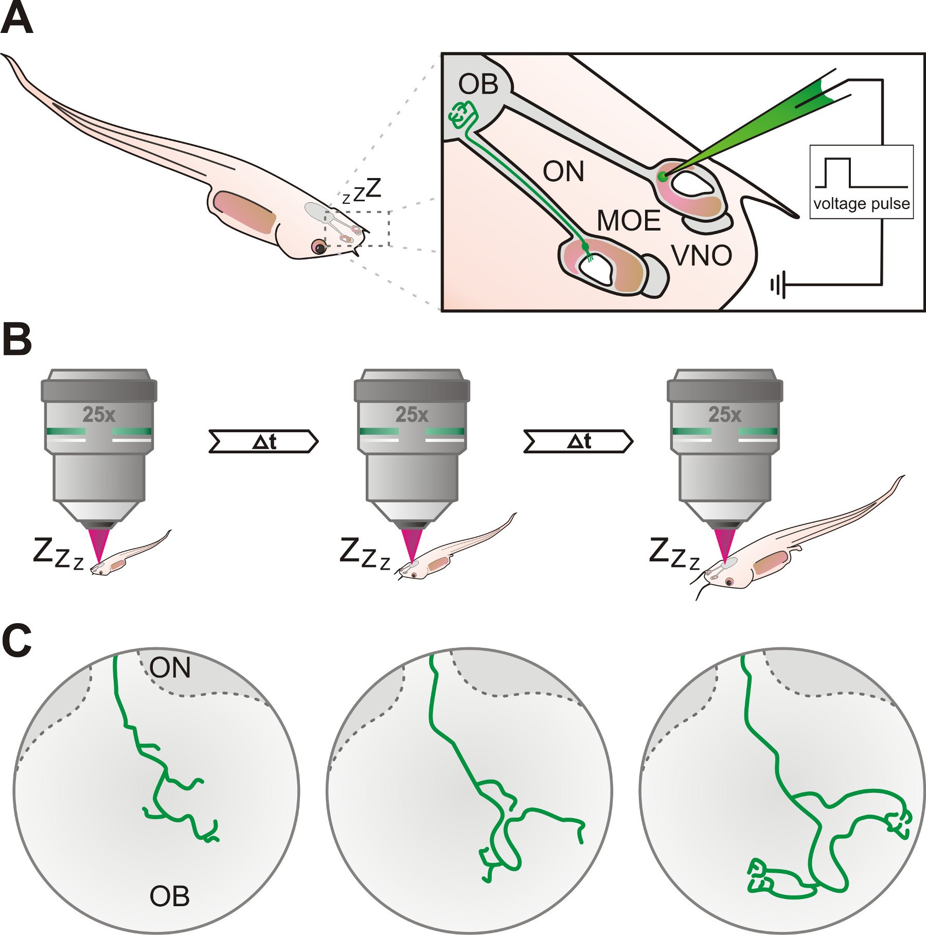
所述协议可以成功地应用于电穿孔和麻醉X的嗅觉系统的感觉神经元的轴突过程的体内可视化蟾 ,使用共聚焦激光扫描或者多光子显微术( 图1)。
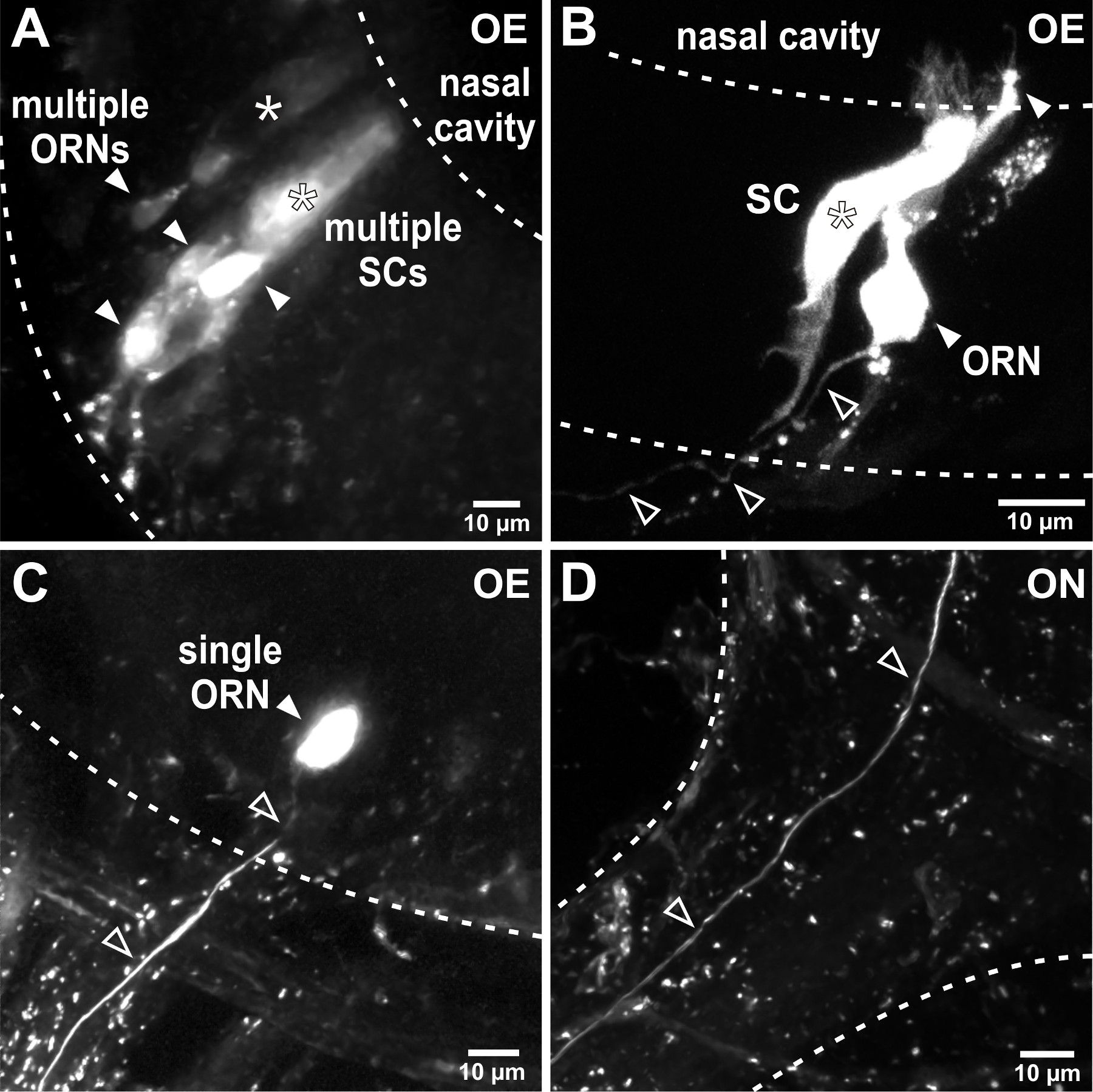
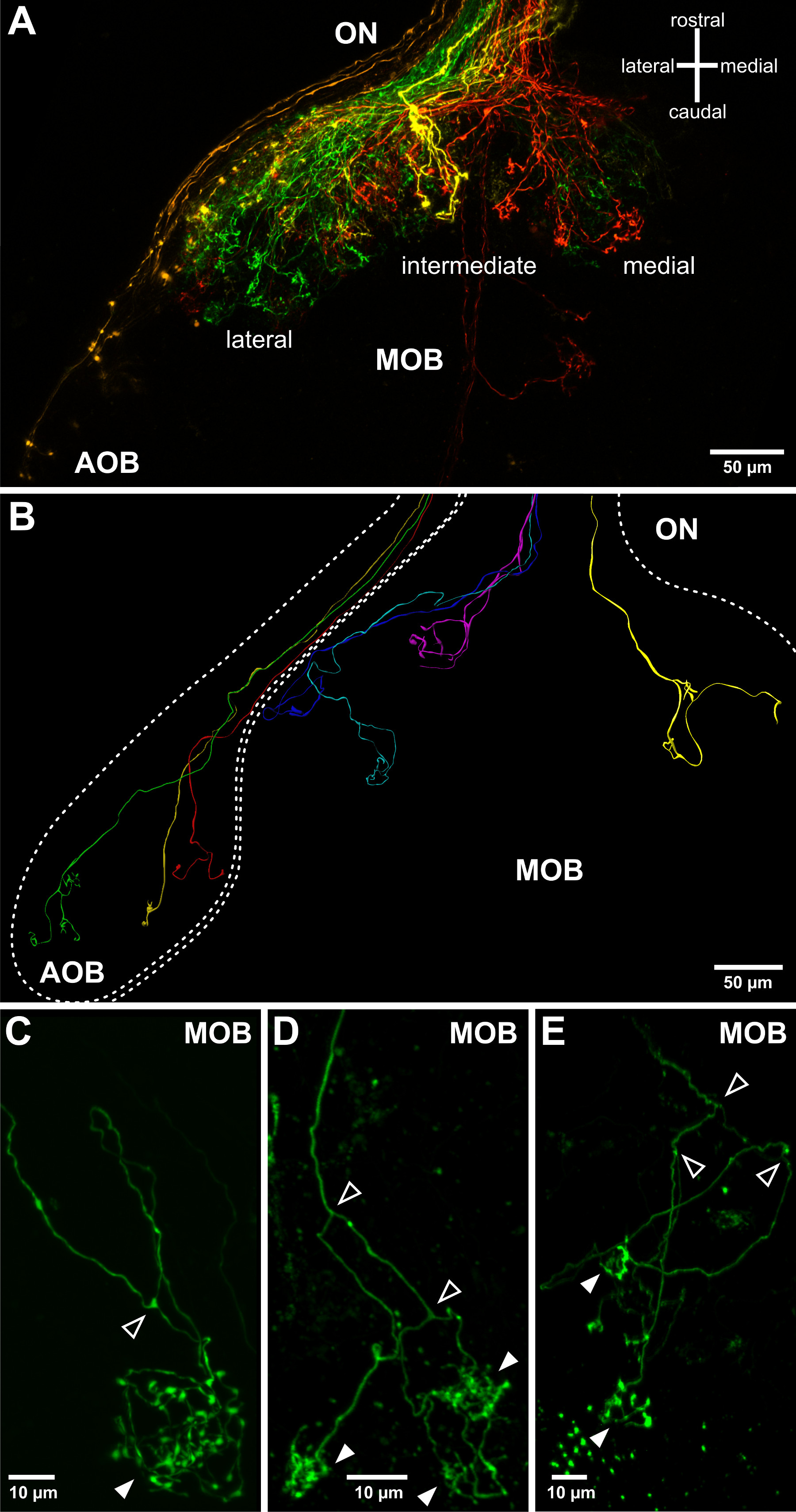
电穿孔使荧光团偶联的葡聚糖进入并迅速传播内的嗅觉器官的细胞。这是有帮助的应用荧光照明的触发电压脉冲后验证成功的标志。根据电穿孔参数, 例如 。,吸液管电阻,将细胞( 图2A,B)或嗅觉器官的单细胞( 图2C)的基团标记。标记的感觉神经元的轴突可以显现在嗅神经( 图2D)和轴突的过程也可以被观察到,在嗅球,成功的电穿孔后,通常24小时( 图3)。感觉神经元群体的电穿孔允许可视化在嗅球( 图3A)的粗配线图案。单个细胞电穿孔可以用于调查个体轴索突起图形,轴突分叉并连接到肾小球的结构在嗅球( 图3C-E)。
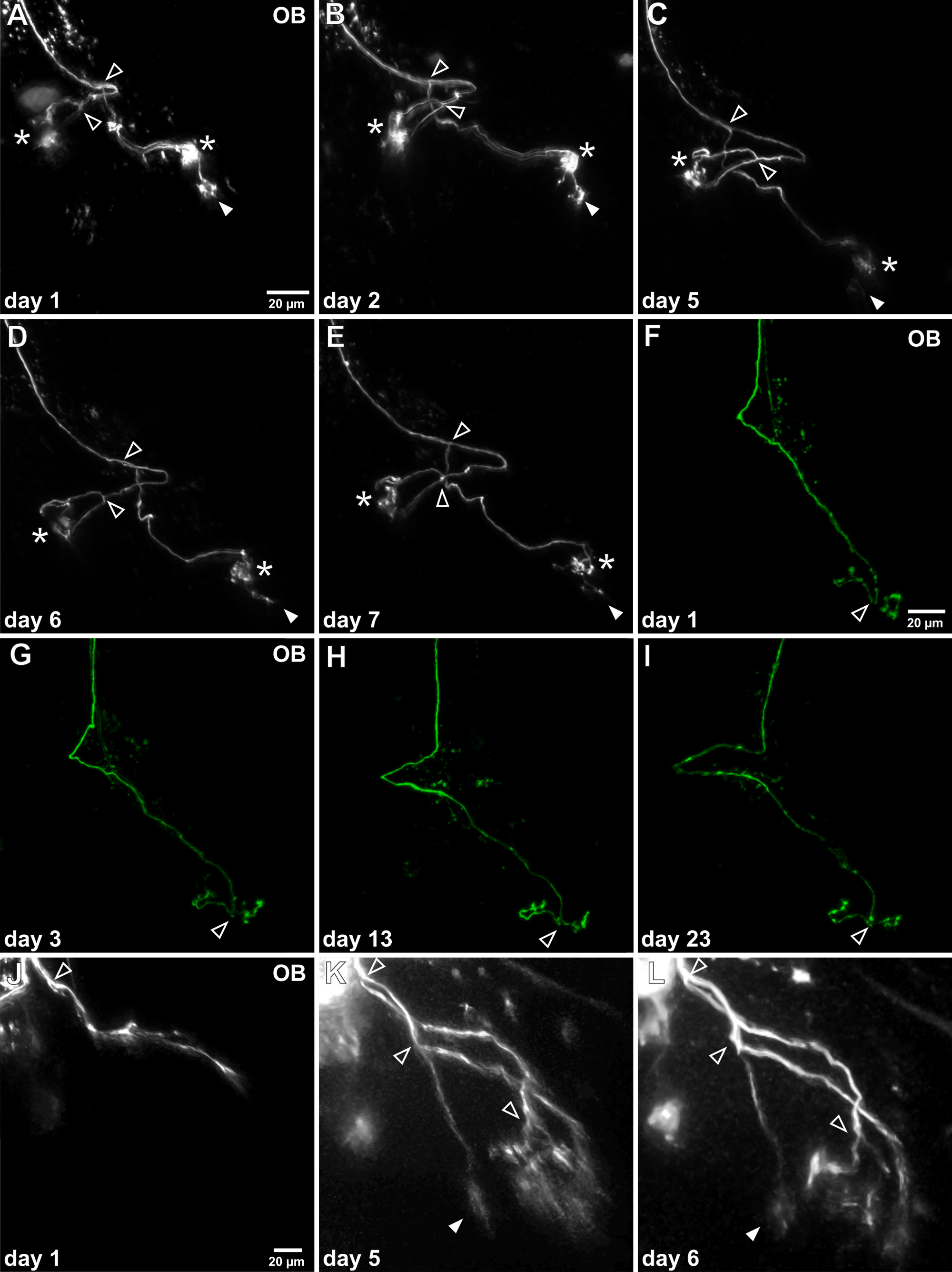
十,透明的白化蝌蚪蟾许可证活体共焦或麻醉蝌蚪的完整的大脑标记的感觉神经元的多光子成像。的轴突生长模式的发展,可以跟踪在数天/周反复在同一标记的感觉神经元( 图4)的体内可视化。根据不同的感觉神经元的电穿孔的时间点,在嗅球轴突图案可以有很大的不同的成熟度。成熟的神经元已经拥有精心设计的轴突分支功能绺连接到肾小球编辑区域。成熟神经元的轴突生长模式是相当稳定的,但轴突分支和肾小球簇改进的伸长率可观察( 图4A-E)。感觉神经元可以在体内观察到的更长的时间内, 例如 。,大于三个周( 图4F-1)。另一方面,未成熟的神经元的轴突仍处于初期生长的过程中,不具有簇绒arborizations和尚未连接到它们的最终肾小球目标。实验协议允许成熟过程中追踪这些神经元的发育, 如:,轴突伸长,分叉和建立簇绒arborizations( 图4J-L)的。更多的例子也看到Hassenklöver和曼齐尼12。

麻醉X的嗅觉器官图1.原理的实验方案概述(一)主嗅上皮感觉神经元(MOE)或犁鼻器(VNO) 蟾幼虫通过电穿孔,使用玻璃移液管填充有荧光葡聚糖溶液进行标记。标记的神经元的轴突可以通过嗅觉神经(ON),随后,最终达到嗅球(OB)。(B)中的蝌蚪被麻醉并标记的神经元是使用在特定的时间间隔的共焦或多光子显微镜反复研究。 (C)的标记细胞的轴突生长模式的渐进发展,可以在后面几天的时间跨度为两周。

图2. ELECtroporation在嗅觉器官的感觉神经元中。(A)电穿孔与低电阻移液器导致多种细胞在嗅觉器官的标签。在这个典型的例子多个嗅觉受体神经元(ORNs,充满箭头)和两个柱状支持细胞(旺,星号)进行染色。虚线划分的嗅觉上皮(OE)。(B)的磨浆电穿孔参数, 例如 ,边界,增加了吸液管电阻,限制了标记细胞的数目。在这个例子中,一个单一的感觉神经元(实心箭头)和一个相邻的支持细胞(星号)电穿孔后进行染色。注意单个轴突离开嗅觉上皮(空心箭头)。(C)的成功单细胞电穿孔导致的个体感觉神经元(实心箭头)并在嗅觉上皮与其连接的轴突(空心箭头)的专属标记。 (D)中的单个标记的轴突(空心箭头)可以通过嗅神经(ON),进入嗅球被遵循。

图3.可视化在切除嗅球轴突突起。(A)中 ,在嗅球的感觉神经元的轴突突起的粗拓扑可以用较低的电阻电吸移管被可视化。一嗅球半球和其相关联的嗅神经(ON)的描绘。四葡聚糖耦合到不同的荧光团进行了电穿孔在嗅觉器官的4远的地方:横向(绿),中间(黄色),内侧弹性模量(红色)和虚拟网络运营商(橙色)。这允许可视化嗅球(AOB)和主嗅球(MOB)的三个主凸域。 (B)的叠加在嗅球的结构单嗅觉感觉神经元的不同的轴突生长型态。示出被合并的来自不同幼虫标本来自多个单细胞荧光染色的三维重建。单嗅觉轴突伸入嗅球和球面肾小球(实心箭头)形成簇绒arborizations /突触(CE)的例子。另外,在X中蟾嗅神经轴突连接到一个,两个或多个肾小球(实心箭头,也看到Hassenklöver和曼齐尼12)之前经常分叉(空心箭头)。

图4 中的单个嗅觉神经元轴突体内延时成像。(AE)成功的单个细胞电穿孔后的标记的轴突可反复可视化在嗅球(OB)。这个例子说明,研究了一个星期的个人轴突。整体的形态不发生变化很大,因此两个主要分支点可被识别(开放箭头)。需要注意的是在时间过程中,1肾小球的绒头经历连续减少(实心箭头),而另外两个肾小球簇保持稳定(星号)。(FI)此代表性示例示出的长期观测的可行性,因为这特定轴突进行了研究三个多星期。可以检测它的生长模式没有明显的变化。(JL)在生长过程中的未成熟的感觉轴突的一个例子被描述。它尚未连接到肾小球和肾小球特征簇的丢失。经过5天的轴突分支被拉长,轴突分叉多次和细arborizations是确立。
讨论
这里描述的实验步骤,允许幼虫X的嗅觉器官的标记的感觉神经元蟾通过的荧光团偶联的葡聚糖和随后的感觉轴突生长在活体动物可视电穿孔。通过改变在体内电穿孔的参数,可以控制标记的感觉神经元的数目。由此,能够以标记大基团的感觉上皮,很少或甚至单个细胞的神经元。
以确保所希望的延长神经元标记的是特别谨慎微量特性和电穿孔脉冲是重要的。更高的吸移管的电阻和减小的电压脉冲的幅度,持续时间,重复次数可以减少标记的细胞的量,而降低吸移管的电阻和较高的电压脉冲的幅度,持续时间和脉冲数可以导致更广泛的标签。日E使用荧光葡聚糖的电,如果提供的应用设置相应的即时视觉反馈。但要注意使用的幅度,持续时间和超过协议中提供的值可能会导致细胞损伤,甚至细胞死亡17脉冲数参数。微量的堵塞或破裂的提示也可以阻碍成功的电穿孔。
在X的嗅觉器官活体电穿孔蟾仅限于幼虫期以来postmetamorphotic青蛙的皮肤是强硬的,不能很容易地穿透用微量。 在体内的可视化神经元突起可通过的激发/发射光在更深的脑区域或通过血管的散射受到阻碍。这个问题在更高的幼虫阶段尤为明显,由于较大的大脑,并可能导致使明确的识别细微的轴突过程的更困难的噪声信号。
所提出的协议允许以可视化的完整的嗅觉系统的感觉神经元,而不解剖动物,标签的过程中损坏的细胞,制备组织切片或根据需要可替代的方法固定组织,如在全细胞膜片标签-clamp实验18。当组合几个或单个的感觉神经元在体内的时间推移成像的标记,能够以可视化单个成熟的感觉神经元的肾小球连接在较长的时间间隔。这种方式还能够监测未成熟的感觉神经元的轴突突起图形的发展在几个星期。这后一种选择是特别有趣,因为它允许监视单个轴突的生长模式,在活的动物。这将打开以调查控制轴突导向和寻路细胞和分子机制的可能性。几个因素,包括气味受体表达,各种轴突ñ导向分子和感觉神经元的加臭剂诱导的/自发性活动已显示调节的感觉神经元的轴突4,5-目标的发现。该协议的应用不限于嗅觉的感觉神经元,但也可应用于研究其他类型的细胞, 例如 。,茎发育的大脑或嗅球的僧帽细胞的神经区域的细胞/祖细胞。此外,表现出技术也可以结合使用钙敏感葡聚糖或注入膜透钙染料来获得关于标记的神经元和/或所连接的电路7,19的功能信息。的大范围的荧光团耦合到葡聚糖的可用性允许多个单个细胞或群体具有不同颜色的标签。还质粒DNA溶液,用于荧光蛋白例如编码,适合于电穿孔,并可以进一步提高versatili一节和技术6的有用性。该协议可以进一步增强,使得葡聚糖和DNA或带电吗啉的组合电操纵基因表达13,17。
所描述的方法,当然代表了一种新的工具,调查复杂,但仍不能完全理解的过程,调节脊椎动物的嗅觉系统轴突的指导。
披露声明
The authors have nothing to disclose.
致谢
This work was supported by DFG Schwerpunktprogramm 1392 (project MA 4113/2-2), cluster of Excellence and DFG Research Center Nanoscale Microscopy and Molecular Physiology of the Brain (project B1-9), and the German Ministry of Research and Education (BMBF; project 1364480).
材料
| Name | Company | Catalog Number | Comments |
| SZX16 | Olympus | Stereomicroscope with fluorescent illumination | |
| Axon Axoporator 800A | Molecular Devices | Single cell electroporator | |
| ELP-01D | npi electronic | Electroporator | |
| MMJ | Märzhäuser Wetzlar | Manual micromanipulator | |
| P-1000 | Sutter | Horizontal micropipette puller | |
| G150F-4 | Warner Instruments | Glass capillaries for electroporation pipette fabrication; internal filament makes backfilling easier | |
| Alexa 488-dextran 10 kD | Life Technologies | D22910 | |
| Alexa 546-dextran 10 kD | Life Technologies | D22911 | |
| Alexa 568-dextran 10 kD | Life Technologies | D22912 | |
| Alexa 594-dextran 10 kD | Life Technologies | D22913 | |
| TMR-dextran 3 kD (micro-Ruby) | Life Technologies | D7162 | |
| Microloader pipette tips | Eppendorf | 930001007 | |
| Tricaine (Ethyl 3-aminobenzoate methanesulfonate) | Sigma-Aldrich | E10521 | Anesthetic; use gloves |
| A1-MP | Nikon | Multiphoton microscope | |
| LSM 780 | Zeiss | Confocal microscope | |
| Imaris | Bitplane | Alternative software for neuronal tracing |
参考文献
- Huard, J. M., Youngentob, S. L., Goldstein, B. J., Luskin, M. B., Schwob, J. E. Adult olfactory epithelium contains multipotent progenitors that give rise to neurons and non-neural cells. J. Comp. Neurol. 400 (4), 486-4810 (1998).
- Schwob, J. E. Neural regeneration and the peripheral olfactory system. Anat. Rec. 269 (1), 33-49 (2002).
- Leung, C. T., Coulombe, P. A., Reed, R. R. Contribution of olfactory neural stem cells to tissue maintenance and regeneration. Nat. Neurosci. 10 (6), 72-726 (2007).
- Lodovichi, C., Belluscio, L. Odorant receptors in the formation of the olfactory bulb circuitry. Physiology (Bethesda. 27 (4), 212-2110 (2012).
- Mori, K., Sakano, H. How is the olfactory map formed and interpreted in the mammalian brain). Annu. Rev. Neurosci. 34, 467-499 (2011).
- De Vry, J., et al. In vivo electroporation of the central nervous system: a non-viral approach for targeted gene delivery. Prog. Neurobiol. 92 (3), 227-244 (2010).
- Hovis, K. R., Padmanabhan, K., Urban, N. N. A simple method of in vitro electroporation allows visualization, recording, and calcium imaging of local neuronal circuits. J. Neurosci. Methods. 191 (1), 1-10 (2010).
- Chen, C., Smye, S. W., Robinson, M. P., Evans, J. A. Membrane electroporation theories: a review. Med. Biol. Eng. Comput. 44 (1-2), 5-14 (2006).
- Haas, K., Jensen, K., Sin, W. C., Foa, L., Cline, H. T. Targeted electroporation in Xenopus tadpoles in vivo--from single cells to the entire brain. Differentiation. 70 (4-5), 148-154 (2002).
- Hewapathirane, D. S., Haas, K. Single cell electroporation in vivo within the intact developing brain. J. Vis. Exp. (17), (2008).
- Gliem, S., et al. Bimodal processing of olfactory information in an amphibian nose: odor responses segregate into a medial and a lateral stream. Cell. Mol. Life Sci. 70 (11), 1965-1984 (2013).
- Hassenklöver, T., Manzini, I. Olfactory wiring logic in amphibians challenges the basic assumptions of the unbranched axon concept. J. Neurosci. 33 (44), 17252-1710 (2013).
- Bestman, J. E., Ewald, R. C., Chiu, S. -. L., Cline, H. T. In vivo single-cell electroporation for transfer of DNA and macromolecules. Nat. Protoc. 1 (3), 1272-1210 (2006).
- Nieuwkoop, P. D., Faber, J., Nieuwkoop, P. D., Faber, J. . Normal table of Xenopus laevis. , (1994).
- Coupé, P., Munz, M., Manjón, J. V., Ruthazer, E. S., Collins, D. L. A CANDLE for a deeper in vivo insight. Med. Image Anal. 16 (4), 849-864 (2012).
- Peng, H., Ruan, Z., Long, F., Simpson, J. H., Myers, E. W. V3D enables real-time 3D visualization and quantitative analysis of large-scale biological image data sets. Nat. Biotechnol. 28 (4), 348-353 (2010).
- Falk, J., et al. Electroporation of cDNA/Morpholinos to targeted areas of embryonic CNS in Xenopus. BMC Dev. Biol. 7 (107), (2007).
- Nezlin, L. P., Schild, D. Individual olfactory sensory neurons project into more than one glomerulus in Xenopus laevis tadpole olfactory bulb. J. Comp. Neurol. 481 (3), 233-239 (2005).
- Nagayama, S., et al. In vivo simultaneous tracing and Ca2+ imaging of local neuronal circuits. Neuron. 53 (6), 789-803 (2007).
转载和许可
请求许可使用此 JoVE 文章的文本或图形
请求许可探索更多文章
This article has been published
Video Coming Soon
版权所属 © 2025 MyJoVE 公司版权所有,本公司不涉及任何医疗业务和医疗服务。