
Method Article
对调查信息处理的神经药理学清醒行为猕猴皮质压力喷射系统
摘要
Here, we show the pressure injection of neuropharmacological substances during single-cell recording in an awake, behaving macaque monkey. This procedure allows pharmacological manipulation in the direct vicinity of a cortical recording site.
摘要
The top-down modulation of feed-forward cortical information processing is functionally important for many cognitive processes, including the modulation of sensory information processing by attention. However, little is known about which neurotransmitter systems are involved in such modulations. A practical way to address this question is to combine single-cell recording with local and temporary neuropharmacological manipulation in a suitable animal model. Here we demonstrate a technique combining acute single-cell recordings with the injection of neuropharmacological agents in the direct vicinity of the recording electrode. The video shows the preparation of the pressure injection/recording system, including preparation of the substance to be injected. We show a rhesus monkey performing a visual attention task and the procedure of single-unit recording with block-wise pharmacological manipulations.
引言
In cortical and subcortical areas, neuronal activity is affected by various neuromodulators, for example acetylcholine1. These modulatory effects on neuronal responses have been reported in in vitro studies2, as well as in electrophysiological recordings from anesthetized animals3 and systemic pharmacological manipulations in humans4. Nevertheless, the exact role of different neuromodulators and the involvement of various receptor subtypes are largely unknown. To measure the effects of specific neuromodulators on the activity of single neurons, it is desirable to induce a temporary neuromodulator change as close as possible to the recording electrode. Furthermore, it is important that those manipulations are done in awake animals, as cognitive functions are only present in the absence of anesthesia. Additionally, anesthesia interacts with cholinergic and GABAergic systems5,6 and can lead to changes in neural activity3.
Within the last decades, two main methods of local drug delivery have been developed and refined: iontophoresis and pressure injection. In both methods drugs are delivered through micropipettes made of either glass or steel. With iontophoresis, an electrical current regulates the release of the drug7. Additionally, there is a significant contribution of electro-osmosis to the total amount of ejected molecules8, correlating with the tip diameter9 of the micropipette as well as with the concentration8 of the substance used. Iontophoresis is a powerful tool to quickly and precisely manipulate small volumes of nervous tissue. For iontophoretic injections, multi-barrel micropipettes are usually used10, with one acting as a recording device while the other positions serve as delivery pipettes. A limitation of this method is that only charged molecules can be used, severely limiting the selection of drugs.
Pressure injection uses either air compression or mechanical pressure to eject a substance from a micropipette. Using this method any soluble substance, charged or uncharged, can be used, including large molecules. The method of pressure injection was first described by Reyniers in 1933 and further refined in the 1950s (see Lalley11 for a review). In the 1980s the method was further refined to allow delivery of amounts in the nanoliter range (mainly lidocain12) to a defined brain area13 while simultaneously performing single-cell recording. The ejected volume was usually monitored by observing the movement of a marker, such as the meniscus in the upper part of the pipette13. Pressure injection was first used in the 1990s in awake animals, both extracellularly14 and intracellularly15,16. Based on the cumulative expertise gained in these studies it is now possible to reliably record from different brain structures in combination with pharmacological manipulation (see17 for a comparison of recent pressure injection systems).
An enduring open issue for both drug delivery methods is the difficulty in determining the precise volume injected. This is an even bigger challenge for experiments with awake, behaving rhesus monkeys where the animal performs the experimental task in a separate room. This can be alleviated by the use of a software-controlled system instead of relying on a visual marker to continuously monitor an injection.
The system described here is an extension of a well-established electrophysiological recording system (Mini Matrix System) and combines an injection pipette with multiple parallel-oriented recording electrodes at defined distances in a customizable arrangement. Pharmacological manipulation of the tissue near the recording electrode is possible using only a small amount of substance, ensuring a fast recovery and allowing multiple blocks of injection and control/recovery within the limited time window offered by the behavioral task of the animal.
研究方案
动物保健和所有试验程序都按照德国法律管辖动物护理进行,由不伦瑞克,德国下萨克森州的区政府批准。
注意:作为实验是在体内进行的,关键的是要保持尽可能高的卫生标准。只要有可能,在无菌条件下工作。
1.准备注入/记录系统
- 消毒,该微量与注射器连接管。使用管可能的最短长度在喷射泵和电生理记录系统之间的实验设置。
- 清洁用清洁线记录系统的导向管中。它们浸在无菌硅油,并通过单独的导管喂它们几次。
- 插入石英玻璃微到记录系统的一个导向管。参见图1A。
- 在固定的微后记录系统,附加的无菌管到微量的金属销。保重;虽然微量被固定在系统中安装的管时,它可以很容易破裂。用两个镊子消毒适用于针管等压。
- 使用液体强力胶密封管和微管之间的交界处。等待至少3小时的胶水充满液体微量之前变硬。
- 之前或微量插入之后插入微电极( 例如 ,石英玻璃绝缘铂钨)到记录系统的其它位置。
2.准备物质
- 消毒1.5毫升为使用高压釜或其它可靠的过程注射溶液以后存储的微量离心管。
- 权衡的相应物质东莨菪碱盐酸盐,制备5mL的0.1摩尔溶液。溶解在无菌盐水(0.9%NaCl)中。
- 在无菌条件下我呐通风柜,过滤用注射器过滤具有足够大的孔径的解决方案, 例如 ,0.2微米。
- 下的通风柜,等分溶液进入足以为单个实验卷, 例如 ,用于东莨菪碱,500微升无菌1.5 ml离心管。使用深色管,以保护该物质由轻;另外,包装铝箔管。对于东莨菪碱,储存在4°C的长达14天的解决方案。
3.注射/记录系统的日常准备
注意:当被装在记录系统中,在电极和微量存储在记录点之间的酶溶液(Tergazyme,用去离子水1%溶液)。下面的步骤必须在每个记录之前进行。
- 收集该物质的管被注入。如果允许它冷藏达到RT。
- 从酶溶液中取出注入/记录系统和冲洗电极和微量用去离子水才能彻底清理酶液。
- 为了目视检查微量和管之间的密封去除记录系统的前盖。
- 为了润滑系统为平稳运动申请无菌硅油到导向管间隙( 见图1A)与电极和微量的提示。
- 检查用显微镜,以确保它们完好无损的电极和微量提示。对准电极和微量在显微镜下,使它们伸出的导向管具有相同的长度。推动他们进入导管,只要他们不再可见停止。这被定义为电极位置为零。在软件中设置的电极的深度和微量为0。
- 填充无菌盐水的无菌注射器和针头插入管,小心不要刺穿管的壁。开车微量出导管的VISU的物质流动的人的控制。
- 冲洗至少2个毫升无菌盐水通过管和微量,以确保没有空气残留在注射器或管。不过大的压力施加到注射器的柱塞。确保管和微量之间的结被密封。如果泄漏是可见的,再粘上交界处(见步骤1.5),并推迟拍摄。
- 填充有所述溶液的新的无菌注射器被注入并与盐水注射器的筒交换它, 也就是说 ,保持在管内的生理盐水填充注射器的针头。确保没有空气被转移到系统中。这最好通过去除生理盐水填充桶后填充盐水针头毂实现。
- 冲洗系统用250微升溶液被注入,以从管完全除去盐水。
- 使用的电动机控制软件,缩回电极和微量成guidetubesto的至少-500微米的深度。
- 降低导向管环( 图1A)到导向管,以保持它们的固定的相对位置的底部。
- 清洁系统用乙醇碱,特别是在将触摸猴子的录音室。
- 通过更换前盖,拧紧螺丝关闭记录系统。
4.喷射系统的验证
注意:虽然公司校准系统时,建议以验证与在实验装置(管,注射器等)所使用的材料的排出量。
- 如步骤3中所述,保持伸出的导管电极和微量准备系统。推荐的至少在7000微米的深度,以避免测量容积的损失,由于沿微量和电极的外表面的粘合性。
- 放置在记录系统中,将在实验过程中使用的位置,并把syrinGE入微量注射泵。固定在使用该橡胶带和可调握持代替注射器( 见图1A)。直到它牢固地在注射器的柱塞后面滑入到位泵的可移动部分。
- 使用软件控制的电动机单元,喷射大到足以精确测量,体积例如 ,1000 NL。它是优选使用一个单一步骤来喷射的总体积,以避免沿微量表面毛细作用的效果。非常低的速度(1升/多个)也可导致在验证过程期间这样的效果。
- 收集在微量下置于一个容器的总容积,或仔细地直接从微量的尖端收集被排出的下降。估计用吸管喷射体积或通过用精密秤称量。
- 重复此步骤几次,以确认测量。
5.急性录音
- 设置坐标位置记录系统的。此定义在该导向管到达长期植入记录室中的硬膜的点。请确保导向管完全缩回(导管Z位置0)。
- 使记录系统到位置,并放置在注射器中的微量注射泵。固定在使用该橡胶带和可调握持代替注射器( 见图1A)。直到它牢固地在注射器的柱塞后面滑入到位泵的可移动部分。如果物质的下降是在引导管的前端可见,用无菌棉签小心取出。
- 准备的动物记录根据实验室的程序(见18例如准则)。
- 牢固地安装在猴的录音室的录音系统。
- 缓慢手动降低导向管放入记录室,直到达到硬脊膜,然后使用马达共同驱动电极ntrol软件。
- 因为它不可能测量微量的阻抗,首先用电极驱动器和在不同深度定期检查它们的阻抗。后不损坏电极成功执行硬脑膜的渗透,促进微管。
- 驱动电极和微量到在该感兴趣的大脑区域有望被发现靶电极的深度。慢慢推进电极,直到其足够接近记录单个单元的活性,通过在所记录的信号中的良好信噪比所证明。重要的是,定位在记录电极和微量在同一深度,以确保电极和微量之间的最小距离。
- 如果可能的话,保持电极和微量该深度在整个记录。然而,如果要保持所记录的细胞的信号质量的唯一方法是将电极,然后驱动电极和微量同时保持它们之间的距离。
6.空间注意任务
- 在一系列的试验中,在屏幕上目前两个移动的圆点图案,一个位于记录的神经元和它的另一外的感受野内,与动物有贯穿每个审判19 foveate中央提出的固定点在一起, 20。
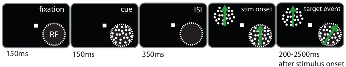
注:猴子被训练在线索点阵图形的方向变化做出反应(目标事件),而忽略了其他的圆点图案的任何方向改变,并奖励与液体进行试19的每一个成功完成的下降, 20。作为感官控制条件,猴子有同时忽略两个移动的点图案(参见图2的任务的一个更详细的说明),以报告该固定点的亮度变化。
7.药理操纵的同时记录
注意:当猴子执行任务,注入物质在逐块的方式。三个连续的块被定义为:控制,其作为一个基线;注射,在这期间的物质被喷射;和恢复,在此期间,细胞中,通过注射返回到基线的目标。
- 在注射块,定期注射物质的预定量例如 ,每分钟以2 NL / s的速率2 NL。对于这个例子,使用东莨菪碱盐酸盐。注射过程是使用软件,该软件提供了各种选项来控制。例如,使用阶梯函数来定义的注射量,并且根据记录的软件的时钟按下注射按钮的每一分钟。
注意:注射块的确切持续时间是物质和实验依赖性, 例如用于东莨菪碱使用2 NL注射每分钟10分钟(20 NL总共)。优选不推进电极和微量杜里纳克注射块。 - 注意时间,并在该物质被注入的试验中,在电极和微量的深度,以及喷射物质的量。
- 按照与恢复块,其中没有物质被注入注入块。恢复块的持续时间是特定物质,需要在预先测试中定义。监控和维护所选单个单元的记录品质,直到恢复块的末尾。
- 重复上述三个区块,只要猴子的录音品质和动机允许。
8.后期录音程序
- 数据记录后,缩回电极和微量到导向管,然后手动缩回导管。从猴子的录音室中取出记录系统。释放从注射泵,注射器和系统传输到制备区域进行清洁。
- 办理动物根据实验室的标准程序,并返回到壳体设施(包括录音室18的清洁)。
- 冲洗导向管与过氧化氢(3%),然后用离子交换水的外部。驱动电极和微量出引导管,冲洗过氧化氢,然后去离子水。
- 交换注射器的筒填充有无菌盐水的注射器的筒,保持针在管。冲洗管,用1-2毫升盐水的微量。冲洗后,取出枪管和空气填充它。重新插入桶进针和干燥管并通过轻轻推动空气从里面微量。
- 存储的引导管,延长电极和微量浸渍在酶溶液,以避免干燥以及确保有机材料的击穿。
结果
图2描绘了当注射过程进行的猴子进行的空间注意任务。猴子被训练参加要么位于记录神经元的感受野内的刺激(参与式),位于感受野外的刺激(参加出)或固定点(参加修复)。这些条件允许神经元活动的不同注意力状态的比较。
图3示出在使用东莨菪碱,毒蕈碱胆碱能拮抗剂的实验样品神经细胞的外周刺激时间直方图。积演示东莨菪碱注入与无注射,当被呈现在神经元的感受野内的细胞的优选的方向上移动的图形,并且由动物出席期间响应抑制。两个第一峰代表的神经元9的响应的在线和空间线索,从而出现了感受野内的偏移量。这后面是响应于出现在屏幕上的提示开始后500毫秒移动图案。灰色阴影区域示出了用于计算每一试验的平均燃烧率的分析期间。绿色区域突出了东莨菪碱注射液对细胞的放电频率的抑制影响。深绿色区域显示分析期内抑制。
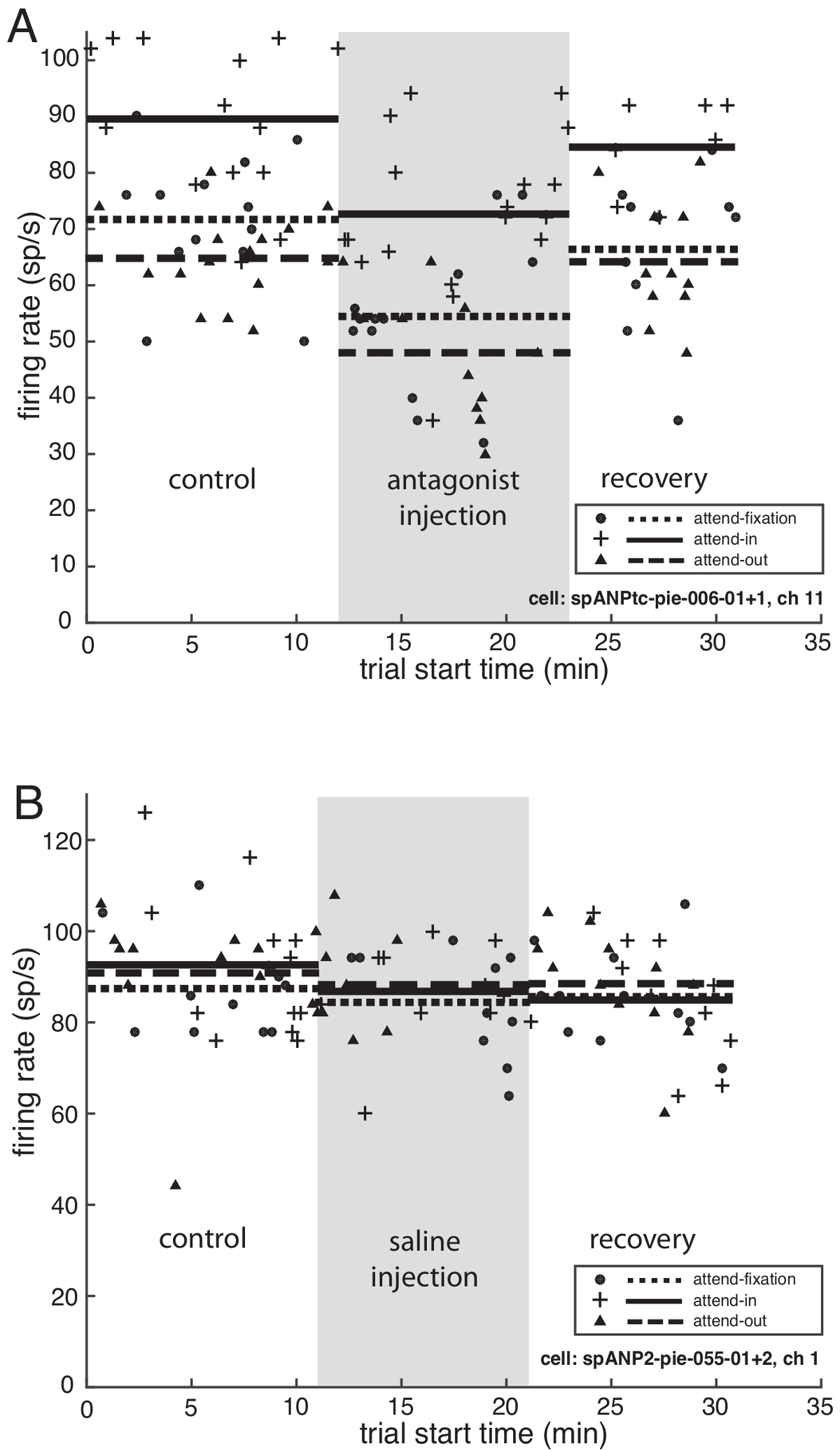
图4A示出了东莨菪碱对每三个注意力条件样品神经元的平均发射率的影响。神经元的放电频率为两个空间注意力条件(内部或外部的记录神经元的感受域的注意力),以及用于感官条件(注意在注视点)在注射块的第一注入(灰色阴影后不久下降是a)和恢复块的延迟之后增加至相同的水平在注射之前,期间。
图4B示出的控制从第二样品神经元记录在其中的盐水(0.9%NaCl)中注入,使用相同的协议作为用于东莨菪碱注入。在注射过程中阻塞,没有观察到神经元的放电频率没有变化相比于对照块。

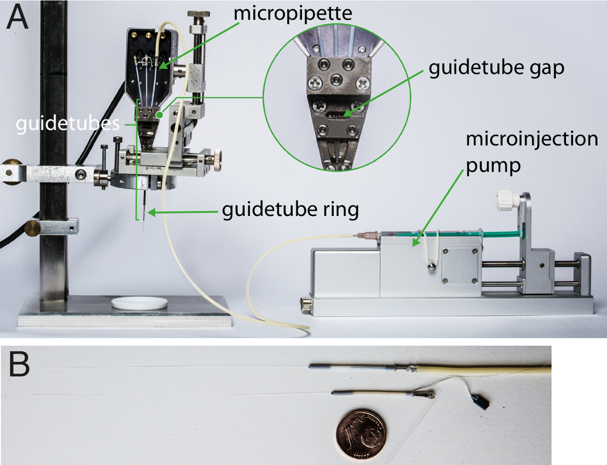
用于药理学处理,同时记录图1的设置。(A)中描述了显微注射泵,并配备有电极和微量的电生理记录系统。的guidetube间隙,在其中硅油被插入润滑电极和微量,示出放大。 (B)显示了一个例子微量(上图)和记录电极(下文)。对于大小比较,一欧分(直径:16毫米)。被置于下方点击此处查看该图的放大版本。

图2.任务设计来引导空间注意。猴子被训练来检测线索点阵图形的运动方向的变化。提示要么放置在神经元的感受野内(出席式),如该图所示,或者在它之外(出席出)。作为传感控制,猴子被训练来检测注视点的亮度变化(参加修复)。 请点击此处查看该图的放大版本。
< IMG ALT ="图3"SRC ="/文件/ ftp_upload / 53724 / 53724fig3.jpg"/>
显示为出席-状况在注射过程中块和控制模块中(所记录的神经元的感受野内的关注) 图3.射速拮抗剂东莨菪碱的影响。对样品神经元围刺激时间直方图。 x轴示出了在球杆发作和y轴之后的毫秒时间显示尖峰燃烧速率/秒。灰色区域描绘分析周期(刺激开始后300-800毫秒)用于计算试验平均射速。绿色阴影区域显示在射击在两个条件率抑制。暗绿色的颜色突出了分析时间内抑制。 请点击此处查看该图的放大版本。
尔斯/ ftp_upload / 53724 / 53724fig4.jpg"/>
图4.影响东莨菪碱和盐的射速。(A)拮抗剂东莨菪碱注射液。示出了用于对所有三个注意力条件的优选刺激从图3在实验的过程中,样品池的试验平均射速。 x轴示出了以分钟试验开始时间和y轴示出了在每秒尖峰部的燃烧速率。符号(  出席式,
出席式,  参加-修复,
  参加-OUT)表示每个成功进行试验分析周期内的神经元的放电频率和水平线条(实线:出席式,虚线:参加修复,虚线:参加-OUT)显示的平均点火率三种不同的实验BLocks(控制,注射,恢复)。灰色阴影区域示出了注入块,与在第一次注射的开始和结束的最后一次注射后1分钟。在注射过程中块2 NL 0.1摩尔东莨菪碱注射用2升/秒的注射速度每分钟。 (B)盐水注射。示出了用于对所有三个注意力条件的优选刺激物比对照实验过程中的样品电池的燃烧率。灰色阴影区域形象化盐水注射块。 请点击此处查看该图的放大版本。
讨论
在这里,我们详细说明了如何与一个"关闭的,现成的"压力喷射系统进行可靠和精确的注射和高品质的单细胞记录。而药物递送的此方法先前已在行为猴(在17中综述)使用,这里提出的系统具有的优点,下面综述。
如图4A中所示,这里描述的系统可以提供有和没有在记录现场的直接附近药理注射单一神经元活动的稳定的测量。 如图4B所示,控制物质,盐水的注射,并没有导致在燃烧率的变化。这个控制表明,注射过程本身对记录神经元的放电性能没有可测量的影响。
神经元,记录电极,以及微量的空间配置是关键在这些实验中的重要性。虽然在记录期间组织它们的相对位置的精确测量是不可能的,我们可以考虑和方差的可能来源的控制。第一,体积注射期间有一个危险,即所关注的神经元可从记录电极位移远,影响记录信号的稳定性。出于这个原因,它是谨慎前和注射块来验证信号稳定后,比较燃烧率。第二,该记录系统的引导管配置定义电极和微量之间的距离( 例如 ,305微米的同心3通道系统在该实验中使用)。由于该系统提供了精确的位置控制为在组织电极和微量的深度,它们之间的距离可以通过仔细校准相对深度的记录(步骤3.5)之前,和录音过程中保持它们在共同的深度被最小化。
ENT"> 潜在的局限性除了在内部质量控制由生产,系统需要在实验室条件下进行验证,因为不同品牌管,注射器等均可使用,并可能导致在喷射量的差异。虽然该系统可被用于注入非常小的体积如这里所示的实验中,这些是可以由于在正常实验室环境进行验证,以实际测量极限的最小体积的下方。然而,更大的注射体积可以用来推断软件定义体积和由硬件喷射的体积之间的关系。如果使用透明管,喷射过程的一个附加的视觉检查可以通过测量一个可视标记的位移。
插入微量进系统比电极插入更为苛刻,因为微量的直径稍大并且材料是更脆弱。此外,加入管到微量的销是具有挑战性,它需要打破微量的上部的高风险。然而,一个成功加载微量的寿命是数月,甚至与日常使用。
在实践中,我们还没有系统的记录后的清洗过程中所遇到的喷射系统的堵塞。尽管如此,没有"在线"检查是可能的,而且是有风险的,一个物理障碍(如在枪头组织)可能会阻止物质注入。因此它可能是可取的保守分析数据,例如仅包括在进一步的分析,显示在烧制该实验的控制和注射块之间率显著变化的那些细胞。
尽管他们的直径小,微电极和吸液管将取代脑组织,并可能导致一些局部组织损伤。这可以通过手动定位日的前端被最小化E导游管正上方的硬脑膜。电极然后穿透硬脑膜及完整性是通过在线测量它们的阻抗推断。然后,将微量被插入。当使用这种方法,定期清除组织硬脑膜以上的建议,以进一步降低电极或吸管断裂的危险。
比较另类的方法
这里使用的系统显示相比其他压力喷射系统明显的优势。一个很强的优势是微量(约100微米),这是其它的探测器17的大小的一半的直径,因此,最大限度地减少神经组织的损伤。与以前的设计中,当前系统使用空间上分离的记录电极和微量。虽然其他系统提供电极和吸管之间的较小的距离,在这里所描述的系统允许电极和移液管,从而permi独立深度变化拟合记录会话中的变量相对距离。重要的是,需要对记录质量没有妥协制成,作为喷射系统是一个既定的记录装置的一个扩展。而只有一个微量,因此一种物质在该协议中使用,它可以在一实验程序内注入几种物质。为了实现这一点,一些微量可以拧入单独的导管和连接到安装在各个喷射泵的注射器。最后,控制该系统是容易的,因为只有一个计算机程序是需要推动电极和微量,并在实验过程中进行压注。
压注入相比离子电渗疗法,有相对的优点和缺点。例如,高压喷射需要被引入到组织中比离子导入,从而增加了神经元的位移的危险性更大的卷。目前原山坳中使用的卷在NL范围内,我们很少经历了一个记录细胞的信号质量显着的变化。该系统还允许被注入更大的体积,这是行为的操作可能有用的,但可能会影响神经元记录的稳定性。注射压力超过离子导入具有明显的优势是规模较大的各种可用物质,因为没有规定使用电荷的物质。然而,pH值下应检查和实验组和对照物质之间进行比较( 例如 ,盐水)。
这个问题可能出现为什么要使用加压注入,而不是新技术的长期建立的方法,如光遗传学操纵的神经活动。虽然还有在啮齿类动物中建立的,光遗传学尚未确立可靠的恒河猴。尤其是,它还不允许选择性地对特定神经递质类型细胞的本地操纵。从长远来看,我们看到用于治疗与optogentic操作的优点药理操作的优点在阐明的认知功能的神经基础组合巨大潜力。
在这里,我们已经表明压喷射如何可用于药理学上操纵的清醒大脑局部禁区,表现恒河猴。我们希望,这种方法激励其他科学家来研究神经元活动的动态神经调节的贡献。
披露声明
作者什么都没有透露。
致谢
这项工作是由通过合作研究中心889"蜂窝机制感觉处理",以ST(项目C04)德意志研究联合会的资助。我们感谢新浪普卢默,雷奥诺拉Burchardt,德克Prüsse,克劳斯Heisig和拉尔夫Brockhausen技术和动物相关的支持和我们的合作者在德国灵长类研究中心,卡塔琳娜Debowski博士和安娜Magerhans的干细胞单元,在技术援助过滤过程。
材料
| Name | Company | Catalog Number | Comments |
| (-)-Scopolamine hydrochloride | Sigma-Aldrich | 55-16-3 | Mr 339.81 g/mol |
| NaCl 0.9% | B. Braun Melsungen AG | 3079870 | 5ml |
| Terg-a-zyme | Sigma-Aldrich | Z273287 | enzyme detergen |
| Hydrogen peroxide | Roth | Used in 3% solution with deionized water | |
| Ethanol | Chemie-Vertieb Hannover | 104642 | 70% |
| Deionized water | |||
| Injekt 40 Duo | B. Braun Melsungen AG | 9166432V | Syringe and needle |
| Eppendorf Safe-Lock microcentrifuge tubes, amber | Eppendorf | 0030 120.191 | 1,5ml |
| Quarzglass micropipette | Thomas Recording | ||
| Recording electrode | Thomas Recording | quartz/platinum-tungsten fiber electrode; impedance value 1-2 MΩ and 0.3-0.5 MΩ | |
| PharmedBPT-Schlauch | Saint-Gobain Performance Plastics | 3702003 | Size: 0,25 x 2,05 mm (Wd: 0,9mm) |
| Loctite 401 | Henkel | 233641 | Superglue |
| Silicon oil | Thomas Recording | M-1000 | |
| Minisart RC15 | Sartorius | 17761----------R | Syringe filter |
| Multichannel Micro Injection System | Thomas Recording | multichannel microelectrode manipulator “System Eckhorn” equipped with microelectrodes and micropipettes and a precision multichannel microinjection pump | |
| McLab | custom | internal lab software to control stimulus presentation |
参考文献
- Noudoost, B., Moore, T. The role of neuromodulators in selective attention. Trends Cogn Sci. 15 (12), 585-591 (2011).
- Jochems, A., Reboreda, A., Hasselmo, M., Yoshida, M. Cholinergic receptor activation supports persistent firing in layer III neurons in the medial entorhinal cortex. Behav Brain Res. 254, 108-115 (2013).
- Thiele, A., Herrero, J. L., Distler, C., Hoffmann, K. P. Contribution of cholinergic and GABAergic mechanisms to direction tuning, discriminability, response reliability, and neuronal rate correlations in macaque middle temporal area. J Neurosci. 32 (47), 16602-16615 (2012).
- Thienel, R., et al. Muscarinic antagonist effects on executive control of attention. Int J Neuropsychopharmacol. 12 (10), 1307-1317 (2009).
- Anthony, B. L., Dennison, R. L., Aronstam, R. S. Disruption of muscarinic receptor-G protein coupling is a general property of liquid volatile anesthetics. Neurosci Lett. 99 (1-2), 191-196 (1989).
- Yamakura, T., Bertaccini, E., Trudell, J. R., Harris, R. A. Anesthetics and ion channels: molecular models and sites of action. Annu Rev Pharmacol Toxicol. 41, 23-51 (2001).
- Herr, N. R., Wightman, R. M. Improved techniques for examining rapid dopamine signaling with iontophoresis. Front Biosci. 5, 249-257 (2013).
- Bevan, P., Bradshaw, C. M., Pun, R. Y., Slater, N. T., Szabadi, E. The relative contribution of iontophoresis and electro-osmosis to the electrophoretic release of noradrenaline from multi barrelled micropipettes [proceedings]. Br J Pharmacol. 67 (3), 478-479 (1979).
- Herr, N. R., Kile, B. M., Carelli, R. M., Wightman, R. M. Electroosmotic flow and its contribution to iontophoretic delivery. Anal Chem. 80, 8635-8641 (2008).
- Thiele, A., Delicato, L. S., Roberts, M. J., Gieselmann, M. A. A novel electrode-pipette design for simultaneous recording of extracellular spikes and iontophoretic drug application in awake behaving monkeys. J Neurosci Meth. 158 (2-4), 207-211 (2006).
- Lalley, P. M., Johansson, H., Windhorst, U. . Microiontophoresis and Pressure Ejection: Modern Techniques in Neuroscience. , 193-209 (1999).
- Malpeli, J. G., Schiller, P. H. A method of reversible inactivation of small regions of brain tissue. J Neurosci Meth. 1 (2), 145-159 (1979).
- Malpeli, J. G. Reversible inactivation of subcortical sites by drug injection. J Neurosci Meth. 86 (2), 119-128 (1999).
- Dias, E. C., Segraves, M. A. A pressure system for the microinjection of substances into the brain of awake monkeys. J Neurosci Meth. 72 (1), 43-47 (1997).
- Szente, M. B., Baranyi, A., Woody, C. D. Effects of protein kinase C inhibitor H-7on membrane properties and synaptic responses of neocortical neurons of awake cats. Brain Res. 506 (2), 281-286 (1990).
- Woody, C. D., Bartfai, T., Gruen, E., Nairn, A. lntracellular injection of cGMP-dependent protein kinase results in increased input resistance in neurons of the mammalian motor cortex. Brain Res. 386 (1-2), 379-385 (1986).
- Noudoost, B., Moore, T. A reliable microinjectrode system for use in behaving monkeys. J Neurosci Meth. 194 (2), 218-223 (2011).
- Association of Primate Veterinarians. . Cranial Implant Care Guidelines for Nonhuman Primates in Biomedical Research. , (2015).
- Treue, S., Martinez-Trujillo, J. C. Feature-based attention influences motion processing gain in macaque visual cortex. Nature. 399, 575-579 (1999).
- Martinez-Trujillo, J. C., Treue, S. Feature-based attention increases the selectivity of population responses in primate visual cortex. Curr Biol. 14 (9), 744-751 (2004).
转载和许可
请求许可使用此 JoVE 文章的文本或图形
请求许可探索更多文章
This article has been published
Video Coming Soon
版权所属 © 2025 MyJoVE 公司版权所有,本公司不涉及任何医疗业务和医疗服务。