
X 射线衍射分析晶体生长
Overview
资料来源: 实验室的博士吉米 · 佛朗哥-梅里马克大学
X 射线晶体学是一种方法通常用于确定中结晶固体,允许一个分子或复杂的三维形状测定的原子的空间排列。确定一种化合物的三维结构是特别重要,因为一种化合物的结构和功能密切相关。一种化合物的结构信息通常用于解释其行为或反应性。这是一个最有用的技术,解决的一种化合物的三维结构或复杂的和在某些情况下它可能确定结构的唯一可行方法。X 射线质量晶体生长是 x 射线晶体学的关键组成部分。大小和晶体质量的往往是化合物的高度依赖宗正由 x 射线晶体学的组成。通常含有重原子的化合物产生了更大的衍射图案,因此需要较小的晶体。一般来说,单晶具有定义良好的面孔是最优的和通常的有机化合物,晶体需要将大于那些含重原子。没有可行的水晶,x 射线晶体学是不可行的。一些分子是天生就比其他人更结晶,因而难以获得 x 射线质量晶体可以不同化合物。X 射线晶体的生长过程是结晶的类似的的常用的纯化化合物,但重点放在生产高质量晶体。通常情况下,可以通过允许结晶过程比较缓慢,可能发生在一天或几个月的课程获得更高质量的晶体。
Procedure
1.编制的水晶管和筛选器
- 在锥形瓶核磁共振管的地方。
- 准备移液器筛选器。
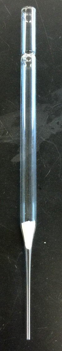
- 构建筛选器通过在移液管,放置一块无绒布擦拭 (由 1 英寸 1 英寸),然后使用杆牢固地楔入 (图 1) 移液管的瓶颈部分的擦拭。
- 使两个吸管筛选器需要每个水晶管。
2.将样品加入水晶管
- 在 0.75 毫升溶剂 (二氯甲烷) 的溶解的化合物 (四苯基卟啉、 10 毫克)。
- 使用吸管,轻轻混合的顶部添加的管,通过筛选器。
- 粒子被过滤掉,以避免会导致小的而不是所需的较大单晶的多个晶体的形核网站创建。
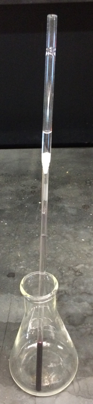
- 一旦样品已放在水晶管,慢慢地,轻轻地,将反溶剂 (1.5 毫升的甲醇) 添加到通过新的筛选器移液管管。允许的反溶剂慢慢层上以前添加的解决方案 (图 2)。请不要使用灯泡推通过吸管溶剂,反而允许溶剂流过该筛选器本身。
- 请确保首先加
Results
Application and Summary
References
- Gilman, J. J., The art and science of growing crystals. Wiley: (1963).
- Orvig, C., A simple method to perform a liquid diffusion crystallization. Journal of Chemical Education 62 (1), 84 (1985).
- Brown, C. S.; Lee, M. S.; Leung, D. W.; Wang, T.; Xu, W.; Luthra, P. et. al. In silico derived small molecules bind the filovirus VP35 protein and inhibit its polymerase co-factor activity. Journal of molecular biology, 426 (10), 2045-2058 (2014)
- Batt, S. M.; Jabeen, T.; Bhowruth, V.; Quill, L.; Lund, P. A.; Eggeling et. al. Structural basis of inhibition of Mycobacterium tuberculosis DprE1 by benzothiazinone inhibitors. Proceedings of the National Academy of Sciences of the United States of America,109 (28), 11354-9 (2012)
- Mortensen, D. S.; Perrin-Ninkovic, S. M.; Shevlin, G.; Elsner, J.; Zhao, J.; Whitefield et al. Optimization of a Series of Triazole Containing Mammalian Target of Rapamycin (mTOR) Kinase Inhibitors and the Discovery of CC-115. Journal of Medicinal Chemistry 58 (14), 5599-5608 (2015)
- Nguyen, T.; Sutton, A. D.; Brynda, M.; Fettinger, J. C.; Long, G. J.; Power, P. P., Synthesis of a Stable Compound with Fivefold Bonding Between Two Chromium(I) Centers. Science310 (5749), 844-847 (2005).
- Chen, K.; Nenzel, M. M.; Brown, T. M.; Catalano, V. J., Luminescent Mechanochromism in a Gold(I)-Copper(I) N-Heterocyclic Carbene Complex. Inorganic Chemistry 54 (14), 6900-6909.(2015).
- Franco, J. U.; Hammons, J. C.; Rios, D.; Olmstead, M. M., New Tetraazaannulene Hosts for Fullerenes. Inorganic Chemistry49 (11), 5120-5125 (2010).
跳至...
此集合中的视频:

Now Playing
X 射线衍射分析晶体生长
Organic Chemistry
32.4K Views

催化导论
Organic Chemistry
34.5K Views

程序集的激烈化学反应回流系统
Organic Chemistry
167.9K Views

进行室温下反应
Organic Chemistry
70.7K Views

溅镀转移的溶剂
Organic Chemistry
41.7K Views

随着冻融泵循环脱气液体
Organic Chemistry
56.3K Views

制备无水试剂和设备
Organic Chemistry
79.4K Views

重结晶法净化化合物
Organic Chemistry
709.4K Views

通过沉淀混合物的分离
Organic Chemistry
157.8K Views

固-液萃取
Organic Chemistry
238.0K Views

旋转蒸发来去除溶剂
Organic Chemistry
212.9K Views

分馏
Organic Chemistry
334.7K Views

Performing 1D Thin Layer Chromatography
Organic Chemistry
289.9K Views

柱层析法
Organic Chemistry
360.5K Views

核磁共振 (NMR)
Organic Chemistry
248.4K Views
版权所属 © 2025 MyJoVE 公司版权所有,本公司不涉及任何医疗业务和医疗服务。