Untersuchung der Atemwege I: Perkussion und Auskultation
Überblick
Quelle: Suneel Dhand, MD, Teilnahme an Arzt, Innere Medizin, Beth Israel Deaconess Medical Center
Lernen die richtige Technik für Perkussion und Auskultation des respiratorischen Systems ist von entscheidender Bedeutung und kommt mit Praxis auf realen Patienten. Percussion ist eine nützliche Fähigkeit, die im klinischen Alltag oft übersprungen wird, aber wenn richtig durchgeführt, kann es helfen, den Arzt, die zugrunde liegende Pathologie der Lunge zu identifizieren. Auskultation bieten eine fast sofortige Diagnose für eine Reihe von akuten pulmonalen Bedingungen, einschließlich chronisch obstruktiver Lungenerkrankung (COPD), Asthma, Lungenentzündung und Pneumothorax.
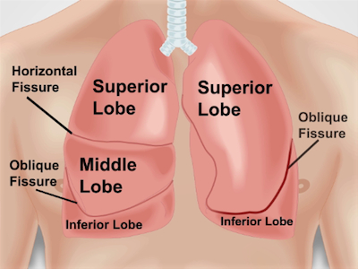
Die Bereiche für die Lunge auskultieren entsprechen die Lunge Zonen. Jeder Lungenflügel kann unterhalb der Brustwand während Perkussion und Auskultation ()Abbildung 1im Bild). Die rechte Lunge hat drei Lappen: die überlegene, Mittel- und minderwertigeren Lappen. Die linke Lunge hat zwei Lappen: die oberen und unteren Lappen. Der überlegene Lappen der linken Lunge hat auch eine separate Projektion, bekannt als die Lingual.

Abbildung 1: Anatomie der Lunge in Bezug auf die Brustwand. Eine ungefähre Projektion der Lungen und deren Lappen auf die Brust und Risse Wand Körpertiefe. RUL - rechten oberen Lappen; RML - rechten mittleren Lappen; RLL - rechten unteren Lappen; LUL - linken Oberlappens; LLL - Unterlappen links.
Verfahren
1. Positionierung
- Stellen Sie sicher, dass der Patient bis auf die Taille entkleidet ist.
- Der Patient auf dem Untersuchungstisch in einem 30 bis 45 Grad Winkel und Ansatz von der rechten Seite zu positionieren. Prüfung den hinteren der Lunge benötigt der Patient nach vorne lehnen oder sitzt auf dem Rand des Bettes.
(2) percussion
- Percuss beide nach hinten und vorn, beginnend auf der Rückseite.
- Geben Sie nicht-dominanten Hand mit Mittelfinger (
Anwendung und Zusammenfassung
Perkussion und Auskultation sollte immer nacheinander durchgeführt werden wenn eine volle Atemwege Untersuchung durchführen. Lernen, wie man richtig percuss braucht Zeit und Übung (Praxis kann über sich selbst oder andere Oberflächen, z. B. eine Tabelle durchgeführt werden). Beachten Sie, wie die Perkussion Hinweis ändert sich natürlich über luftgefüllte Lunge, Rippen und solide Organe wie das Herz.
Auskultation muss über jede Lunge Zone geben dem Arzt die beste Chance zur Identifiz...
pringen zu...
Videos aus dieser Sammlung:

Now Playing
Untersuchung der Atemwege I: Perkussion und Auskultation
Physical Examinations I
212.6K Ansichten

Allgemeines Vorgehen: Die körperliche Untersuchung
Physical Examinations I
116.6K Ansichten

Beobachtung und Inspektion
Physical Examinations I
93.9K Ansichten

Abtasten
Physical Examinations I
83.4K Ansichten

Perkussion
Physical Examinations I
100.5K Ansichten

Auskultation
Physical Examinations I
61.1K Ansichten

Richtiges Anpassen der Patientenkleidung während der körperlichen Untersuchung
Physical Examinations I
83.3K Ansichten

Messung des Blutdrucks
Physical Examinations I
107.8K Ansichten

Messung der Vitalzeichen
Physical Examinations I
114.3K Ansichten

Untersuchung der Atemwege I: Inspektion und Palpation
Physical Examinations I
157.0K Ansichten

Kardiologische Untersuchung I: Inspektion und Palpation
Physical Examinations I
176.2K Ansichten

Kardiologische Untersuchung II: Auskultation
Physical Examinations I
139.7K Ansichten

Kardiologische Untersuchung III: Abnormale Herztöne
Physical Examinations I
91.7K Ansichten

Periphere vaskuläre Untersuchung
Physical Examinations I
68.2K Ansichten

Periphere Gefäßuntersuchung mit der Continuous-Wave (CW-) Dopplersonographie
Physical Examinations I
38.5K Ansichten
Copyright © 2025 MyJoVE Corporation. Alle Rechte vorbehalten