
Examen de los oídos
Visión general
Fuente: Richard Glickman-Simon, MD, profesor asistente, Departamento de salud pública y medicina comunitaria, Tufts University School of Medicine, MA
Este video describe el examen de la oreja, comenzando con una revisión de su anatomía de superficie y el interior (figura 1). La aurícula cartilaginosa consta de la helix, antihelix, lóbulo de la oreja y trago. La apófisis mastoides se coloca justo detrás de la oreja. El ligeramente curvado auditivo canal termina en la membrana timpánica, que transmite las ondas sonoras recogidas por el oído externo al oído medio lleno de aire. La trompa de Eustaquio conecta el oído medio con la nasofaringe. Las vibraciones de la membrana timpánica se transmiten a los tres conectados osículos del oído medio (martillo, yunque y estribo). Las vibraciones se transforma en señales eléctricas en el oído interno y después llevadas al cerebro por el nervio coclear. Audiencia, por lo tanto, comprende una fase conductora que implique el externo y el oído medio y una fase sensorineural que consiste en el oído interno y nervio coclear.
El canal auditivo y la membrana timpánica se examinan con el otoscopio, un instrumento portátil con una fuente de luz, una lupa y un espéculo en forma de cono. Es importante estar familiarizado con las señales de la membrana timpánica (figura 2). Normalmente se observan sólo dos de los tres huesecillos - malleus y incus; el martillo está cerca del centro, y el uncus es sólo posterior. Se aprecia un cono de luz emanando hacia abajo y hacia delante desde el punto de contacto entre la membrana y la punta del martillo o umbo. El proceso corto aproximadamente demarca el límite entre las dos regiones de la membrana timpánica: la pars flaccida, mentira superior y posterior y el más grande pars tensa, mentira anterior e inferior. Normalmente, la membrana timpánica es color de rosa-gris en color y fácilmente refleja la luz del otoscopio.
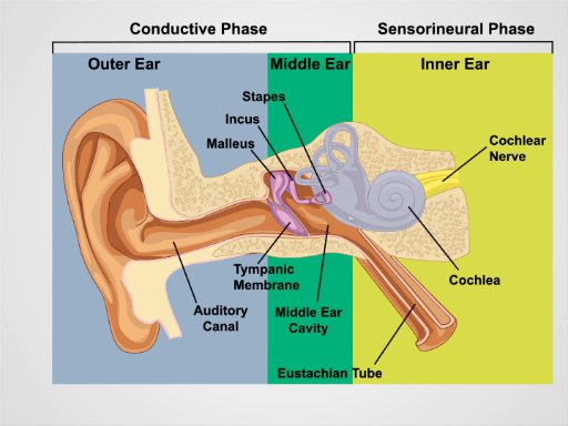
Figura 1. Anatomía del oído. Un dibujo esquemático del oído humano en sección frontal con externo, medio y las estructuras del oído interno con la etiqueta.
Procedimiento
1. examen y audición del oído
- Inspeccione las aurículas y el tejido circundante para cambios en la piel, nódulos y deformidades.
- Sujete la hélice superior entre los dedos pulgar e índice uno en un momento y tire suavemente hacia arriba y hacia atrás para verificar la molestia en el oído externo.
- Palpe el trago y la apófisis mastoides de ternura.
- Realizar el test de voz susurrada de agudeza auditiva.
- Asegúrese de que la habitación es tranquila.
- Soporte 2 pies
Aplicación y resumen
Evaluación adecuada del oído requiere una verificación de la audiencia y el examen otoscópico. Pérdida de oído conductor resulta de trastornos del oído medio y externo. Impactación de cerumen, otitis externa, trauma, cuerpos extraños y (menos comúnmente) Exostosis pueden conducir a pérdida de la audición por obstruir el canal auditivo. Oído medio causas de pérdida de la audición incluyen otitis media, disfunción de la trompa de Eustaquio, barotrauma y otosclerosis. Pérdida auditiva neurosensorial es debi...
Saltar a...
Vídeos de esta colección:

Now Playing
Examen de los oídos
Physical Examinations II
55.4K Vistas

Examen de la vista
Physical Examinations II
77.4K Vistas

Oftalmoscopía
Physical Examinations II
68.2K Vistas

Exploración de la nariz, senos paranasales, cavidad oral y faringe
Physical Examinations II
65.9K Vistas

Exploración de la tiroides
Physical Examinations II
105.3K Vistas

Exploración de ganglios linfáticos
Physical Examinations II
388.3K Vistas

Exploración abdominal I: Inspección y auscultación
Physical Examinations II
203.0K Vistas

Exploración abdominal II: Percusión
Physical Examinations II
248.5K Vistas

Exploración abdominal III: Palpación
Physical Examinations II
138.6K Vistas

Exploración abdominal IV: Evaluación de dolor abdominal agudo
Physical Examinations II
67.4K Vistas

Tacto rectal masculino
Physical Examinations II
114.8K Vistas

Exploración mamaria
Physical Examinations II
87.9K Vistas

Exploración pélvica I: Evaluación de los genitales externos
Physical Examinations II
308.4K Vistas

Exploración pélvica II: Examen con espéculo
Physical Examinations II
150.7K Vistas

Exploración pélvica III: Bimanual y examen rectovaginal
Physical Examinations II
148.1K Vistas
ACERCA DE JoVE
Copyright © 2025 MyJoVE Corporation. Todos los derechos reservados