
Examen des oreilles
Vue d'ensemble
Source : Richard Glickman-Simon, MD, professeur adjoint, département de santé publique et médecine sociale, Tufts University School of Medicine, MA
Cette vidéo décrit l’examen de l’oreille, en commençant par un examen de son anatomie de surface et l’intérieur (Figure 1). Le pavillon cartilagineux se compose de l’hélice, antihelix, lobe de l’oreille et tragus. L’apophyse mastoïde est placé juste derrière le lobe de l’oreille. L’auditif légèrement incurvée du canal se termine à la membrane tympanique, qui émet des ondes sonores perçus par l’oreille externe de l’oreille moyenne remplis d’air. La trompe d’Eustache se connecte à l’oreille moyenne avec le nasopharynx. Vibrations de la membrane tympanique transmettent les trois osselets connectés de l’oreille moyenne (le marteau, enclume et étrier). Les vibrations sont transformées en signaux électriques dans l’oreille interne et ensuite transportées au cerveau par le nerf cochléaire. Audition, par conséquent, comprend une phase conductrice qui implique l’externe et l’oreille moyenne et une phase neuro-sensorielle qui implique l’oreille interne et le nerf cochléaire.
Le conduit auditif et le tympan sont examinés avec l’otoscope, un instrument portatif avec une source lumineuse, une loupe et un spéculum à usage unique en forme de cône. Il est important de se familiariser avec les repères de la membrane tympanique (Figure 2). Seulement deux des trois osselets : le marteau et enclume - peut normalement être vu ; le marteau est près du centre, et l’uncus est juste postérieure. Un cône de lumière peut être vu émanant l’umbo, ou point de contact entre la membrane et la pointe du marteau vers le bas et vers l’avant. Le processus abrégé environ délimite la frontière entre les deux régions de la membrane tympanique : la pars flaccida, couché supérieure et postérieure et la beaucoup plus grande pars tensa, située à antérieure et inférieure. Normalement, la membrane tympanique est rose-gris en couleurs et facilement reflète la lumière de l’otoscope.
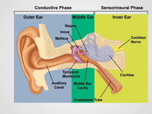
Figure 1. Anatomie de l’oreille. Un dessin schématique de l’oreille humaine en section frontale avec externe, intermédiaire et structures de l’oreille interne marqués.
Procédure
1. examen et audition de l’oreille
- Inspecter les oreillettes et les tissus environnants pour les modifications de la peau, des nodules et des déformations.
- Saisir l’hélice supérieurement entre le pouce et l’index l’un à la fois et tirer doucement vers le haut et vers l’arrière pour vérifier les malaises n’importe où dans l’oreille externe.
- Palper le tragus et l’apophyse mastoïde de tendresse.
- Effectuez le test de la voix chuchotée d’acuité auditive.
- S’assurer que la cha
Applications et Résumé
Une évaluation adéquate de l’oreille nécessite une vérification de l’audition et l’examen otoscopique. Perte auditive de conduction résulte de troubles de l’externe et l’oreille moyenne. Impaction de cérumen, otite externe, traumatisme, corps étrangers et (moins souvent) exostoses peuvent conduire à une perte auditive en obstruant le conduit auditif. Oreille moyenne causes de perte auditive comprennent une otite, dysfonctionnement de la trompe d’Eustache, barotraumatisme et otospongiose. Perte auditiv...
Passer à...
Vidéos de cette collection:

Now Playing
Examen des oreilles
Physical Examinations II
55.1K Vues

Examen des yeux
Physical Examinations II
77.1K Vues

Examen ophtalmologique
Physical Examinations II
67.9K Vues

Examen du nez, des sinus, de la cavité orale et du pharynx
Physical Examinations II
65.7K Vues

Examen de la thyroïde
Physical Examinations II
105.0K Vues

Examen des ganglions lymphatiques
Physical Examinations II
387.3K Vues

Examen abdominal I: Inspection et auscultation
Physical Examinations II
202.6K Vues

Examen abdominal II: Percussion
Physical Examinations II
248.2K Vues

Examen abdominal III: Palpation
Physical Examinations II
138.5K Vues

Examen abdominal IV: Évaluation de la douleur abdominale aiguë
Physical Examinations II
67.3K Vues

Toucher rectal chez l'homme
Physical Examinations II
114.4K Vues

Examen général des seins
Physical Examinations II
87.6K Vues

Examen pelvien I: Évaluation des organes génitaux externes
Physical Examinations II
306.9K Vues

Examen pelvien: Examen au spéculum
Physical Examinations II
150.4K Vues

Examen pelvien III: Examen bi-manuel et toucher rectal
Physical Examinations II
147.7K Vues
À PROPOS DE JoVE
Copyright © 2025 MyJoVE Corporation. Tous droits réservés.