Exame de orelha
Visão Geral
Fonte: Richard Glickman-Simon, MD, Professor Assistente, Departamento de Saúde Pública e Medicina Comunitária, Tufts University School of Medicine, MA
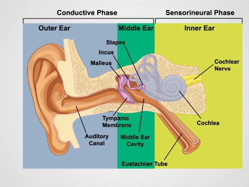
Este vídeo descreve o exame do ouvido, começando com uma revisão de sua superfície e anatomia interior (Figura 1). O auricle cartilaginoso consiste na hélice, antihlix, lóbulo da orelha e trago. O processo mastoide está posicionado logo atrás do lóbulo da orelha. O canal auditivo ligeiramente curvo termina na membrana timpânica, que transmite ondas sonoras coletadas pelo ouvido externo para o ouvido médio cheio de ar. O tubo eustáquio conecta-se ao ouvido médio com a nasofaringe. Vibrações da membrana timpânica transmitem aos três ossículos conectados da orelha média (o malleus, incus e stapes). As vibrações são transformadas em sinais elétricos no ouvido interno, e depois levadas para o cérebro pelo nervo coclear. A audição, portanto, compreende uma fase condutiva que envolve o ouvido externo e médio, e uma fase sensorial que envolve o ouvido interno e o nervo coclear.
O canal auditivo e a membrana timpânica são examinados com o otoscópio, um instrumento portátil com uma fonte de luz, uma lupa e um espéculo descartável em forma de cone. É importante estar familiarizado com os marcos da membrana timpânica(Figura 2). Apenas dois dos três ossículos - o malleus e o incus - normalmente podem ser vistos; o malleus está perto do centro, e o uncus é apenas posterior. Um cone de luz pode ser visto emanando para baixo e anteriormente a partir do umbo, ou ponto de contato entre a membrana e a ponta do malleus. O processo curto demarca aproximadamente a fronteira entre as duas regiões da membrana timpânica: o pars flaccida, deitado superior e posterior, e os pars muito maiores, deitadosanteriores e inferiores. Normalmente, a membrana timpânica é cor-de-rosa-cinza e reflete prontamente a luz do otoscópio.

Figura 1. Anatomia da Orelha. Um desenho esquemático do ouvido humano na seção frontal com estruturas externas, médias e internas do ouvido rotuladas.
Procedimento
1. Exame de Ouvido e Audição
- Inspecione as aurículos e o tecido circundante em busca de alterações de pele, nódulos e deformidades.
- Segure a hélice superiormente entre o polegar e o indicador um de cada vez e puxe suavemente para cima e para trás para verificar se há desconforto em qualquer lugar do ouvido externo.
- Palpa o processo de tragus e mastoide para ternura.
- Realize o teste de voz sussurrado para acuidade auditiva.
- Certifique-se de que o quarto está quieto. <
Aplicação e Resumo
A avaliação adequada do ouvido requer uma verificação auditiva e exame otoscópico. A perda auditiva condutiva resulta de distúrbios do ouvido externo e médio. A impacto cerumen, otite externa, trauma, corpos estranhos e (menos comumente) exostoses podem levar à perda auditiva, obstruindo o canal auditivo. As causas da perda auditiva incluem otite media, disfunção do tubo eustáquio, barotrauma e otosclerose. A perda auditiva neurosensorial é devido a distúrbios do ouvido interno. Presbycusis e traumas sonolic...
Pular para...
Vídeos desta coleção:

Now Playing
Exame de orelha
Physical Examinations II
55.1K Visualizações

Exame ocular
Physical Examinations II
77.1K Visualizações

Exame oftalmoscópico
Physical Examinations II
67.9K Visualizações

Exame de Nariz, Seios da face, Cavidade Oral e Faringe
Physical Examinations II
65.7K Visualizações

Exame de Tireóide
Physical Examinations II
105.0K Visualizações

Exame do Linfonodo
Physical Examinations II
387.3K Visualizações

Exame Abdominal I: Inspeção e Auscultação
Physical Examinations II
202.6K Visualizações

Exame Abdominal II: Percussão
Physical Examinations II
248.2K Visualizações

Exame Abdominal III: Palpação
Physical Examinations II
138.5K Visualizações

Exame Abdominal IV: Avaliação da Dor Abdominal Aguda
Physical Examinations II
67.3K Visualizações

Exame Retal Masculino
Physical Examinations II
114.4K Visualizações

Exame Abrangente das Mamas
Physical Examinations II
87.6K Visualizações

Exame Pélvico I: Avaliação da Genitália Externa
Physical Examinations II
306.9K Visualizações

Exame Pélvico II: Exame Especular
Physical Examinations II
150.4K Visualizações

Exame Pélvico III: Exame Bimanual e Retovaginal
Physical Examinations II
147.7K Visualizações
Copyright © 2025 MyJoVE Corporation. Todos os direitos reservados