
Fluorescencia de rayos x (XRF)
Visión general
Fuente: Laboratorio del Dr. Lydia Finney, laboratorio nacional de Argonne
Fluorescencia de rayos x es una radiación inducida, emitida que puede utilizarse para generar la información espectroscópica. Microscopía de fluorescencia de rayos x es una técnica no destructiva que utiliza la emisión de fluorescencia inducida de metales para identificar y cuantificar su distribución espacial.
Procedimiento
1. preparación de las ventanas de nitruro de silicio
- Use pinzas atrás para recoger una ventana (windows se rompen si cayó del nitruro de silicio).
- Coloque la ventana en un portaobjetos de vidrio, cara plana hacia arriba.
- Se adhieren pequeños trozos de cinta adhesiva a los lados de la ventana y utilizar éstos para cumplir las ventanas en la parte inferior de la placa de cultivo.
- Esterilizar las ventanas en los platos de la cultura con la radiación UV. Esto se puede lograr con el a
Resultados
El mapa de fluorescencia de rayos x de una célula adherente se muestra en la figura 1. Cada panel muestra la distribución de un elemento determinado (por ejemplo, cobre, hierro, zinc, etcetera) en la célula. El panel con la etiqueta 's_a' muestra la absorción de rayos x.
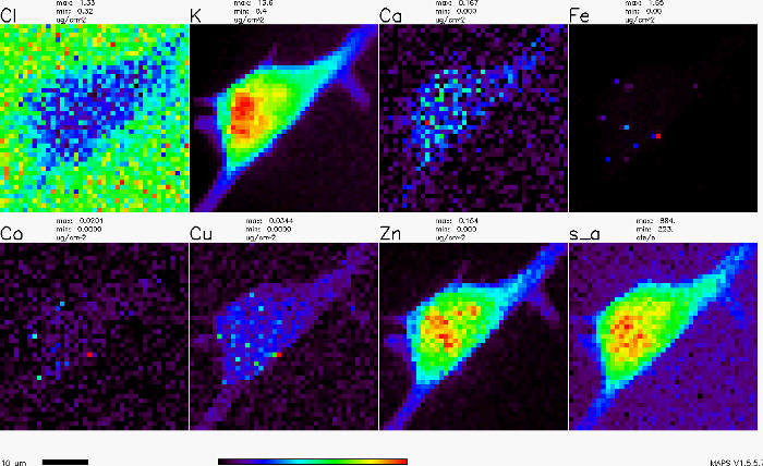
Figura 1. Mapa de fluorescencia de rayos x de una célula adhere...
Aplicación y resumen
Imágenes de fluorescencia de rayos x pueden ser una herramienta útil en muchos campos, incluyendo ciencia forense, ciencia de materiales, Geociencias, biología y aún en el estudio de nuestro patrimonio cultural. En ciencia de materiales, puede ayudar a encontrar defectos en chips y catalizadores con metales. En la obra de patrimonio cultural, se ha utilizado para identificar los metales tóxicos en los cabellos de las personas famosas (por ejemplo, Beethoven) y a identificar la fuente de pinturas utilizadas en el art...
Tags
Saltar a...
Vídeos de esta colección:

Now Playing
Fluorescencia de rayos x (XRF)
Analytical Chemistry
25.9K Vistas

Preparación de muestras para la caracterización analítica
Analytical Chemistry
85.2K Vistas

Estándares internos
Analytical Chemistry
205.3K Vistas

Método de adición estándar
Analytical Chemistry
320.7K Vistas

Curvas de calibración
Analytical Chemistry
798.5K Vistas

Espectroscopía ultravioleta-visible (UV-Vis)
Analytical Chemistry
625.1K Vistas

Espectroscopía de Raman para el análisis químico
Analytical Chemistry
51.4K Vistas

Cromatografía de gases (CG) con detección de ionización de llama
Analytical Chemistry
283.0K Vistas

Cromatografía de líquidos de alto rendimiento (HPLC)
Analytical Chemistry
385.8K Vistas

Cromatografía de intercambio iónico
Analytical Chemistry
265.0K Vistas

Electroforesis capilar (EC)
Analytical Chemistry
94.4K Vistas

Introducción a la espectrometría de masas
Analytical Chemistry
112.8K Vistas

Microscopía electrónica de barrido (MEB)
Analytical Chemistry
87.6K Vistas

Mediciones electroquímicas de catalizadores soportados utilizando un potenciostato/galvanostato
Analytical Chemistry
51.8K Vistas

Voltametría cíclica (CV)
Analytical Chemistry
125.9K Vistas
ACERCA DE JoVE
Copyright © 2025 MyJoVE Corporation. Todos los derechos reservados