
Single-Molecule Imaging of Lateral Mobility and Ion Channel Activity in Lipid Bilayers using Total Internal Reflection Fluorescence (TIRF) Microscopy
February 17th, 2023
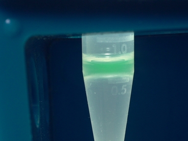
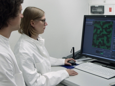
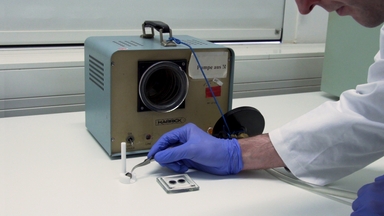
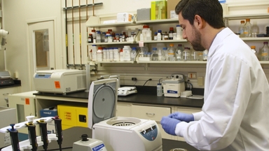
•This protocol describes how to use TIRF microscopy to track individual ion channels and determine their activity in supported lipid membranes, thereby defining the interplay between lateral membrane movement and channel function. It describes how to prepare the membranes, record the data, and analyze the results.
Videos relacionados
Detergent-free Ultrafast Reconstitution of Membrane Proteins into Lipid Bilayers Using Fusogenic Complementary-charged Proteoliposomes.
In Vitro Reconstitution of Self-Organizing Protein Patterns on Supported Lipid Bilayers
Dissipative Microgravimetry to Study the Binding Dynamics of the Phospholipid Binding Protein Annexin A2 to Solid-supported Lipid Bilayers Using a Quartz Resonator
Single-Molecule Tracking Microscopy - A Tool for Determining the Diffusive States of Cytosolic Molecules
Production of Dynein and Kinesin Motor Ensembles on DNA Origami Nanostructures for Single Molecule Observation
High-Throughput Total Internal Reflection Fluorescence and Direct Stochastic Optical Reconstruction Microscopy Using a Photonic Chip
Conventional BODIPY Conjugates for Live-Cell Super-Resolution Microscopy and Single-Molecule Tracking

An In Vitro Single-Molecule Imaging Assay for the Analysis of Cap-Dependent Translation Kinetics
Single Cell Analysis Of Transcriptionally Active Alleles By Single Molecule FISH
Single Molecule Fluorescence Energy Transfer Study of Ribosome Protein Synthesis
ACERCA DE JoVE
Copyright © 2024 MyJoVE Corporation. Todos los derechos reservados