Method Article
4D הדמיה של צבירת חלבון בתאים חיים
In This Article
Summary
כדאיות ניידת תלויה בניהול הזמן והיעיל של misfolding חלבון. כאן אנו מתארים שיטה להמחשת הגורל הפוטנציאלי השונה של חלבון misfolded: קפל שוב, שפלה, או תפיסה בתכלילים. אנו מדגימים את השימוש בחיישן מתקפל, Ubc9 Ts, לproteostasis ניטור ובקרת איכות צבירה בתאים חיים באמצעות מיקרוסקופ 4D.
Abstract
One of the key tasks of any living cell is maintaining the proper folding of newly synthesized proteins in the face of ever-changing environmental conditions and an intracellular environment that is tightly packed, sticky, and hazardous to protein stability1. The ability to dynamically balance protein production, folding and degradation demands highly-specialized quality control machinery, whose absolute necessity is observed best when it malfunctions. Diseases such as ALS, Alzheimer's, Parkinson's, and certain forms of Cystic Fibrosis have a direct link to protein folding quality control components2, and therefore future therapeutic development requires a basic understanding of underlying processes. Our experimental challenge is to understand how cells integrate damage signals and mount responses that are tailored to diverse circumstances.
The primary reason why protein misfolding represents an existential threat to the cell is the propensity of incorrectly folded proteins to aggregate, thus causing a global perturbation of the crowded and delicate intracellular folding environment1. The folding health, or "proteostasis," of the cellular proteome is maintained, even under the duress of aging, stress and oxidative damage, by the coordinated action of different mechanistic units in an elaborate quality control system3,4. A specialized machinery of molecular chaperones can bind non-native polypeptides and promote their folding into the native state1, target them for degradation by the ubiquitin-proteasome system5, or direct them to protective aggregation inclusions6-9.
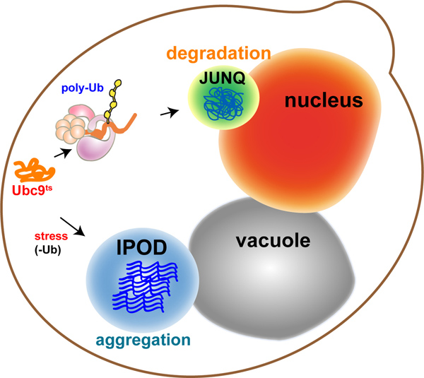
In eukaryotes, the cytosolic aggregation quality control load is partitioned between two compartments8-10: the juxtanuclear quality control compartment (JUNQ) and the insoluble protein deposit (IPOD) (Figure 1 - model). Proteins that are ubiquitinated by the protein folding quality control machinery are delivered to the JUNQ, where they are processed for degradation by the proteasome. Misfolded proteins that are not ubiquitinated are diverted to the IPOD, where they are actively aggregated in a protective compartment.
Up until this point, the methodological paradigm of live-cell fluorescence microscopy has largely been to label proteins and track their locations in the cell at specific time-points and usually in two dimensions. As new technologies have begun to grant experimenters unprecedented access to the submicron scale in living cells, the dynamic architecture of the cytosol has come into view as a challenging new frontier for experimental characterization. We present a method for rapidly monitoring the 3D spatial distributions of multiple fluorescently labeled proteins in the yeast cytosol over time. 3D timelapse (4D imaging) is not merely a technical challenge; rather, it also facilitates a dramatic shift in the conceptual framework used to analyze cellular structure.
We utilize a cytosolic folding sensor protein in live yeast to visualize distinct fates for misfolded proteins in cellular aggregation quality control, using rapid 4D fluorescent imaging. The temperature sensitive mutant of the Ubc9 protein10-12 (Ubc9ts) is extremely effective both as a sensor of cellular proteostasis, and a physiological model for tracking aggregation quality control. As with most ts proteins, Ubc9ts is fully folded and functional at permissive temperatures due to active cellular chaperones. Above 30 °C, or when the cell faces misfolding stress, Ubc9ts misfolds and follows the fate of a native globular protein that has been misfolded due to mutation, heat denaturation, or oxidative damage. By fusing it to GFP or other fluorophores, it can be tracked in 3D as it forms Stress Foci, or is directed to JUNQ or IPOD.
Protocol
1. הכנות שמרים
- להפוך זני שמרים עם פלסמיד שנשא GAL1-GFP-Y68L (UBC9 ts) קלטת.
- לגדול שמרים בתקשורת סינטתית המכילה raffinose 2% עבור 24 שעות וחוזרים לדילול של 2% מדיה המכילה גלקטוז עבור 16 שעות או O / נ דגירת תאים ב 30 מעלות צלזיוס בזמן רועד ב 200 סל"ד.
- בבוקר שלמחרת, מדלל את מתח השאילתה לOD 600 = 0.2. Shake עבור 4-6 שעות של 30 ° C (200 סל"ד) עד שהתרבות מגיעה OD 600 = 0.8-1.0.
- להדחיק ביטוי (כדי לפקח על הברכה תורגמה-כבר מקופלת ורק של ה-GFP-Ubc9 ts), תקשורת ישתנה לתקשורת הסינטתית עם תוספת סוכר 2% (SD) 30 דקות לפני ההדמיה.
- טיפול - הלם חום התאים ל20 דקות ב 37 ° C כדי לגרום misfolding, או באופן רציף במיקרוסקופ חממה לעקוב צבירת חלבון בתנאי חום הלם. הערה - אם אתה משתמש במיקרוסקופ בכל טמפרטורה מעל RT, מראש חום for כ 2 שעות לאזן את הטמפרטורה לאורך הגוף של המיקרוסקופ ואת המטרות (ראה להלן).
2. צלחת \ הכנות שקופיות
- בחר צלחת מתאימה. השיקול העיקרי הוא היכולת לשמור על מיקוד ברכישת הדמיה. הנקודות הבאות הן קריטיות עבור 4D הדמיה ולא לפערי הזמן 2D או 3D הדמיה.
- מרובה בארות צלחת תחתית הבארות השונות לא יכולה להיות הומוגנית (כלומר הבארות יהיו בגבהים יחסית למטרה שונים). זה יעשה את זה קשה לשמור על פוקוס לאורך נקודתי XY ונקודתי זמן, ללא קשר לשימוש בטכניקת autofocusing.
- חומרי שקופיות: זכוכית לעומת פלסטיק. שקופיות זכוכית תחתיות יותר Z-הומוגני בין הבארות, אך הם יקרים יותר.
- עובי של תחתית של הצלחת: אנחנו בדרך כלל להשתמש בצלחות coverslip-תחתונת מדד 1.5, אבל 1 מדד הוא גם מקובל בהתאם למטרה.
- Covאה תחתון של צלחת \ שקופית עם ConA (0.25 מ"ג / מ"ל) למשך 10 דקות. ConA משמש לדבוק תאים לשקופית, המאפשרת בעקבות תא בודד לאורך זמן.
- הסר ConA ודגירת השקופיות בברדס כימי כך שConA העודף יתאדה.
- 200 μl צלחת מדגם שמרים (OD 600 = 0.5) לConAed היטב.
- דגירה במשך 15 דקות, על מנת לאפשר לתאים להיצמד למשטח של הצלחת.
- חלץ את המדיום, ולשטוף שלוש פעמים עם מדיום חדש כדי לקבל שכבה אחת של תאים. הערה: אם פקיעת זמן מתוכנן, את תאי הזרע בדלילות, כך שהניצנים חדשים לא ימלא ולקטוע את האזור של עניין.
3. הכנות מיקרוסקופ
- אנו משתמשים בניקון A1Rsi confocal מיקרוסקופ עם כמה שינויים שאינם סטנדרטיים. עבור הדמית שמרים אנו משתמשים עד 4 לייזרים (405 ננומטר, 50 mW ליזר CUBE; 457-514 ננומטר, 65 mW ארגון יון הליזר; 561 ננומטר, 50 mW ליזר ספיר; ו640 ננומטר, 40 mW CUBE ליזר), ועד 4 PMTs מצוידיםמסננים. רוב ההדמיה שלנו נעשה עם קבוצה ירוקה מסנן לEGFP ולהגדיר מסנן אדום לmCherry וtdTomato. עם GFP וmCherry אין כמעט bleedthrough, ולכן לעתים קרובות אנו משתמשים סריקה בו זמנית. עם זאת, קו לסריקה יכולה לשמש גם כדי למנוע bleedthrough כשהוא מתרחש. confocal מצויד גם בשלב PInano Piezo (MCL), אשר יכול להפוך 3 שלבי msec בz, המאפשר 2-3 Z-ערימות (30-50 חלקים) לשנייה. המערכות יש גם רוח רפאי גלאים, פוקוס מושלם (ליזר קיזוז), ושני סורקים - סורק galvano וסורק תהודה.
- בחר מטרה המתאימה. שיקולים עיקריים:
- מטרת מים / שמן / אוויר: השיקול העיקרי הוא מקדם שבירה של מדיום ההדמיה לעומת הבינוני של המדגם.
- יתרונות אובייקטיביים שמן:
- מטרות נפט יכולות להיות פתחים מספריים גבוהים יותר (אור שמן הפסקות יותר ממים, ולכן פוטונים יותר לחזור לאובייקטיבי). מטרות נפט יכולות להיות עדלNA 1.49, לעומת 1.27 למים. (עם זאת, זה לא ממש הבדל עצום ברזולוציה).
- מקדם השבירה של שמן תואם את מקדם השבירה של זכוכית, ולכן אין פוטונים אבדו הולכים מהמדגם, מבעד לזכוכית, למטרה. (הערה - בחרה שמן טבילה שיש שבירה שמתאימה למטרה שלך וcoverslip שלך).
- שמן מאפשר ארוכה ללא טרחה בפעם שמעידות שכן הוא לא מתאדה.
- הרזולוציה הגבוהה מופעלת על ידי NA הגבוה יותר של מטרות נפט מדרדרת במהירות אם יש האור לנסוע דרך מדיום מימי (כגון תא).
- תמונות בדרך כלל יש סטיות כדוריות ויותר "מתוח" אפקט 3D.
- נפט הוא דביק ומבולגן, וסכנה ליעדים.
- יתרונות אובייקטיביים מים
- התא עצמו הוא מימי, ולכן יש פחות עיוותים בZ, and הרזולוציה נותרת גבוה עמוקה לתוך מדגם מימי.
- נקה וידידותי למשתמש. חסרונות: מים מתאדה במהירות אובייקטיבי, ולכן אינה מתאימים לסרטים ארוכים ללא הסדרים מיוחדים שנעשים כדי לשאוב מים ברציפות למטרה.
- יתרונות אוויר אובייקטיבי: 1. אין חומר הולך לאיבוד / מתאדה במהלך רכישת נקודה מרובה או סרט ארוך. 2. נקה וידידותי למשתמש. חסרון: רזולוציה נמוכה ורגישות אות נמוכה.
- יתרונות אובייקטיביים שמן:
- טמפרטורת חדר ובחממה: טמפרטורה משתנית משפיעה על תכונות חומר, ובמערכות עדינות משנה את הפוקוס. הטמפרטורה צריכה להיות מותאמת לפני בחירת נקודות עבור רכישה. שינוי הטמפרטורה במהלך הרכישה ישבש הדמיה ויגרום לאובדן מיקוד. אנו נותנים למערכת מיקרוסקופ לאזן עבור 2 שעות בטמפרטורה הרצויה לפני ההדמיה.
- קולרי תיקון: יעדים רבים מגיעים עם טבעת המאפשרת תיקוןהמטרה להיות מותאמת לעובי נתון כיסוי להחליק. אנו ממליצים להתאים את צווארון התיקון תוך שימוש בחרוזי ניאון לדמיין פונקצית התפשטות הנקודה. זה יבטיח הגדרות נכונות עבור כל מדגם.
- מחזיקי מדגם יציבים: אנו מוצאים כי מחזיקי מדגם מסחרי הזמינים ביותר הם חלק החלש ביותר של המיקרוסקופ. הם לעתים קרובות מתנדנדים ולא ישר. זה אסון לרזולוציה גבוהה, הדמית 4D ריבוי נקודות. אנו מעצבים מחזיקי המדגם שלנו, כי הם ישרים ונתאים היטב לבמה. הם יכולים גם להיות מחוזקים בברגים בעת צורך.
- רכישת ריבוי נקודות: מערכת ההדמיה שלנו מצוידת במערכת ניקון "המושלמת המיקוד", שהוא בעצם מנגנון מבוסס ליזר פוקוס אוטומטי. עם זאת, autofocusing הוא לא בהכרח צורך ב4D הדמיה, במיוחד אם המערכת לא נסחפת הרבה.
- מטרת מים / שמן / אוויר: השיקול העיקרי הוא מקדם שבירה של מדיום ההדמיה לעומת הבינוני של המדגם.
4. הדמיה
- הפעל את המערכת: לייזרים, במה, בקר, מצלמה וcompuתוכנת ter.
- צלחת מניח על בעל במה, בצורה נאותה והתייצבה.
- השתמש ביציאת עין כדי לקבוע את המיקום וכיוון של שמרים.
- שימוש brightfield, להעריך את בריאות תא ואת כדאיות על פי צורה ומרקם.
- הפעל אור epifluorescent פי fluorophore הרלוונטי (למשל FITC מסנן לGFP), ולהתמקד בתאים המציגים את הפנוטיפ אשר כפופים למחקר שלך. תאים אשר הניאון ביותר באורך גל אחד או יותר יכולים להיות מתים ולכן autofluorescent. אנו לדמיין גרעין עם fluorophore tdTomato התמזג SV40 אות NLS (NLS-TFP). פריון הכולל הוא כפול בגודלו של ה-GFP, ולכן מעל גבול דיפוזיה של הגרעין, ולכן זה עובד טוב מאוד כסמן גרעיני. אנחנו גם יכולים לרגש TFP עם ליזר ירוק (488 ננומטר) בו זמנית כGFP, אבל לאסוף את הפליטות הירוקות ואדומות לשתי PMTs הנפרד לרזולוציה ספקטרלית.
- התאם את ההגדרות הבאות כדי למזער את הרעשוoversaturation:
- כוח הליזר - photobleaching וphototoxicity לעומת בהירות של תמונה צריך להיחשב.
- רווח: משפיע על רגישות מצלמה, ובכך יחס אות לרעש.
- קוטר חריר - צריך להיות מותאם על פי הליזר עם אורך גל הקצר יותר. קוטר החריר מקובע את העובי (גובה) של הסעיף האופטי צלם. לפיכך, פתיחת החריר תאסוף יותר אור (נותן יותר פוטונים ב), אבל תקטין רזולוציה בz (confocality פחות).
- עצות שימושיות:
- אם fluorophores השונה פולט אורכי גל חופפים, השתמש בתכונת רפאי הגלאים המאפשרת הבחירה של מסננים וירטואליים. זכור כי גלאי רפאים פחות רגישים מאשר PMTs הרגיל.
- אם fluorophores השונה נרגש על ידי אותו הליזר, השתמש בתכונת סריקת הקו.
- סורק מאפשר Galvano יותר רגיש, אבל יש סיכון גבוה יותר לphotobleaching. סורק תהודה מאפשר מהר יותרcquisition, ובכך להקטין את הסיכון לphotobleaching, אך הוא פחות רגיש.
- ממוצע בין 2 ל 16 תמונות, זה משפר את יחס אות לרעש, אבל עושה את הרכישה איטית יותר, וכמובן, כרוך בחשיפה רבה יותר של המדגם לליזר.
- משך הרכישה תלויה בשאלות הביולוגיות (למשל JUNQ והיווצרות IPOD לוקחת ~ 2 שעות). אנחנו צלמנו שמרים עם סורק התהודה לתקופה של עד 30 שעות בזמן לשגות 3D.
- מרווחי זמן לשגות מרווחים קטנים יותר ליצור סרט עקבי יותר אך עלול לגרום לתמונת הלבנה ולכן אובדן האות.
תוצאות

איור 1. דגם:. מידור תת תאי של חלבוני misfolded מכונה בקרת האיכות מכוון את החלבונים לתאי misfolded שונים עם פונקציות שונות: חלבונים מסיסים, המיועדים לפירוק, לעבור-ubiquitination פולי, ונשלחים לתא Ju XTA-N uclear ש uality השליטה (JUNQ ). חלבונים מסיסים שלא ניתן ubiquitinated נשלחים לתפיסה הגנתית כדי שPr otein D eposit nsoluble (IPOD), בסמוך לvacuole, שם הם עוברים צבירה פעילה.
es.jpg "src =" / files/ftp_upload/50083/50083fig2.jpg "/>
איור 2. misfolding חלבון דוגמנות עם GFP-Ubc9 ts. בתנאים נורמלים, GFP-Ubc9 TS (ירוק) מקופל באופן מקורי, ומותאם diffusely בגרעין וcytosol. הגרעין מתויג על ידי NLS-TFP (אדום). הביטוי של Ubc9 ts היה סגור בתוספת של 2% גלוקוז לפני הדמיה בכל הניסויים. לחץ כאן לצפייה בדמות גדולה.
- עם שינוי הטמפרטורה עד 37 המעלות צלזיוס, GFP-Ubc9 TS (ירוק) misfolded ויוצר מוקדי מתח cytosolic. הגרעין מתויג על ידי NLS-TFP (אדום).
- לאחר ההתאוששות מהלם חום בשעת 23 ° C, GFP-Ubc9 denaturated תרמית TS הוא מושפל, כפי שצוין על ידי רמת פלואורסצנטי ירד.
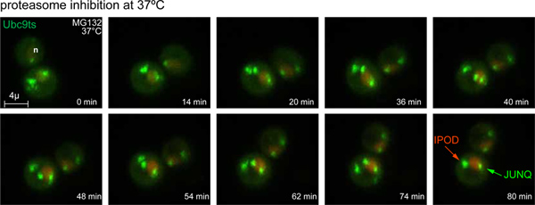
- עם שינוי הטמפרטורה ל ° C 37 עכבות והפרוטאזום עם 80 מ"מ MG132, GFP-U bc9 TS הוא misfolded ומעובד לJUNQ ותכלילים IPOD. הגרעין מתויג על ידי NLS-TFP (אדום).
- עם שינוי הטמפרטורה ל 37 מעלות צלזיוס ועיכוב ubiquitination, GFP-Ubc9 TS הוא misfolded ומעובד להכללת IPOD. הגרעין מתויג על ידי NLS-TFP (אדום). פרוטאז יוביקוויטין 4 (Ubp4) הוא overexpressed לחסום ubiquitination ts Ubc9.

איור 3. זמן פקיעה של JUNQ והיווצרות IPOD. עם שינוי הטמפרטורה ל 37 מעלות צלזיוס ועיכוב הפרוטאזום עם 80 מ"מ MG132, GFP-Ubc9 מוקדי מתח ts מעובדים לJUNQ ותכלילים IPOD. הגרעין מתויג על ידי NLS-TFP (אדום). תמונות 3D נרכשו במרווחי 4 דקות. (ראה גם בסרט 1).com/files/ftp_upload/50083/50083fig3large.jpg "target =" _blank "> לחץ כאן לצפייה בדמות גדולה.
Discussion
האינטואיציות שלנו על תהליכים ביוכימיים נובעות מניסויים גבי ספסל שבו פתרון מעורב ושל מגיבים ומוצרים רשאי להגיע לשיווי משקל בכוס. בהגדרה כזו, הריכוז של תרכובות כימיות הניתנות עלול להתבטא במספר אחד, שהוא היחס בין כמות טוחנת של מולקולות לנפח מקרוסקופית. הרבה ממה שאנו יודעים על מבנה חלבון ותפקוד נובע משימוש בשיטות המשקפות את התמונה הקלסית, עיקר תגובה: כתמים מערביים, centrifugations ומידות spectrophotometric בוצעו על תמציות מhomogenates של אוכלוסיות שלמות של תאים.
ככל שהטכנולוגיה שאנו משתמשים להסתכל על תאים בהגדלה משפר בקפיצות, הוא הופך להיות ברור יותר מתמיד כי תנאים שבתגובות ביוכימיות ביותר מתרחשות בגוף חי נושאים רק דמיון הקלוש לאלה של התרחיש גבי הספסל הקלסי. לא רק הפנים של celלה צפוף סביבה, שבו צפיפות השפעות משמעותיות לשנות את פעילותם של מגיבים שונים, זה גם די ההפך ממעורב ו. דבר זה מסביר את הפער התדיר בין במבחנה ביעילות vivo של מגוון רחב של תגובות macromolecular מורכבות.
בשום מקום הן נובעות מאינטואיציות קלסיות בניסויי מבחנה ביוכימיים נוטים יותר להטעות כמו בשאלות הנוגעות לקיפול vivo, misfolding, וצבירה של חלבונים. בעוד שמחקרים של כימית חלבון בתגובות בתפזורת יכולים לטפל בנושא של קיפול לחלבון ניתן כפשוטים כן או לא שאלה, כל ניסיון לעקוב אחר הדינמיקה של אוכלוסיות שלמות של מקרומולקולות בתא חי חייב להיות רגיש לכל ההפצה אפשרית תוצאות קונפורמציה זמינות לשרשרת פוליפפטיד, ובפרט לסיכון של misfolding וצבירה. לדוגמה, אנו יכולים לבחון ly תא תפזורתלהשביע של חלבון על ידי צבירה מערבית סופג, ולקבוע כי החלבון הוא בעיקר מסיס ולא ubiquitinated. עם זאת, בתא החי תת אוכלוסייה דיסקרטית של החלבון, קשה לזהות כאשר הממוצע מעל תאים רבים, עשוי להיות מסיס וubiquitinated בתא מסוים שבו הריכוז המקומי של המינים הוא גבוה ביותר. התרחיש זה האחרון עשוי להיות השלכות חשובות יותר לחיוניות של התא מתת אוכלוסיית התפזורת הגדולה יותר. יתרה מכך, בעוד מלווים להציג מגוון רחב של התנהגויות ותפקודים במבחנה pleiotropic, זה הופך ברור כי בתא הפונקציות הבדידות מוגבלות מרחב ובזמן.
בפרדיגמה החדשה הלהבנת הביוכימיה, ריכוז הופך לרכושו של כל משתנה ננו סביבה מסוימת בתא, ואת האירועים המולקולריים שבבסיס תהליכים ביולוגיים יש assayed לא רק בזמן, אלא גם בSPACדואר. גישת הדמית 4D הוצגה כאן מאפשרת מודלים רגישים של misfolding חלבון בתאים חיים, אם כי ניתן להשתמש בו כדי ללמוד על כל מספר התהליכים ביולוגיים אחרים וכיצד הם מוסדרים במרחב, זמן, ובעקבות שינויים בתנאי הסביבה. במאמר זה אנו משתמשים בחיישן Ubc9 ts מתקפל, אשר למעשה מדגים את השלבים ואפשרויות להתמודדות עם ההתפרצות של צבירת חלבון בcytosol. בנוסף לממחיש את הביולוגיה של התא של בקרת איכות צבירה, גישה זו יכולה לשמש ככלי רב עצמה לפענוח ההשפעה של הפרעות ספציפיות או מוטציות גנטיות בproteostasis (לדוגמא Ubc9 ts יכול לשמש למדידת לחץ קיפול חלבונים בתגובה לחמצון, הביטוי לצבירה רעילה, או מוטציות במסלול בקרת האיכות).
4D הדמיה חיונית גם לקביעת לוקליזציה חלבון או החלבון colocalization בין שתי שונים במדויקs, ולאיתור תופעות שאולי להיות חולף אך חשוב. לדוגמה, במיוחד באורגניזם כדורי קטן כמו שמרים, זה עשוי להיראות במקרה שמבנה או המצרפי יש לוקליזציה juxtanuclear, בעוד 4D הדמיה יכולה לגלות שזה פשוט תוצר של הזווית של בדיקה.
בניסוי לדוגמא אנו מציגים כאן, אנו מדגימים את השימוש במודל misfolded חלבון, Ubc9 TS, לעקוב בקרת איכות הצטברות לאורך זמן ובמרחב בcytosol. בטמפרטורה המתירנית, Ubc9 ts מקופל ומתפזר בגרעין וcytosol. על misfolding נגרם עקב החום, זה יוצר תחילה במהירות מוקדי diffusing קטנים מצטברים מתח שעוברים עיבוד לפירוק proteasomal. כאשר הפרוטאזום חלקית עכבות, מוקדי מתח אלה מומרים לJUNQ ותכלילים IPOD במשך כ 2 שעות. אם שפלת יוביקוויטין בתיווך אינה זמינה כאפשרות בקרת איכות, UBC9 ts מחדש מנותב מייד להכללת IPOD לצבירת מגן. כלים אלה מציעים הזדמנויות מדהימות לגלות גורמים גנטיים מעורבים ברומן בקרת איכות צבירה, ולחקור הסדרת המרחב ובזמנם בתא.
Disclosures
אין ניגודי האינטרסים הכריזו.
Materials
| Name | Company | Catalog Number | Comments |
| MG132 | Mercury | mbs474790 | |
| con A | Sigma | C2010 | |
| Glass bottom plates | ibidi | ibd81158 | |
| 4D Fluorescence Imaging of Protein Aggregation Confocal 3D movies were acquired using a Nikon A1R-si microscope equipped with a PInano Piezo stage (MCL), using a 60x water objective NA 1.27, 0.3 micron slices, 0.5% laser power (from 65 mW 488 laser and 50 mW 561 laser). z-stacks were acquired every 5 min for 90 min. Each z-series was acquired with 0.5 micron step size and 30 total steps. Image processing was performed using NIS-Elements software. | |||
References
- Gershenson, A., Gierasch, L. M. Protein folding in the cell: challenges and progress. Current opinion in structural biology. 21, 32-41 (2011).
- Aguzzi, A., Calella, A. M. Prions: protein aggregation and infectious diseases. Physiological reviews. 89, 1105-1152 (2009).
- Morimoto, R. I. Proteotoxic stress and inducible chaperone networks in neurodegenerative disease and aging. Genes & development. 22, 1427-14 (2008).
- Cohen, E., Dillin, A. The insulin paradox: aging, proteotoxicity and neurodegeneration. Nature reviews. Neuroscience. , (2008).
- Hershko, A., Ciechanover, A. The ubiquitin system. Annual review of biochemistry. 67, (1998).
- Tyedmers, J., Mogk, A., Bukau, B. Cellular strategies for controlling protein aggregation. Nature reviews. Molecular cell biology. 11, 777-788 (2010).
- Treusch, S., Cyr, D. M., Lindquist, S. Amyloid deposits: Protection against toxic protein species?. Cell cycle (Georgetown, Tex.). 8, 1668-1674 (2009).
- Spokoini, R., et al. Confinement to Organelle-Associated Inclusion Structures Mediates Asymmetric Inheritance of Aggregated Protein in Budding Yeast. Cell Rep. , (2012).
- Weisberg, S. J., et al. Compartmentalization of superoxide dismutase 1 (SOD1G93A) aggregates determines their toxicity. Proc Natl Acad Sci U S A. 109, 15811-15816 (2012).
- Kaganovich, D., Kopito, R., Frydman, J. Misfolded proteins partition between two distinct quality control compartments. Nature. 454, 1088 (2008).
- Betting, J., Seufert, W. A yeast Ubc9 mutant protein with temperature-sensitive in vivo function is subject to conditional proteolysis by a ubiquitin- and proteasome-dependent pathway. The Journal of biological chemistry. 271, 25790 (1996).
- Tongaonkar, P., Beck, K., Shinde, U. P., Madura, K. Characterization of a temperature-sensitive mutant of a ubiquitin-conjugating enzyme and its use as a heat-inducible degradation signal. Analytical biochemistry. 272, 263 (1999).
Reprints and Permissions
Request permission to reuse the text or figures of this JoVE article
Request PermissionExplore More Articles
This article has been published
Video Coming Soon
Copyright © 2025 MyJoVE Corporation. All rights reserved