
Histological Sample Preparation for Light Microscopy
Panoramica
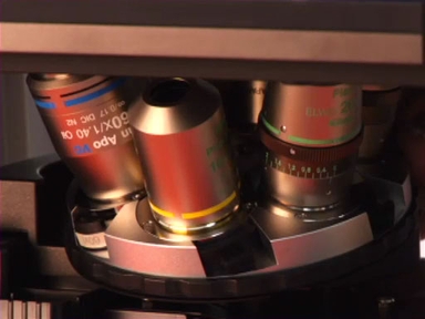
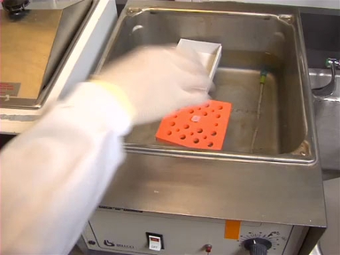
Histology is the study of cells and tissues, which is typically aided by the use of a light microscope. The preparation of histological samples can vary greatly based on the inherent properties of the samples such as size and hardness as well as expected post-processing which includes planned staining techniques or other down-stream applications. As described in this video, specimen preparation typically begins with a fixation procedure to prevent degradation of the sample by naturally occurring enzymes that are released by the cells upon death. Once fixed, samples are placed into an embedding medium that is able to sufficiently support the sample. Most commonly this is paraffin wax, but other materials such as a glycerin based freezing medium and agars are also used to surround the sample during sectioning. Sectioning then takes place on a microtome or other cutting device that allows the user to shave the sample into thin slices ranging from a few microns to a few millimeters in thickness. Once cut, sections are mounted on a glass slide and stained to bring out specific features of the sample before being imaged on a microscope.
Procedura
Histology is a term that refers to the study of the microscopic anatomy of tissues and cells. Proper histological sample preparation for light microscopy is essential for obtaining quality results from tissue samples.
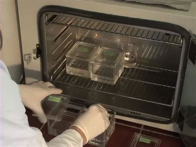
There are 3 main steps common to nearly all histological procedures. First, the sample is fixed, in order to preserve the tissue and slow down tissue degradation. Next, the sample is immersed in a material, or embedding media, that has similar mechanical properties to itself. Once embedded, the sample
Vai a...
Video da questa raccolta:
Now Playing
Histological Sample Preparation for Light Microscopy
General Laboratory Techniques
240.3K Visualizzazioni
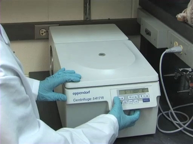
Un'introduzione alla centrifuga
General Laboratory Techniques
488.3K Visualizzazioni
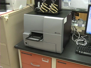
Introduzione al lettore di micropiastre
General Laboratory Techniques
126.7K Visualizzazioni
Comprendere la concentrazione e misurare i volumi
General Laboratory Techniques
216.0K Visualizzazioni
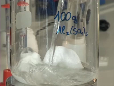
Realizzare soluzioni in laboratorio
General Laboratory Techniques
211.4K Visualizzazioni
Introduzione al micropipettor
General Laboratory Techniques
584.2K Visualizzazioni
Introduzione alle pipette sierologiche e ai pipettatori
General Laboratory Techniques
219.1K Visualizzazioni
Introduzione al Bunsen Burner
General Laboratory Techniques
207.2K Visualizzazioni
Un'introduzione al lavoro sotto cappa
General Laboratory Techniques
151.3K Visualizzazioni
Misurazione della massa in laboratorio
General Laboratory Techniques
170.9K Visualizzazioni
Introduzione allo spettrofotometro
General Laboratory Techniques
518.2K Visualizzazioni
Introduzione alla microscopia a fluorescenza
General Laboratory Techniques
349.9K Visualizzazioni
Introduzione alla microscopia ottica
General Laboratory Techniques
815.1K Visualizzazioni

Regolazione della temperatura in laboratorio: conservazione dei campioni utilizzando il freddo
General Laboratory Techniques
65.7K Visualizzazioni
Regolazione della temperatura in laboratorio: applicazione del calore
General Laboratory Techniques
81.3K Visualizzazioni
Personale delle biblioteche
Copyright © 2025 MyJoVE Corporation. Tutti i diritti riservati