Histology is a term that refers to the study of the microscopic anatomy of tissues and cells. Proper histological sample preparation for light microscopy is essential for obtaining quality results from tissue samples.
There are 3 main steps common to nearly all histological procedures. First, the sample is fixed, in order to preserve the tissue and slow down tissue degradation. Next, the sample is immersed in a material, or embedding media, that has similar mechanical properties to itself. Once embedded, the sample is sectioned, or cut, into thin slices using a precise cutting tool known as a microtome. This video describes these general steps and some of the concepts that relate to them.
Tissue fixation is a critical step that preserves cell and tissue components and maintains their structure. Following cell death, naturally occurring enzymes are released from cellular organelles and begin to degrade the proteins throughout the cell and extracellular matrix, thereby destroying the structure of the cell. Fixation prevents such degradation by directly inhibiting the ability of these enzymes digest protein and by making the enzyme cleavage sites unrecognizable.
The two main mechanisms of fixation are cross-linking and coagulation. Cross-linking involves covalent bond formation both within proteins and between them, which causes tissue to stiffen and therefore resist degradation. Coagulation is caused by the dehydration of proteins through the use of alcohols or acetone, which deform protein tertiary structure, so that hydrophobic, or water fearing, regions move the protein surface. Coagulative fixation can help embedding media, like paraffin wax, penetrate tissue.
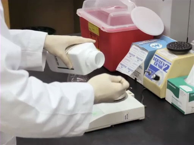
One of the most commonly-used fixatives is Formalin, which is formaldehyde dissolved in a water. During fixation, formaldehyde attaches to primary amines, such as those found on the side chains of the amino acids lysine and glutamine to form a stable crosslink called a methelene bridge. This process is inherently slow and can take up to 1-2 days.
Prior to fixation a few considerations should be made. First, consider the diffusivity of the sample. Fixatives will diffuse a distance through tissues at a rate related to the coefficient of diffusion for the fixative multiplied by the square root of time. A fixative that takes 1 hour to penetrate 1 mm into the sample will take 25 hours to penetrate 5 mm. Once formalin has reached the center of the tissue, the crosslinking reaction sill needs to occur. Thus, sample size should be limited to 4 mm in thickness for thorough fixation in a reasonable amount of time.
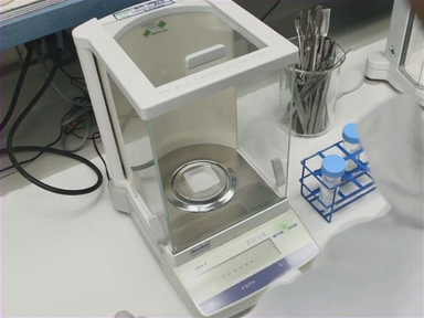
Second, consider the volume and pH of the fixative. The fixative to sample volume ratio should be at least 40:1 so that the reagent is not depleted easily over time. While some fixatives are designed to work at an acidic pH, formalin works best when buffered with phosphate to maintain a neutral pH, so that excess acid is not produced, which causes artifacts in tissue.
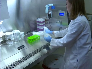
The next step in histological preparation is embedding, which involves supporting specimens in media that has similar mechanical rigidity to the specimen itself. Choosing the right embedding medium is critical, because if it is too stiff or too weak, defects may occur while sectioning. By far, the most common media used for embedding is paraffin wax.
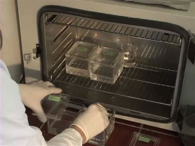
Prior to paraffin-embedding, samples must be dehydrated by replacing the water in the tissue with ethanol, first, followed by xylenes, and then finally warmed paraffin wax. Once wax has infiltrated the sample, it is carefully positioned and surrounded with additional wax using a mould to form a block. Next, samples are attached to a tissue cassette for sectioning.
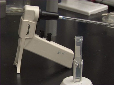
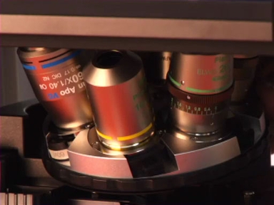
Sectioning is the process of cutting thin slices of a sample from an embedding block. The sections are usually on the order of 4-10 µm thick for use with light microscopy.
To cut sections, a metal, glass, or diamond blade is first secured on a microtome. The sample is then placed in a sample holder. Next, the sample is advanced to the cutting surface and drawn across the blade to create a slice of desired thickness. Sections will amass as thin ribbons which can be placed on glass slides.
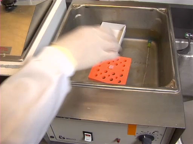
Following sectioning, samples are placed on slides. For paraffin embedded samples, slices are first placed into a warmed water bath and are then lifted out of the water onto the slide and allowed to dry.
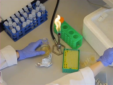
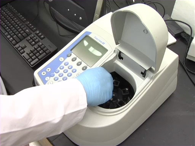
Biological tissue has very little inherent contrast, so after histology, slides are usually stained with dyes or antibodies that highlight morphology or specific proteins. The most common stain performed is hematoxylin and eosin, or H&E. Because it is so common, many automated machines have been made to reproducibly stain numerous sections with H&E. Hematoxylin stains the nuclei of cells blue and eosin stains the cytoplasm pink revealing cellular morphology of tissues.
One drawback to fixation is that the cross-linking of proteins can make it more for labeling antibodies to find their binding sites. Rather than using fixation to preserve samples, rapid freezing of tissue, or snap freezing can be used, which is followed by a sectioning technique called cryosectioning
Tissue samples are embedded and oriented in a special freezing media called OCT, or optimal cutting temperature medium and then rapidly frozen. Like other embedding media, OCT matches the rigidity of the sample.
A cryomicrotome or cryostat, is used to cut frozen sections and this instrument maintains an internal temperature of -20°C to keep the cutting blade and samples cool. In contrast to paraffin-embedded sections, frozen sections can be directly lifted onto a positively charged glass slide immediately after sectioning.
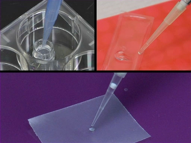
Another way to avoid fixation is via agar embedding, in which samples are covered with freshly prepared liquid agarose. As the agarose cools, it locks the tissue into place and excess agarose can be trimmed away.
Vibrotomes, another alternative to the microtome, have a blade that vibrates and moves across the agarose-embedded sample.Vibrotomes are used to create thick sections on the order of 50-1000 µm from agarose embedded samples.
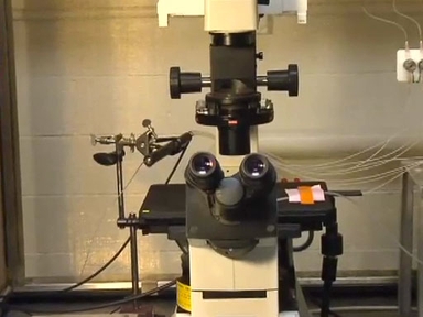
Samples are typically removed from the agarose before staining , and then placed onto a slide surrounded by a substance such as vacuum grease that keeps the coverslip from squishing the sample. They are best viewed using confocal microscopy in order to obtain high resolution images of each thick section.
You’ve just watched JoVE’s video on histological sample preparation for light microscopy.
You should now understand the steps involved for sample preparation to get you from a piece of tissue to a stainable section. As always, thanks for watching!