
検眼鏡検査
概要
ソース: リチャード Glickman サイモン、MD、アシスタント教授、公衆衛生およびコミュニティ薬の部門のタフツ大学医学部、マサチューセッツ州
最も簡単な ophthalmoscopes は、目を通すに絞り、視度インジケーター、およびレンズを選択するためのディスクで構成されます。検眼は、眼底や脈絡膜、網膜、黄斑、黄斑、視神経乳頭、網膜血管(図 1から成っている後の眼の内側の壁をみるために主に)。球形の眼球を収集し、麻痺、網膜細胞の光を焦点を当てています。順番に、レンズと角膜、硝子体を通過する際、光が屈折します。
眼底検査の時に観測された最初のランドマークは視神経は視神経と網膜血管が目 (図 2) の後ろを入力します。ディスクには通常、血管を入力; 中央の白っぽい生理カップが含まれていますこれは通常、ディスク全体の半分以下の径を占めています。ちょうど外側とやや劣るが中心窩、暗い円形領域中心視のポイントを設定します。これは黄斑です。ブラインド スポット約 15 ° の視線の行に時間は視神経乳頭に光受容細胞の欠如に起因します。
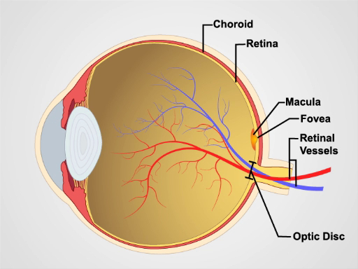
図 1。目の解剖学します。標識構造物と人間の目の矢状ビューを示す図。

図 2: 正常な網膜。正常な網膜に眼科ビューを表示写真。
手順
Mydriatic 目が値下がりしましたが通常は使用されていないので一般に練習、眼底のビューは網膜のセクションのみに限定されます。患者を検討する前に、これらの機能に精通していること。
- 患者の屈折異常は、網膜上にピントが合いにくい、場合を除き、試験のため、自分の眼鏡を削除するが最善です。
- 部屋を暗くした後、検眼鏡をオンにし、あなたの手や壁に光を当てます。
- レンズ ディスクを回して光の最大の白い円を見ることができると視度インジケーター読み取り 0、検眼レンズを意味が収束しても光を分散させます。
- ように人差し指レンズ ディスク上、試験中にジオプトリーを調整ことができますに応じて網膜の構造に集中します。
- 患者の右目を調べるあなたの右手で、検眼鏡を保持して、右の目と絞り値に目を通す試験の患者の左目、左手と左眼で絞りを見て検眼鏡を保持します。患者の鼻をバンプを回避できます。
- 自分の目の高さで患者から足の位置、あなたの肩越しにだけ壁にスポットで凝視する患者を求めます。
- あなたは開口部をのぞく、保つ両方の目を開いて、あなたの骨の軌道に対してしっかり
申請書と概要
スキップ先...
このコレクションのビデオ:

Now Playing
検眼鏡検査
Physical Examinations II
68.0K 閲覧数

目の試験
Physical Examinations II
77.2K 閲覧数

耳の検査
Physical Examinations II
55.2K 閲覧数

鼻、副鼻腔、口腔、咽頭の検査
Physical Examinations II
65.8K 閲覧数

甲状腺検査
Physical Examinations II
105.1K 閲覧数

リンパ節試験
Physical Examinations II
387.6K 閲覧数

I: の腹部の試験検査・聴診
Physical Examinations II
202.8K 閲覧数

腹部の試験 II: パーカッション
Physical Examinations II
248.2K 閲覧数

腹部の試験 III: 触診
Physical Examinations II
138.5K 閲覧数

IV の腹部の試験: 急性腹痛評価
Physical Examinations II
67.3K 閲覧数

男性の直腸の検査
Physical Examinations II
114.6K 閲覧数

包括的な乳房検査
Physical Examinations II
87.7K 閲覧数

性器の内診 i: 評価
Physical Examinations II
307.3K 閲覧数

内診 II: 鏡試験
Physical Examinations II
150.4K 閲覧数

内診 III: 両手と直腸試験
Physical Examinations II
147.8K 閲覧数
Copyright © 2023 MyJoVE Corporation. All rights reserved