Ophthalmoscopic Examination
Source: Richard Glickman-Simon, MD, Assistant Professor, Department of Public Health and Community Medicine, Tufts University School of Medicine, MA
The simplest ophthalmoscopes consist of an aperture to look through, a diopter indicator, and a disc for selecting lenses. The ophthalmoscope is primarily used to examine the fundus, or the inner wall of the posterior eye, which consists of the choroid, retina, fovea, macula, optic disc, and retinal vessels (Figure 1). The spherical eyeball collects and focuses light on the neurosensory cells of the retina. Light is refracted as it passes sequentially through the cornea, the lens, and the vitreous body.
The first landmark observed during the funduscopic exam is the optic disc, which is where the optic nerve and retinal vessels enter the back of the eye (Figure 2). The disc usually contains a central whitish physiologic cup where the vessels enter; it normally occupies less than half the diameter of the entire disc. Just lateral and slightly inferior is the fovea, a darkened circular area that demarcates the point of central vision. Around this is the macula. A blind spot approximately 15° temporal to the line of gaze results from a lack of photoreceptor cells at the optic disc.
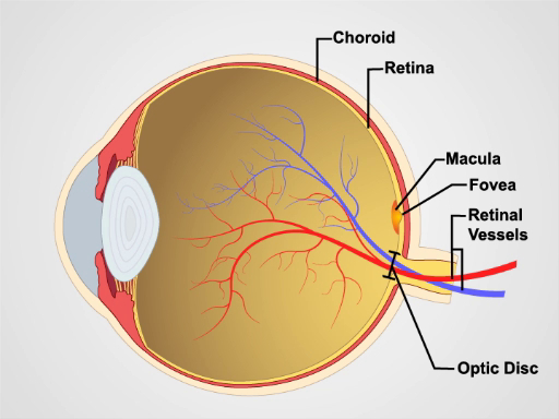
Figure 1. Anatomy of the eye. A diagram showing a sagittal view of the human eye with the structures labeled.

Figure 2: Normal retina. A photograph showing an ophthalmoscopic view on the normal retina.
Since mydriatic eye drops are typically not used in general practice, the view of the fundus is limited to only a section of the posterior retina. Be familiar with these features before attempting to examine the patient.
- Unless the patient's refractive errors make it difficult to focus on the retina, it is usually best to remove your own eyeglasses for the exam.
- After darkening the room, turn on the ophthalmoscope and shine the light on your hand or on the wall.
- Turn the lens disc until the largest white
The ophthalmologic exam is probably the most challenging for students to master. With time, however, it becomes routine. It is also one of the most productive parts of the physical exam, as it not only offers a window into the condition of the eye, but also provides evidence of disease elsewhere in the body. Elevated intracranial pressure from a variety of causes may lead to swelling of the optic nerve, which appears as papilledema on a funduscopic exam. In papilledema, the optic disc is swollen, its margins are blurred,
跳至...
此集合中的视频:

Now Playing
Ophthalmoscopic Examination
Physical Examinations II
66.0K Views

眼科检查
Physical Examinations II
74.7K Views

耳朵考试
Physical Examinations II
52.8K Views

鼻子、 鼻窦、 口腔和咽部考试
Physical Examinations II
63.9K Views

甲状腺考试
Physical Examinations II
102.5K Views

淋巴结考试
Physical Examinations II
377.0K Views

腹部考试 i: 检查和听诊
Physical Examinations II
198.8K Views

腹部考试 II: 打击乐
Physical Examinations II
243.5K Views

腹部考试 III: 触诊
Physical Examinations II
136.5K Views

腹部考试四: 急性腹痛评估
Physical Examinations II
65.7K Views

男性直肠检查
Physical Examinations II
111.1K Views

全面的乳房检查
Physical Examinations II
84.0K Views

外生殖器的盆腔检查 i: 评估
Physical Examinations II
293.5K Views

骨盆 II: 窥镜考试成绩
Physical Examinations II
146.8K Views

盆腔检查三: 双手和直肠阴道考试
Physical Examinations II
143.7K Views
版权所属 © 2025 MyJoVE 公司版权所有,本公司不涉及任何医疗业务和医疗服务。