
Examen ophtalmologique
Vue d'ensemble
Source : Richard Glickman-Simon, MD, professeur adjoint, département de santé publique et médecine sociale, Tufts University School of Medicine, MA
Les plus simples ophtalmoscopes consistant en une ouverture de regarder à travers un indicateur de dioptrie et un disque de sélection des lentilles. L’ophtalmoscope est principalement utilisé pour examiner le fond de l’oeil, ou la paroi interne de le œil postérieur, qui comprend la choroïde, rétine, fovéa, macula, disque optique et des vaisseaux rétiniens ()Figure 1). Le globe oculaire sphérique recueille et focalise la lumière sur les cellules de la rétine neurosensorielles. Lumière est réfractée lorsqu’il passe dans l’ordre dans la cornée, le cristallin et le corps vitré.
Le premier repère observé lors de l’examen de fond est du disque optique, qui est où le nerf optique et les vaisseaux rétiniens entrer l’arrière de le œil (Figure 2). Le disque contient généralement une coupe physiologique blanchâtre centrale où les navires entrer ; normalement, elle occupe moins de la moitié du diamètre du disque entier. Juste latéral et légèrement inférieure est la fovéa, une zone circulaire sombre qui délimite le point de la vision centrale. Autour de cela est la macula. Un angle mort environ 15 ° temporelle à la ligne du regard résulte d’un manque de cellules photoréceptrices au disque optique.
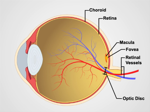
Figure 1. Anatomie de le œil. Un diagramme montrant une vue sagittale de le œil humain avec les structures marquées.

Figure 2 : rétine normale. Une photo montrant une vue ophtalmoscopique sur la rétine normale.
Procédure
Depuis mydriatique eye drops sont en général pas utilisé en général pratique, l’affichage du fond de le œil est limitée à seulement une partie de la rétine postérieure. Être familier avec ces caractéristiques avant d’examiner le patient.
- À moins que les erreurs de réfraction du patient, il est difficile de se concentrer sur la rétine, il est généralement préférable de supprimer vos propres lunettes à l’examen.
- Après obscurcissement de la pièce, allumez l’ophtalmoscope et briller la lumière sur votre main ou sur le mur
Applications et Résumé
L’examen ophtalmologique est probablement le plus difficile pour les étudiants de master. Avec le temps, cependant, il devenait une routine. Il est également une des régions plus productives de l’examen physique, car il offre non seulement une fenêtre dans l’état de le œil, mais fournit également des signes de maladie ailleurs dans le corps. Hypertension intracrânienne d’une variété de causes peut-être conduire à un gonflement du nerf optique, qui apparaît comme le œdème papillaire sur un examen de...
Passer à...
Vidéos de cette collection:

Now Playing
Examen ophtalmologique
Physical Examinations II
68.1K Vues

Examen des yeux
Physical Examinations II
77.3K Vues

Examen des oreilles
Physical Examinations II
55.3K Vues

Examen du nez, des sinus, de la cavité orale et du pharynx
Physical Examinations II
65.9K Vues

Examen de la thyroïde
Physical Examinations II
105.2K Vues

Examen des ganglions lymphatiques
Physical Examinations II
387.9K Vues

Examen abdominal I: Inspection et auscultation
Physical Examinations II
202.8K Vues

Examen abdominal II: Percussion
Physical Examinations II
248.4K Vues

Examen abdominal III: Palpation
Physical Examinations II
138.6K Vues

Examen abdominal IV: Évaluation de la douleur abdominale aiguë
Physical Examinations II
67.3K Vues

Toucher rectal chez l'homme
Physical Examinations II
114.7K Vues

Examen général des seins
Physical Examinations II
87.8K Vues

Examen pelvien I: Évaluation des organes génitaux externes
Physical Examinations II
307.9K Vues

Examen pelvien: Examen au spéculum
Physical Examinations II
150.6K Vues

Examen pelvien III: Examen bi-manuel et toucher rectal
Physical Examinations II
147.9K Vues
À PROPOS DE JoVE
Copyright © 2025 MyJoVE Corporation. Tous droits réservés.