
サインイン
スプリット ブレイン
概要
ソース: ジョナス ・ t. カプラン サラ I. ギンベル所-南カリフォルニア大学
脳の損傷により認知機能がどのような影響を与える研究認知神経科学の最も重要なツールの一つとされてきました。脳は体の最もよく保護された部分の 1 つで、脳の機能に影響を与えることができる多くのイベントがあります。血管の問題、腫瘍、変性疾患、感染症、鈍力外傷、脳神経外科は脳損傷、脳のさまざまな方法で機能に影響を与える組織の損傷のさまざまなパターンを生成可能性がありますすべての根本的な原因のほんの一部です。
神経心理学の歴史は、脳の理解の進歩につながったいくつかのよく知られている例で示されます。例えばで 1861 ポール ブローカー様子が観察された失語症、獲得した言語障害で起因した前頭葉左への損傷。別の例として多大なメモリについてきた神経心理学の文献の多くの年のため「陛下、「特定種類の新しい記憶の形成に深刻な赤字につながった-が側頭葉手術として知られているヘンリー Molaison の有名な事件など、記憶喪失の患者から。
観察および焦点の脳損傷患者のテスト中には、細心の注意、脳の機能に洞察力と神経科学は、赤字の特定の性質を明らかにするテストの設計で取られなければならない提供をしています。また、脳が相互接続されたニューロンの複雑なネットワークには、1 つの脳の領域への損傷は損傷から遠く離れた地域で機能している影響ことができます。脳の損傷が脳の領域間の接続に与える影響を示すため、このビデオは、いわゆるスプリット ブレインの場合を調べます。
脳の左右の半球を接続する線維の大きな束である脳梁脳の最大の白質路の一つであり脳の正中線の矢状ビューに簡単に認識することができます。1960 年代、脳神経外科医は、脳梁を切断はできる特定の種類のてんかんは、脳に広がる手に負えないの神経活動を含む治療の成功を発見しました。ブレインの手術を受けた人に分離手術の 2 つの半球が持っていたは、左と右半球が通信することが不要になったようなもの。この条件は、左と右の脳半球の機能を相対的な能力およびそれらの間の通信の性質について学習する独立して、プローブに実験者を許可しました。
このビデオは、スプリットブレイン患者脳の 2 つの半球間の相違のいくつかを明らかにするため、このような切断のいくつかの劇的な結果を参照してくださいにテストする方法を示します。これらの実験の元のバージョンはマイケル ・ ガッザニガと同僚1、2によって開発され、後に他人によって練られました。3バージョンがここには、方法論の最近の近代化が組み込まれています。
手順
1. 患者とコントロールの募集
- 完全および部分的な手術 callosotomies と、脳梁が完全に発達していなかった脳梁 (ACC) 欠損症などの先天性の条件を含む切断症候群患者のさまざまながあります。2 つの半球を接続する複数の管があります。最大の脳梁は、しかし、いくつかの繊維は前交連、海馬交連、後交連で交差します。
切断のこれらの異なる品種がこのテストの別の行動の結果につながる可能性がありますに注意してください。 - この実験のため接続ファイバーの有無を確認する神経イメージング研究を使用して患者を事前に選択します。
- 標準的な MRI や画像白質路に使用できます、拡散イメージングは、特に便利です。接続する繊維が患者の存在を知ることは、結果の解釈に役立ちます。このデモでは、完全な脳梁離断症例が選択されています。
- 患者研究手順の完全に通知されている、すべての適切な同意書を締結したことを確認します。
- 同じ年齢や性別、患者としての 20 の参加者を募集する知能、ウェクスラー成人知能スケール (WAIS) のスコアを使用して一致します。
2. データの収集
- だけ左または右半球に視覚刺激を提示するために、1 つの視覚野に刺激を適切に提示する必要があります。これが 1 つ目に刺激と同等ではないことに注意してください。それぞれの目は、脳の両半球にプロジェクトします。たとえば、左の視野を見る左目の部分は右半球によって処理されますが、左半球で処理右視野の左目の部分を見ています。したがって、左半球にイメージを提示する患者が見ている場所の右側にある右視野内に完全紹介します。
- この左右差を達成するため、コンピューターの画面からおよそ 22 インチ目を維持するために、あごを使用します。画面に直面している、あご内快適患者の顎を配置します。
- 患者が彼らの目を凝視するための場所を提供するために画面の中央に残っている小さな十字があります。
- 左またはそれの右側に画像が表示されるとしても、実験を通してこのクロスの固定を維持するために患者に指示します。
- 画像が表示されたら、オブジェクトの名前を声に出して言う必要があります彼らは患者へ説明します。
- 脳の左右の半球にそれぞれ投影画面の左右に簡単によく知られている物体の画像を提示します。50 個のイメージは、リンゴ、ボール、ほうき、鶏など、わかりやすい図面を含むオブジェクトの集合からランダムな順序で掲載されています。
- 適切な左右差を確保するため 150 ミリ秒未満の画像を提示します。これは、刺激を見る十分な時間が、十分高速なので患者が中心視野に刺激を参照してくださいに目を移動することができるではないです。
- 大声で、画面に表示されるオブジェクトの名前に患者をお願いし、その応答を記録します。これは口頭言語能力のテスト、スピーキング能力の半球間の相違点を明らかにする必要があります。
- せれば (上と同じ側) と同側の手で、紙を見もせず、オブジェクトを描画する患者の患者は任意のオブジェクトに名前を付けることができる、刺激。これは刺激の知識の非言語的手段として提供しています。
- 刺激と同側の手は、刺激を見た半球によって制御されます。たとえば、左視野の刺激策を発表すると、それは右半球によって処理されます。右半球は左の手の制御を主担当です。
- それが 1 つの半球に刺激の分離を維持するために描画中に患者は自分の手を見ていないことを確認します。
- 患者は、オブジェクトの描画を終了したときは、オブジェクトを見て、大声だと言ってもらいます。これは、患者を知っているとオブジェクトの名前は、中心の展望で提示彼らはそれが 1 つの半球に提示されるときに名前を付けることができない場合でも確認できます。
- 各コントロールの参加者に対して手順を繰り返します。
3. データ分析
- 患者さんのパフォーマンスを解析するには、互いの左側と右側の視覚の半分フィールドからのデータを比較します。これを行うには、各視野の正しいと不適切な回答数を集計し、独立性のカイ二乗検定を用いて観察 1 つと同じ大きさの差を得られる可能性をテストします。
- 年齢、性別、および欠損患者の行動を決定するための知能をマッチさせた対照人口のデータを患者からデータを比較します。これを行うにを個別に、それぞれの人の左視野と右視野の平均スコアをコンパイルおよび比較するには、繰り返し対策分析の分散の使用分布は (ANOVA) をテストします。
結果
通常、脳梁離断の患者は左視覚半分フィールドでオブジェクトの合併した健忘を展示します。合併した健忘は、名前オブジェクトにできないです。右の視野に提示されるただし、名前付け高精度 (図 1)。
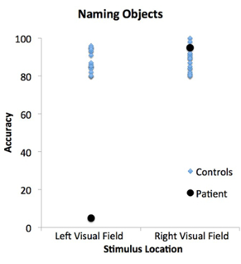
図 1:左と右の視野に刺激の名前付けのオブジェクトのタスクで患者とコントロール性能。患者 (黒丸) 口頭で名前オブジェクトの左視野に提示することができるではない右の視野に名前オブジェクトすることが。対照的に、コントロール人口 (ブルー ダイヤモンド) 左と右の両方の visual フィールドでオブジェクトに名前をすることができます。
何人かの患者は彼らことができない口頭で名前を付けます(図 2でも左視野に提示される正常に描画できる場合があります)。
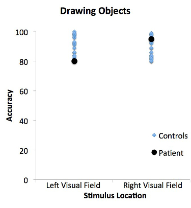
図 2:左と右の視野に刺激の図面オブジェクトのタスクで患者とコントロール性能。患者 (黒丸) と対照群 (ブルー ダイヤモンド)、左と右の両方の visual フィールドでオブジェクトを描画することができます。患者さんのパフォーマンスは、一致するコントロールから違いはありません。
この場合、患者は通常、彼らは何かを見ていない言います。音声認識の制御は、左の脳半球は視覚的なイメージを見ていないためにです。しかし、オブジェクトを見ている、右の脳半球はそれを認識することができます、音声を生成することができません。右半球は主に左手のコントロールは、患者は左の手でオブジェクトを描画することができます。この結果は、オブジェクトを認識する能力と口頭でオブジェクトに名前を付ける機能との間の乖離を示しています。
制御の人口、そのままコーパス callosa 両方名前でき左または右視野でオブジェクトを描画できます。これについては他の脳部位間の情報の共有を可能にする 1 つの半球から自由に渡すことができるためです。
申請書と概要
分離脳の患者のケースでは、2 つの大脳半球の相対的な特殊化を明らかにします。これらの特殊化の多くも、同様の技術を使用してそのまま交連と健康な人で示されることができます。たとえば、人々 は、右視野に比べて左視野に提示されたときに簡潔に提示されたとき高速単語を認識する傾向があります。この実験では、2 つの脳領域が健康な場合でも異なる地域間の接続への損傷することができます動作に影響することも示しています。
しかし、そのまま脳の 2 つの大脳半球間の違いを示して分割脳のテストことを覚えていることが重要です、2 つの半球は継続的にお互いの対話し、コンサートでの作業します。1 つの視覚野に刺激を分離するには、非常に簡単と中央固定から刺激を提示することができます特殊な機器が必要です。中心視野は両半球によって処理されますので、目は通常環境をスキャンこれは日常生活の中で遭遇する可能性のある状況ではないです。
参考文献
- Gazzaniga, M. S., Bogen, J. E., & Sperry, R. W. (1962). Some functional effects of sectioning the cerebral commissures in man. Proc Natl Acad Sci U S A, 48, 1765-1769.
- Gazzaniga, M. S., Bogen, J. E., & Sperry, R. W. (1965). Observations on visual perception after disconnexion of the cerebral hemispheres in man. Brain, 88(2), 221-236.
- Zaidel, E., Zaidel, D., & Bogen, J. E. (1990). Testing the commussurotomy patient. In A. Boulton, G. Baker, & M. Hiscock (Eds.), Neuromethods (pp. 147-201). Clifton, NJ: Humana Press.
タグ
スキップ先...
このコレクションのビデオ:

Now Playing
スプリット ブレイン
Neuropsychology
68.3K 閲覧数
モーター マップ
Neuropsychology
27.5K 閲覧数
神経心理学の視点
Neuropsychology
12.0K 閲覧数
意思決定とギャンブル課題アイオワ
Neuropsychology
32.6K 閲覧数
自閉症スペクトラム障害の実行機能
Neuropsychology
17.8K 閲覧数
前向性健忘
Neuropsychology
30.3K 閲覧数
感情認識の生理学的相関
Neuropsychology
16.3K 閲覧数
事象関連電位とオドボール課題
Neuropsychology
27.5K 閲覧数
言語: 意味の違和感で N400
Neuropsychology
19.6K 閲覧数
学習と記憶: 覚えて知っているタスク
Neuropsychology
17.2K 閲覧数
ボクセル ベース形態計測と灰白質の違いを測定: 音楽脳
Neuropsychology
17.3K 閲覧数
Multivoxel パターン分析で聴覚イメージをデコード
Neuropsychology
6.4K 閲覧数
視覚的注意: fMRI 調査のオブジェクト ベースの注意制御
Neuropsychology
41.9K 閲覧数
外傷性脳損傷の拡散テンソル画像を用いた
Neuropsychology
16.8K 閲覧数
TMS を使用して行動観察中にモーターの興奮性を測定するには
Neuropsychology
10.2K 閲覧数
Copyright © 2023 MyJoVE Corporation. All rights reserved
当社はcookieを使用しています。
「続行」をクリックすることで、当社のcookieへの同意となります。