
視覚的注意: fMRI 調査のオブジェクト ベースの注意制御
概要
ソース: ジョナス ・ t. カプラン サラ I. ギンベル所-南カリフォルニア大学
人間の視覚システムは非常に高度な非常に迅速に大量の情報を処理できます。ただし、情報を処理する脳の容量は無限の資源ではありません。注意、選択的に情報を処理する能力を現在の目標に関連するではない、情報を無視する視覚認知の重要な部分。注目のいくつかの側面は、自発的、意識的なコントロールは予告なく他、自動。この実験では、視覚情報処理に自発的なまたは「トップダウン」の注意制御のメカニズムを探る。
トップダウン方法注意を確認する視覚野の整然とした組織に選択的にすることができますこの実験の活用は、視覚刺激の処理を調整します。特定の視覚的項目を処理するために特化する視覚皮質の特定の領域が表示されます。具体的には、カンウィッシャーらによって動作します。1科目表示面を比較した他の一般的なオブジェクトを観察する場合は、有意に活発下側頭葉の紡錘状回内の領域を識別しています。このエリアは、紡錘顔領域 (FFA) として知られているになってきた。家や場所、顔には、海馬傍場所エリア (PPA) として知られている別の脳領域は強く応答します。2我々 はこれらの地域が特定の種類の刺激に応答する方法を知っていることを考える彼らの活動さらに探索できますビジョン visual 注目の主要コンポーネントを識別します。
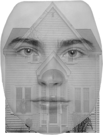
このビデオは、fMRI を使用して遊離脂肪酸と脳の PPA をローカライズする方法を示しています、注意制御の方法オブジェクト ベースを調べますこれらの領域の活動を調節します。後続の仮説テストを制限する機能ローカライザーの使用は機能イメージングの強力な技法です。参加者は顔と家のオーバーレイ イメージが表示されている間の機能的 MRI を受けます。にもかかわらず、顔と家は、各刺激の提示が、我々 は彼らの遊離脂肪酸および PPA の活動のパターンがどの項目に出席されているに基づいて変更されます予測します。3
手順
1. 参加者募集
- 20 人の参加者を募集します。
- 参加者は右利きで、神経学的または精神的な障害の歴史があります。
- 参加者に視覚的な合図を正しく表示できるように正常または正常に修正のビジョンが必要です。
- 参加者は、自分の体で金属をなりません。これ高磁場 fMRI の関与のための重要な安全要件です。
- 参加者する必要があります閉所恐怖症に苦しむ、fMRI スキャナーの小さなスペースで横になっている必要があるためにの穴はありません。
2. 事前スキャン手順
- スキャン前書類を記入します。
- 参加者は、fMRI スキャンのために来る、彼らは MRI、カウンター表示がない確認する金属スクリーン フォーム、放射線科、見てする彼らのスキャンのために同意を与える偶発的所見フォームおよびリスクと研究の利点について詳述した同意書の記入を最初に教えます。
- ベルト、財布、携帯電話、ヘアー クリップ、
結果
申請書と概要
参考文献
- Kanwisher N.G, McDermott J, Chun M.M. (1997). The fusiform face area: a module in human extrastriate cortex specialized for face perception. J. Neurosci., 17, 4302-4311.
- Epstein, R., & Kanwisher, N. (1998). A cortical representation of the local visual environment. Nature, 392, 598-601.
- Serences, J. T., Schwarzbach, J., Courtney, S. M., Golay, X., & Yantis, S. (2004). Control of Object-based Attention in Human Cortex. Cerebral Cortex, 14, 1346-1357.
タグ
スキップ先...
このコレクションのビデオ:

Now Playing
視覚的注意: fMRI 調査のオブジェクト ベースの注意制御
Neuropsychology
42.2K 閲覧数

スプリット ブレイン
Neuropsychology
68.5K 閲覧数

モーター マップ
Neuropsychology
27.7K 閲覧数

神経心理学の視点
Neuropsychology
12.2K 閲覧数

意思決定とギャンブル課題アイオワ
Neuropsychology
32.8K 閲覧数

自閉症スペクトラム障害の実行機能
Neuropsychology
18.0K 閲覧数

前向性健忘
Neuropsychology
30.5K 閲覧数

感情認識の生理学的相関
Neuropsychology
16.4K 閲覧数

事象関連電位とオドボール課題
Neuropsychology
27.6K 閲覧数

言語: 意味の違和感で N400
Neuropsychology
19.7K 閲覧数

学習と記憶: 覚えて知っているタスク
Neuropsychology
17.3K 閲覧数

ボクセル ベース形態計測と灰白質の違いを測定: 音楽脳
Neuropsychology
17.4K 閲覧数

Multivoxel パターン分析で聴覚イメージをデコード
Neuropsychology
6.5K 閲覧数

外傷性脳損傷の拡散テンソル画像を用いた
Neuropsychology
16.9K 閲覧数

TMS を使用して行動観察中にモーターの興奮性を測定するには
Neuropsychology
10.3K 閲覧数
Copyright © 2023 MyJoVE Corporation. All rights reserved