
Dissection and Immunohistochemistry of Larval, Pupal and Adult Drosophila Retinas
November 14th, 2012
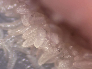
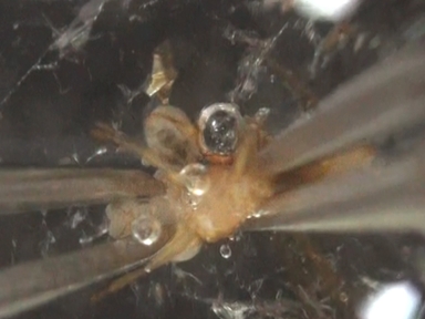
•The Drosophila retina is a crystal-like lattice composed of a small number of cell types that are generated in a stereotyped manner 1. Its amenability to sophisticated genetic analysis allows the study of complex developmental programs. This protocol describes dissections and immunohistochemistry of retinas at three discrete developmental stages, with a focus on photoreceptor differentiation.
Tags
Related Videos

Mapping and Application of Enhancer-trap Flippase Expression in Larval and Adult Drosophila CNS

Single Drosophila Ommatidium Dissection and Imaging

Dissection of Adult Mouse Utricle and Adenovirus-mediated Supporting-cell Infection
Functional Analysis of the Larval Feeding Circuit in Drosophila

Live Imaging of Drosophila Larval Neuroblasts

Acquisition of High-Quality Digital Video of Drosophila Larval and Adult Behaviors from a Lateral Perspective

Whole Mount Dissection and Immunofluorescence of the Adult Mouse Cochlea

Using Linear Agarose Channels to Study Drosophila Larval Crawling Behavior
A Simple One-step Dissection Protocol for Whole-mount Preparation of Adult Drosophila Brains

Metabolic Analysis of Drosophila melanogaster Larval and Adult Brains
Copyright © 2024 MyJoVE Corporation. 판권 소유