
The Use of Primary Human Fibroblasts for Monitoring Mitochondrial Phenotypes in the Field of Parkinson's Disease
Invalid date
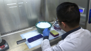
•Fibroblasts from patients carrying mutations in Parkinson's disease-causing genes represent an easily accessible ex vivo model to study disease-associated phenotypes. Live cell imaging gives the opportunity to study morphological and functional parameters in living cells. Here we describe the preparation of human fibroblasts and subsequent monitoring of mitochondrial phenotypes .
Related Videos

Formation of Covalent DNA Adducts by Enzymatically Activated Carcinogens and Drugs In Vitro and Their Determination by 32P-postlabeling

Generation of Self-assembled Vascularized Human Skin Equivalents

Using Retinal Imaging to Study Dementia

Quantification of three DNA Lesions by Mass Spectrometry and Assessment of Their Levels in Tissues of Mice Exposed to Ambient Fine Particulate Matter
A Luciferase-fluorescent Reporter Influenza Virus for Live Imaging and Quantification of Viral Infection

Abbiategrasso Brain Bank Protocol for Collecting, Processing and Characterizing Aging Brains

The Lublin Protocol of the Uterine Arteries Embolization in the Treatment of Symptomatic Uterine Fibroids

Basophil Activation Test for Allergy Diagnosis

Recording Electrical Currents across the Plasma Membrane of Mammalian Sperm Cells

Rodent Estrous Cycle Monitoring Utilizing Vaginal Lavage: No Such Thing As a Normal Cycle
Copyright © 2024 MyJoVE Corporation. 판권 소유