
Immunofluorescence Analysis of Endogenous and Exogenous Centromere-kinetochore Proteins
March 3rd, 2016
•Here we report protocols to detect endogenous and exogenous centromere-kinetochore proteins in human cells and quantify these protein levels at centromeres-kinetochores by indirect immunofluorescent staining through the use of fixation (paraformaldehyde, acetone, or methanol fixation).
Related Videos
A Neuronal and Astrocyte Co-Culture Assay for High Content Analysis of Neurotoxicity

ReAsH/FlAsH Labeling and Image Analysis of Tetracysteine Sensor Proteins in Cells
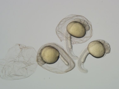
Analysis of Apoptosis in Zebrafish Embryos by Whole-mount Immunofluorescence to Detect Activated Caspase 3

Enrichment of Extracellular Matrix Proteins from Tissues and Digestion into Peptides for Mass Spectrometry Analysis

Use of Enzymatic Biosensors to Quantify Endogenous ATP or H2O2 in the Kidney

Removal of Exogenous Materials from the Outer Portion of Frozen Cores to Investigate the Ancient Biological Communities Harbored Inside

Co-immunoprecipitation Assay Using Endogenous Nuclear Proteins from Cells Cultured Under Hypoxic Conditions

Visualizing and Tracking Endogenous mRNAs in Live Drosophila melanogaster Egg Chambers

Quantitative Immunoblotting of Cell Lines as a Standard to Validate Immunofluorescence for Quantifying Biomarker Proteins in Routine Tissue Samples

Sequential Immunofluorescence and Immunohistochemistry on Cryosectioned Zebrafish Embryos
Copyright © 2024 MyJoVE Corporation. 판권 소유