
A Co-Culture Method to Study Neurite Outgrowth in Response to Dental Pulp Paracrine Signals
February 14th, 2020
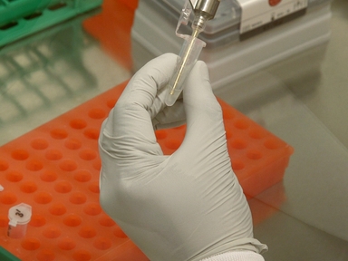
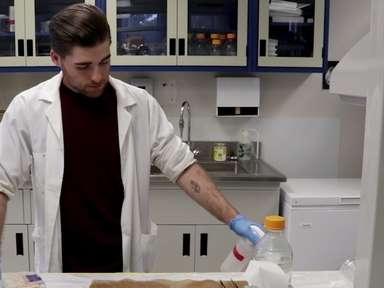
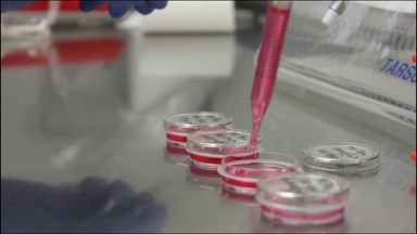
•We describe the isolation, dispersion and plating of dental pulp (DP) primary cells with trigeminal (TG) neurons cultured atop overlying transwell filters. Cellular responses of DP cells can be analyzed with immunofluorescence or RNA/protein analysis. Immunofluorescence of neuronal markers with confocal microscopy permits the analysis of neurite outgrowth responses.
Related Videos

An Optogenetic Approach for Assessing Formation of Neuronal Connections in a Co-culture System
Generating CRISPR/Cas9 Mediated Monoallelic Deletions to Study Enhancer Function in Mouse Embryonic Stem Cells

Using Touch-evoked Response and Locomotion Assays to Assess Muscle Performance and Function in Zebrafish

Application of Impermeable Barriers Combined with Candidate Factor Soaked Beads to Study Inductive Signals in the Chick

The Aortic Ring Co-culture Assay: A Convenient Tool to Assess the Angiogenic Potential of Mesenchymal Stromal Cells In Vitro
Organotypic Culture Method to Study the Development Of Embryonic Chicken Tissues

An In Vivo Method to Study Mouse Blood-Testis Barrier Integrity

Isolation, Propagation, and Prion Protein Expression During Neuronal Differentiation of Human Dental Pulp Stem Cells
Assessment of Oxidative Damage in the Primary Mouse Ocular Surface Cells/Stem Cells in Response to Ultraviolet-C (UV-C) Damage

An Enhanced Green Fluorescence Protein-based Assay for Studying Neurite Outgrowth in Primary Neurons
Copyright © 2024 MyJoVE Corporation. 판권 소유