
Dual-Dye Optical Mapping of Hearts from RyR2R2474S Knock-In Mice of Catecholaminergic Polymorphic Ventricular Tachycardia
December 22nd, 2023
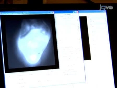
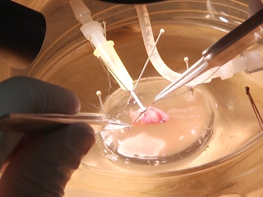
•This protocol introduces dual-dye optical mapping of mouse hearts obtained from wild-type and knock-in animals affected by catecholaminergic polymorphic ventricular tachycardia, including electrophysiological measurements of transmembrane voltage and intracellular Ca2+ transients with high temporal and spatial resolution.
Related Videos

Proper Care and Cleaning of the Microscope

Major Components of the Light Microscope

Phase Contrast and Differential Interference Contrast (DIC) Microscopy
Optical Mapping of Langendorff-perfused Rat Hearts

Semi-automated Optical Heartbeat Analysis of Small Hearts
Isolation of Embryonic Ventricular Endothelial Cells

Voltage and Calcium Dual Channel Optical Mapping of Cultured HL-1 Atrial Myocyte Monolayer

Optical Mapping of Intra-Sarcoplasmic Reticulum Ca2+ and Transmembrane Potential in the Langendorff-perfused Rabbit Heart
High-resolution Optical Mapping of the Mouse Sino-atrial Node

Subtype-specific Optical Action Potential Recordings in Human Induced Pluripotent Stem Cell-derived Ventricular Cardiomyocytes
Copyright © 2024 MyJoVE Corporation. 판권 소유