
Aby wyświetlić tę treść, wymagana jest subskrypcja JoVE. Zaloguj się lub rozpocznij bezpłatny okres próbny.
Method Article
Formulating and Characterizing an Exosome-based Dopamine Carrier System
W tym Artykule
Podsumowanie
Here, we aimed to obtain a formulation by dopamine loading of the isolated exosomes of stem cells from Wharton's jelly mesenchymal stem cells. Exosome isolation and characterization, drug loading into the resulting exosomes, and the cytotoxic activity of the developed formulation are described in this protocol.
Streszczenie
Exosomes between 40 and 200 nm in size constitute the smallest subgroup of extracellular vesicles. These bioactive vesicles secreted by cells play an active role in intercellular cargo and communication. Exosomes are mostly found in body fluids such as plasma, cerebrospinal fluid, urine, saliva, amniotic fluid, colostrum, breast milk, joint fluid, semen, and pleural acid. Considering the size of exosomes, it is thought that they may play an important role in central nervous system diseases because they can pass through the blood-brain barrier (BBB). Hence, this study aimed to develop an exosome-based nanocarrier system by encapsulating dopamine into exosomes isolated from Wharton's jelly mesenchymal stem cells (WJ-MSCs). Exosomes that passed the characterization process were incubated with dopamine. The dopamine-loaded exosomes were recharacterized at the end of incubation. Dopamine-loaded exosomes were investigated in drug release and cytotoxicity assays. The results showed that dopamine could be successfully encapsulated within the exosomes and that the dopamine-loaded exosomes did not affect fibroblast viability.
Wprowadzenie
Exosomes, bioactive vesicles with significant features, range in size from 40 nm to 200 nm. Exosomes originate from the cell membrane and are formed because of the release of the endosomes1. These structures serve as cell-to-cell communicators and interact with neighboring cells to facilitate the transfer of active molecules. Exosomes can be isolated from many different sources. These include body fluids such as plasma, urine, cerebrospinal fluid, saliva, as well as cell lines cultured under in vitro conditions. Exosomes have an important role in the elimination of nerve damage, thanks to the biomacromolecules they contain, such as lipids, proteins, and nucleic acids2. Glia, which are the supporting cells of the nervous system3, transfer proteins and micro RNAs to the axons of neurons via exosomes4.
Lipids forming the myelin sheath, which are a characteristic feature in nerve conduction, are also released from oligodendrocytes via exosomes4,5. Exosomes are also involved in processes such as synaptic plasticity, neuronal stress response, cell-cell communication, and neurogenesis in the brain6,7. The fact that exosomes possess nano-dimensions enables them to pass through the BBB. There is a special transition route from the interstitial fluid to the cerebrospinal fluid after penetrating this membrane8. Thanks to their surface properties, exosomes can interact efficiently with target cells as a drug delivery system and actively deliver the loaded drugs.
Due to the expression of various adhesive proteins (tetraspanins and integrins) on the surface of exosomes, these extracellular vesicles can easily interact and fuse with host cell membranes9. It is thought that exosomes can be used as a drug delivery system, especially in the treatment of central nervous system diseases due to their ability to penetrate the BBB and their surface properties. Mesenchymal stem cell (MSC)-derived exosomes have a lower risk of immune rejection compared to allogeneic cellular therapies, and in this respect, they can be an important component of cell-free treatment applications10.
Dopamine is a molecule whose deficiency in the brain is the characteristic feature of Parkinson's disease (PD), worsening day by day11,12,13. It is known that PD is associated with degeneration of dopaminergic neurons in the substantia nigra of the mesencephalon and loss of motor neuron functions14,15. The death of dopaminergic neurons prevents the supply of the neurotransmitter dopamine to the brain striatum. This, in turn, results in the emergence of PD-specific symptoms16. These symptoms of PD are bradykinesia, postural instability, rigidity, and especially resting tremor12,13. Although PD was first described more than two centuries ago, studies to understand the pathology and etiology of the disease are still ongoing and it is currently accepted that PD is a complex systemic disease17. It is predicted that dopamine deficiency occurs, and clinical PD symptoms are observed when more than 80% of neurons degenerate18. In the treatment of the disease, incomplete dopamine supplementation is preferred to reduce motor symptoms. In vivo studies have shown that direct infusion of dopamine into the brain significantly reduces symptoms in animals19. Dopamine precursors such as L-DOPA (L-3,4-dihydroxyphenylalanine) and dopamine receptor drugs are used in the clinic because the direct infusion of dopamine into the brain is not possible in humans and dopamine entering the system cannot cross the BBB20. These types of drugs lose their effectiveness over time. However, there is still no curative treatment approach for PD. Hence, there is a huge necessity to develop new therapeutic strategies and treatment modalities to reveal the pathophysiology of the disease and reduce the impact of PD on patients.
Exosome-based studies have recently attracted attention for gathering information about both therapeutic approaches and pathologies of nervous system diseases. MSC-derived exosomes have been shown to reduce inflammation in nerve damage and contribute to neuronal regeneration21,22,23. In addition, it has been reported that MSC-derived exosome secretomes reduce apoptosis by showing neurotrophic and neuroprotective effects, especially on dopaminergic neurons24,25. Research on platforms in which exosomes are used as therapeutic drug delivery systems have intensively accelerated in recent years. In numerous studies, it has been observed that relevant drugs can be easily encapsulated into exosomes and delivered safely into target cells, tissues, and organs26,27. Different methods such as incubation, freeze/thaw cycles, sonication, and extrusion could be used for drug loading into exosomes28.
Coincubation with exosomes or exosome-like vesicles allows lipophilic small molecules to be passively encapsulated into these delivery systems28,29,30. In particular, various molecules such as curcumin31, catalase30, doxorubicin32, and paclitaxel33 were effectively loaded into exosomes. It has been observed that catalase-containing exosomes, which have antioxidant activity, efficiently accumulated in the neurons and microglial cells in the brain and exhibited strong neuroprotective activities30. In the same study, saponin, added into the complex to increase the loading efficiency, was found to increase the drug loading percentage during incubation30,34. However, further studies are needed to establish the standards for drug loading into exosomes.
This paper describes the development of a nanocarrier system by encapsulation of dopamine into exosomes that were isolated from WJ-MSCs. All steps, including the cultivation of WJ-MSCs, isolation, and characterization of exosomes, drug loading experiments, characterization of dopamine-loaded exosomes with various techniques, and in vitro cytotoxicity analysis are explained in detail.
Access restricted. Please log in or start a trial to view this content.
Protokół
NOTE: See the Table of Materials for details related to all materials and equipment used in this protocol.
1. Culturing and cryopreservation of Wharton's jelly mesenchymal stem cells
- Remove the WJ-MSCs (from ATCC) from the -80 °C freezer. Seed the cells into a flask containing DMEM-F12 medium supplemented with 10% fetal bovine serum (FBS). Incubate them at 37 °C in an incubator containing 5% CO2.
- Change the culture medium every 2 days. Passage the cells when they reach 80% confluence. Wash the cells to be passaged with 5 mL of phosphate-buffered saline (PBS).
NOTE: Washing with PBS before adding trypsin ensures easy separation of cells from the flask surface. - Remove the PBS and add 5 mL of trypsin-EDTA solution to the flask. Incubate the flask for 5 min at 37 °C in an incubator containing 5% CO2.
- Collect the supernatant into the tube and centrifuge at 1,500 x g for 5 min. After centrifugation, remove the supernatant and add 1 mL of DMEM-F12 + 10% FBS to the tube by pipetting.
- Transfer the cells to a larger flask and continue the culture by adding DMEM-F12 + 10% FBS until sufficient exosomes are obtained35.
NOTE: Cultured cells are frozen at a density of 1 million/mL with 10% dimethyl sulfoxide (DMSO) and stored at -80 °C.
2. Production of exosomes from Wharton's Jelly mesenchymal stem cells
NOTE: Exosomes are isolated from cultured WJ-MSCs. Exosome isolation is performed when cells cover the flask surface and reach approximately 80% density.
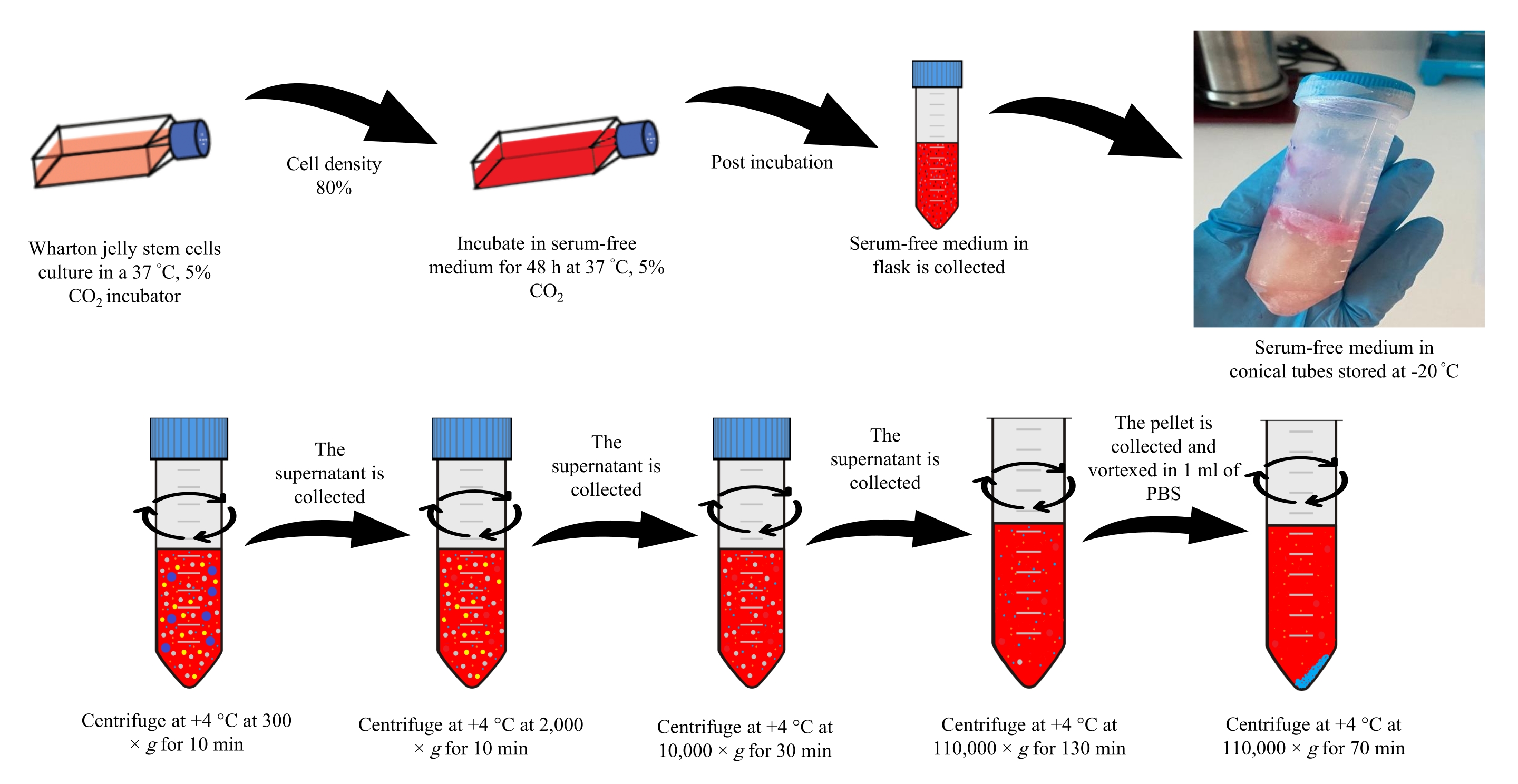
- Remove the supernatant medium containing 10% FBS from the flask and wash the cells with 5 mL of PBS. Add serum-free DMEM-F12 medium to the cells after rinsing with PBS. Incubate flasks for 48 h at 37 °C in a 5% CO2 incubator. Following incubation, collect the serum-free medium in the tube and store at -20 °C.
NOTE: In step 2.1, 180 mL of serum-free medium is collected and stored at -20 °C. After thawing, a batch centrifugation process is started, as described below. - Isolation of exosomes
- Centrifuge the collected serum-free medium at 300 x g for 10 min at +4 °C. Carefully withdraw the supernatant and transfer it to another tube.
- Centrifuge the collected supernatant at 2,000 × g for 10 min at +4 °C. Carefully withdraw the supernatant and transfer it to another tube.
- Centrifuge the collected supernatant at 10,000 × g for 30 min at +4 °C. Carefully withdraw the supernatant and transfer it to another tube.
- Transfer the collected supernatant to ultracentrifuge tubes and ultracentrifuge at 110,000 × g for 130 min. Slowly remove the supernatant and add 1 mL of PBS to the pellet.
- Vortex the pellet and ultracentrifuge at 110,000 × g for 70 min at +4 °C36 (Figure 1). Slowly remove the supernatant and add 1 mL of PBS to the pellet.
NOTE: After the ultracentrifugation steps are completed, the exosomes obtained can be stored at -80 °C. The obtained pellet is now ready for characterization.

Figure 1: Exosome isolation. Please click here to view a larger version of this figure.
3. Characterization of exosomes
- Nanoparticle Tracking Analysis (NTA)
NOTE: Nanoparticle tracking analysis is performed to determine the size and concentration of the isolated exosomes. Tablets used for 1x PBS solution must be in the range of pH 7.3-7.5. 1x PBS Solution contains 10 mM phosphate buffer, 137 mM sodium chloride, and 2.7 mM potassium chloride. The solution is prepared by adding 1 PBS tablet in 100 mL of distilled water. This prepared solution is sterilized by autoclaving.- Dilute the exosomes 100-fold by adding 990 µL of PBS to 10 µL of exosomes. Take the diluted suspension into a disposable syringe.
- Turn on the NTA device and connect the computer. Open the software.
- Inject the sample in the syringe into the tubing in the cassette section of the device. Close the cassette cover of the device.
- In the software that opens, click on the Start Analysis button. Save the results obtained by pressing the Record button.
- Dynamic light scattering analysis
NOTE: Exosomes isolated for zeta potential and size measurement are also diluted in the same manner as for NTA analysis.- Add 980 µL of PBS to 20 µL of the exosomes.
- Open the zeta sizing device and the connected computer. Open the software.
- Add the suspension from step 3.2.1 to the disposable cuvette. Open the lid of the device, place the cuvette in the cassette, and close the lid.
- Select the cuvette type in the device's software. Click on the Start Analysis button for zeta potential and dimensional analysis.
NOTE: Analyses are performed separately for dimensional analysis and zeta potential.
4. Dopamine loading into exosomes
NOTE: After the characterization of WJ-MSCs exosomes is completed, dopamine-loaded exosomes were obtained as a drug delivery system. Drug loading into exosomes is performed using the incubation method.
- Add 3 mL of PBS to the exosome suspension from step 2.2.5. Sterilize the diluted suspension by filtration using 0.22 µm filters. Transfer 500 µL of the sterilized exosome suspension to another tube.
- Prepare dopamine solution (0.5 mg/mL) with distilled water. Add 500 µL of dopamine solution into the tubes containing the exosomes.
- Add saponin to the suspension in step 4.2. Incubate the prepared exosome-dopamine suspension for 24 h at 37 °C.
NOTE: Saponin concentration should not exceed 0.002% of the total solution. - Ultracentrifuge the suspension at 90,000 × g for 70 min to remove free dopamine and saponin from the medium. Store the isolated exosomes at -20 °C until use for further analysis37.
NOTE: Add 0.02% ascorbic acid for the stability of the prepared dopamine solution.
5. Characterization of dopamine loaded exosomes
- Nanoparticle tracking analysis (NTA)
NOTE: Nanoparticle tracking analysis is performed to determine the size and concentration of isolated exosomes.- Follow steps 3.1.1-3.1.4 to dilute the exosomes and perform NTA.
- Dynamic light scattering analysis
NOTE: Exosomes isolated for zeta potential and size measurement are also diluted similarly to NTA analysis.- Follow steps 3.2.1-3.2.4 to dilute the exosomes and perform dynamic light scattering analysis.
NOTE: Size and zeta potentials for each solution (saponin, dopamine, exosome-dopamine) are analyzed separately.
- Follow steps 3.2.1-3.2.4 to dilute the exosomes and perform dynamic light scattering analysis.
6. High-Performance Liquid Chromatography (HPLC)
NOTE: The amounts of dopamine loaded into exosomes are measured by the high-performance liquid chromatography (HPLC) method. To detect the presence of dopamine within the obtained formulation, exosomes are detonated by a special process.
- Put the formulation in a 75 °C heater to evaporate. Add acetonitrile (50:50 ratio) to the suspension and vortex. Sonicate the solution for 10 min.
- Centrifuge the solution for 10 min at 10,000 × g. Filter the supernatant through a 0.22 µm filter.
- Analyze all analytes using a C18 column at 30 °C at a flow rate of 1 mL/min (the mobile phase H2O/acetonitrile). Measure the absorbance at 230 nm38.
7. Drug loading capacity (DL) measurement and in vitro drug release kinetics
- Drug loading capacity (DL)
NOTE: The amount of dopamine loaded into exosomes is quantified using UV-Vis spectroscopy. The absorbance is read at 280 nm. The collected supernatants during synthesis are used to measure the amount of unloaded drug. The dopamine concentration in the supernatant is determined via a standard calibration curve for dopamine.- Prepare a stock solution of 1 mg/mL of dopamine. Prepare concentrations of 0.05, 0.1, 0.2, 0.3, 0.4, and 0.5 mg/mL from the stock solution using distilled water.
NOTE: Add 0.02% ascorbic acid for the stability of the prepared dopamine solution. - Generate a standard calibration curve by measuring the absorbance of each dilution of dopamine at 280 nm in a UV-Spectrophotometer.
- Measure the absorbance of the supernatant at 280 nm.
- Calculate the drug loading capacity for dopamine using Eq (1)39.
DL (%) = 100 (1)
NOTE: The analysis is performed in triplicate.
- Prepare a stock solution of 1 mg/mL of dopamine. Prepare concentrations of 0.05, 0.1, 0.2, 0.3, 0.4, and 0.5 mg/mL from the stock solution using distilled water.
- In vitro drug release kinetics
NOTE: Drug release kinetics of dopamine-loaded exosomes are performed using a dialysis membrane. PBS, pH 7.4, is used as the release medium to simulate a physiological microenvironment.- Add 1 mL of dopamine-loaded exosomes to the dialysis membrane. Place the membrane in a beaker. Add 15 mL of PBS to the beaker.
- Sample 1 mL of release medium in a beaker at 0.5, 1, 2, 4, 6, and 8 h. At each time point, replace the volume of the sample with fresh PBS. Analyze the samples taken at specified intervals at 280 nm using a UV-Vis spectrometer.
- Calculate the results using Eq (2)40.
Release (%) = 100 (2)
NOTE: A calibration curve is prepared for the determination of in vitro drug release kinetics. Dopamine solution is prepared at 0.5, 1, 1.5, 2, 2.5, 3, 3.5, 4, 4.5, and 5.0 mg/mL concentrations. The UV absorbance value of each concentration is determined at 280 nm with a UV-Vis spectrophotometer.
8. In vitro cytotoxicity test
- Revitalization and culture of fibroblasts
- Remove the fibroblasts from the -80 °C freezer. Seed the cells into a flask containing DMEM-F12 + 10% FBS.
- Incubate the cells at 37 °C in an incubator containing 5% CO2. Change the culture medium every 2 days.
- Passage the cells until they reach 80% confluence. Follow steps 1.3-1.5. After centrifugation at 1,500 × g for 5 min, remove the supernatant, add 1 mL of DMEM-F12 + 10% FBS, and seed 10,000 cells per well in a 96 well plate.
- Add DMEM-F12 medium + 10% FBS and incubate the cells at 37 °C for 18 h in an incubator containing 5% CO2. Change the medium of the wells at the end of the incubation.
- Prepare the stock of dopamine-loaded exosome suspension. Dilute the dopamine-loaded exosome formulation with medium to obtain concentrations of 100 µL/mL, 250 µL/mL, 500 µL/mL, and 1,000 µL/mL.
- Add the prepared concentrations onto the cells within each well of the 96-well plate. Incubate the plates at 37 °C for 18 h in an incubator containing 5% CO241.
- MTT cell viability assay
NOTE: At the end of the incubation, the viability ratios of cells are determined by the MTT test.- Prepare the MTT solution (5 mg/mL) with PBS. Add 10 µL of MTT solution to each well. Incubate for 4 h at 37 °C.
- At the end of the incubation, add 100 µL of DMSO to each well. Incubate the plates for 30 min at room temperature in the dark. Measure the absorbance values in ELISA reader at 570 nm42,43.
NOTE: The MTT viability test is performed in triplicate.
Access restricted. Please log in or start a trial to view this content.
Wyniki
Exosome isolation and characterization
Wharton jelly stem cells are cultured and incubated in a serum-free medium for 48 h when the culture reaches sufficient density. After the end of the incubation, the supernatant is stored at -20 °C. The collected supernatants are diluted with PBS and subjected to ultracentrifugation (Figure 1). The obtained solution is analyzed by NTA and DLS analyses. The exosomes are sterilized by passing through a 0.22 µm filter. The ...
Access restricted. Please log in or start a trial to view this content.
Dyskusje
Exosomes are small membrane vesicles with dimensions of 40-200 nm secreted by most cell types, e.g., MSCs1. Capable of enabling communication between cells, exosomes can enter cells in different ways such as endocytosis, phagocytosis, micropinocytosis, lipid-mediated internalization, and fusion33,44. Compared to other nanocarrier systems, the lipids and cholesterol found on the exosome surface confer the ability to carry both hydrophilic a...
Access restricted. Please log in or start a trial to view this content.
Ujawnienia
The authors declare that they have no competing financial interests.
Podziękowania
The work was primarily supported by research funding provided by Yıldız Technical University Scientific Research Projects (TSA-2021-4713).
Access restricted. Please log in or start a trial to view this content.
Materiały
| Name | Company | Catalog Number | Comments |
| 0.22 µm membrane filter | Aisimo | Used for the sterilization process | |
| 0.45 µm syringe filter | Aisimo | Used for the sterilization process | |
| 15 mL Falcon tube | Nest | Used in cell culture step | |
| 25 cm2 cell culture flasks (Falcon, TPP tissue culture flasks | Nest | Used in cell culture step | |
| 50 mL Falcon tube | Nest | Used in cell culture step | |
| 75 cm2 cell culture flasks (Falcon, TPP tissue culture flasks | Nest | Used in cell culture step | |
| 96 well plates (Falcon, TPP microplates) | Merck Millipore | Used in cell culture step | |
| Acetonitrile | Sigma | 271004-1L | Used for HPLC analysis |
| Autoclave | NUVE-OT 90L | Used for the sterilization process | |
| Cell Culture Cabin | Hera Safe KS | Used for the cell culture process | |
| Centrifugal | Hitachi | CF16RN | Used in the exosome isolation step |
| CO2 incubator with Safe Cell UV | Panasonic | Used for the cell culture process | |
| Dopamine hydrochloride H8502-10G | Sigma | H8502-10G | Used in exosome dopamine loading |
| Dulbecco's Modified Eagle's Medium/Nutrient Mixture-F12 | Sigma | RNBJ7249 | Used as cell culture medium |
| Fetal Bovine Serum-FBS | Capricorn | FBS-16A | It was used by adding to the cell culture medium. |
| Freezer -80 °C | Panasonic | MDF-U5386S-PE | To store cells and the resulting exosomes |
| Fridge | Panasonic | MPR-215-PE | Used to store cell culture and other materials |
| High performance liquid chromatography-HPLC | Agilent Technologies | The presence of dopamine from the content of the obtained formulation was investigated. | |
| Microscope- Primovert | Zeiss | Used to observe cells in cell culture. | |
| MTT Assay | Biomatik | Used to measure cell viability | |
| NanoSight NS300 | Malvern panalytical | Malvern panalytical | Used for exosome characterization |
| Optima XPN-100 Ultracentrifuge | Beckman Coulter | Used in the exosome isolation step | |
| PBS tablet | Biomatik | 43602 | In the preparation of the PBS solution |
| Penicilin/Streptomycin Solution | Capricorn | PB-S | It was added to the medium to prevent contamination in cell culture. |
| Pipette Aid | Isolab | ||
| Precision balance-Kern | Kern-ABJ220-4NM | Used in the preparation of solutions | |
| Q500 Sonicator | Qsonica, LLC | Used to digest exosomes in HPLC analysis | |
| Saponin | Sigma | 47036-50G-F | It was used by adding it to the total solution in the exosome dopamine loading process. |
| Spectrostar-Nano-Spectrophotometry | BMG LABTECH | Used for MTT and drug release analyzes | |
| SPSS 22 | statistical package program | ||
| Vorteks-FinePCR | FinePCR-FineVortex | Used to mix solutions homogeneously | |
| Water Bath 37 °C-Senova | Senova | Used in cell culture step | |
| Wharton’s jelly mesenchymal stem cells | ATCC | ||
| ZetaSizer | Malvern Nano ZS | Malvern Nano ZS | Used for exosome characterization |
Odniesienia
- Ingato, D., Lee, J. U., Sim, S. L., Kwon, Y. L. Good things come in small packages: Overcoming challenges to harness extracellular vesicles for therapeutic delivery. Journal of Controlled Release. 241, 174-185 (2016).
- Riazifar, M., et al. Stem cell-derived exosomes as nanotherapeutics for autoimmune and neurodegenerative disorders. ACS Nano. 13 (6), 6670-6688 (2019).
- Ursavaş, S., Darıci, H., Karaoz, E. Olfactory ensheathing cells: Unique glial cells promising for treatments of spinal cord injury. Journal of Neuroscience Research. 99 (6), 1579-1597 (2021).
- Skog, J., et al. Glioblastoma microvesicles transport RNA and proteins that promote tumour growth and provide diagnostic biomarkers. Nature Cell Biology. 10 (12), 1470-1476 (2008).
- Fruhbeis, C., Frohlich, D., Kramer-Albers, E. M. Emerging roles of exosomes in neuron-glia communication. Frontiers in Physiology. 3 (119), 1-7 (2012).
- Qing, L., Chen, H., Tang, J., Jia, X. Exosomes and their microRNA cargo: new players in peripheral nerve regeneration. Neurorehabil Neural Repair. 32 (9), 765-776 (2018).
- Saeedi, S., Israel, S., Nag, C., Turecki, G. The emerging role of exosomes in mental disorders. Translational Psychiatry. 9 (1), 122(2019).
- Jan, A. T., et al. Perspective insights of exosomes in neurodegenerative diseases: A critical appraisal. Frontiers in Aging Neuroscience. 9, 317(2017).
- Montecalvo, A., et al. Mechanism of transfer of functional microRNAs between mouse dendritic cells via exosomes. Blood. 119 (3), 756-766 (2012).
- Yu, B., Zhang, X., Li, X. Exosomes derived from mesenchymal stem cells. International Journal of Molecular Sciences. 15 (3), 4142-4157 (2014).
- Pahuja, R., et al. Trans-blood brain barrier delivery of dopamine-loaded nanoparticles reverses functional deficits in Parkinsonian rats. ACS Nano. 9 (5), 4850-4871 (2015).
- Rao, S. S., Hofmann, L. A., Shakil, A. Parkinson's disease: Diagnosis and treatment. American Family Physician. 15 (74), 2046-2054 (2006).
- Teves, J. M. Y., et al. Parkinson's disease skin fibroblasts display signature alterations in growth, redox homeostasis, mitochondrial function, and autophagy. Frontiers Neuroscience. 11, 737(2017).
- Kalia, L. V., Lang, A. E. Parkinson disease in 2015: Evolving basic, pathological and clinical concepts in PD. Nature Reviews Neurology. 12 (2), 65-66 (2016).
- Poewe, W., et al. Parkinson disease. Nature Reviews Disease Primers. 3, 17013(2017).
- Carlsson, T., Björklund, T. Restoration of the striatal dopamine synthesis for Parkinson's disease: Viral vector-mediated enzyme replacement strategy. Current Gene Therapy. 7 (2), 109-120 (2007).
- Simon, D. K., Tanner, C. M., Brundin, P. Parkinson disease epidemiology, pathology, genetics, and pathophysiology. Clinics in Geriatric Medicine. 36 (1), 1-12 (2020).
- Salat, D., Tolosa, E. Levodopa in the treatment of Parkinson's disease: Current status and new developments. Journal of Parkinson's Disease. 3 (3), 255-269 (2013).
- Senthilkumar, K. S., et al. Unilateral implantation of dopamine-loaded biodegradable hydrogel in the striatum attenuates motor abnormalities in the 6-Hydroxydopamine model of Hemi-Parkinsonism. Behavioral Brain Research. 184 (1), 11-18 (2007).
- Krishna, R., Ali, M., Moustafa, A. A. Effects of combined MAO-B inhibitors and levodopa vs. monotherapy in Parkinson's disease. Frontiers Aging Neuroscience. 6, 180(2014).
- Armstrong, M. J., Okun, M. S. Diagnosis and treatment of Parkinson disease: A review. JAMA. 323 (6), 548-560 (2020).
- Rivero Vaccari, J. P. Exosome-mediated inflammasome signaling after central nervous system injury. Journal of Neurochemistry. 136, Suppl. S1 39-48 (2016).
- Han, D., Wu, C., Xiong, Q., Zhou, L., Tian, Y. Anti-inflammatory mechanism of bone marrow mesenchymal stem cell transplantation in rat model of spinal cord injury. Cell Biochemistry and Biophysics. 71 (3), 1341-1347 (2015).
- Vilaça-Faria, H., Salgado, A. J., Teixeira, F. G. Mesenchymal stem cells-derived exosomes: A new possible therapeutic strategy for Parkinson's disease? Cells. 8 (2), 118-135 (2019).
- Mendes-Pinheiro, B., et al. marrow mesenchymal stem cells secretome exerts neuroprotective effects in a Parkinson's disease rat model. Frontiers in Bioengineering and Biotechnology. 7, 294(2019).
- Kalani, A., Kamat, P. K., Chaturvedi, P., Tyagi, S. C., Tyagi, N. Curcumin-primed exosomes mitigate endothelial cell dysfunction during hyperhomocysteinemia. Life Sciences. 107 (1-2), 1-7 (2014).
- Kalani, A., Tyagi, A., Tyagi, N. Exosomes: Mediators of neurodegeneration, neuroprotection, and therapeutics. Molecular Neurobiology. 49 (1), 590-600 (2014).
- Jamur, M. C., Oliver, C. Permeabilization of cell membranes. Methods in Molecular Biology. 588, 63-66 (2010).
- Rani, S., Ryan, A. E., Griffin, M. D., Ritter, T. Mesenchymal stem cell-derived extracellular vesicles: Toward cell-free therapeutic applications. Molecular Therapy. 23 (5), 812-823 (2015).
- Haney, M. J., et al. Exosomes as drug delivery vehicles for Parkinson's disease therapy. Journal of Controlled Release. 207, 18-30 (2015).
- Sun, D., et al. A novel nanoparticle drug delivery system: The anti-inflammatory activity of curcumin is enhanced when encapsulated in exosomes. Molecular Therapy. 18 (9), 1606-1614 (2010).
- Tian, Y., et al. A doxorubicin delivery platform using engineered natural membrane vesicle exosomes for targeted tumor therapy. Biomaterials. 35 (7), 2383-2390 (2014).
- Yang, T., et al. Exosome delivered anticancer drugs across the blood-brain barrier for brain cancer therapy in danio rerio. Pharmaceutical Research. 32 (6), 2003-2014 (2015).
- Chen, H. X., et al. Exosomes derived from mesenchymal stem cells repair a Parkinson's disease model by inducing autophagy. Cell Death Disease. 11 (4), 288(2020).
- Yu, F., et al. Olfactory ensheathing cells seeded decellularized scaffold promotes axonal regeneration in spinal cord injury rats. Journal of Biomedical Materials Research. 109 (5), 779-787 (2020).
- Coughlan, C., et al. Exosome isolation by ultracentrifugation and precipitation and techniques for downstream analyses. Current Protocols in Cell Biology. 88 (1), 110(2020).
- Qu, M., et al. Dopamine-loaded blood exosomes targeted to brain for better treatment of Parkinson's disease. Journal of Controlled Release. 287, 156-166 (2018).
- Kim, M. S., et al. Development of exosome-encapsulated paclitaxel to overcome MDR In cancer cells. Nanomedicine: Nanotechnology, Biology, and Medicine. 12 (3), 655-664 (2016).
- Branquinho, R. T., et al. HPLC-DAD and UV-spectrophotometry for the determination of lychnopholide in nanocapsule dosage form: Validation and application to release kinetic study. Journal of Chromatographic Science. 52 (1), 19-26 (2014).
- Karagöz Kutlutürk, I., et al. Producing aflibercept loaded poly (lactic-co-glycolic acid) [PLGA] nanoparticles as a new ocular drug delivery system and its challenges. Fresenius Environmental Bulletin. 30 (2), 1481-1493 (2021).
- Setiawatie, E. M., et al. Viability of nigella sativa toothpaste with SLS compared non-SLS on fibroblast cell culture. Journal of International Dental and Medical Research. 14 (2), 525-528 (2021).
- Jebelli, A., Khalaj-Kondori, M., Rahmati-Yamchi, M. The effect of beta-boswellic acid on the expression of Camk4 and Camk2α genes in the PC12 cell line. Advanced Pharmaceutical Bulletin. 10 (3), 437-443 (2020).
- Cantelmo, R. A., Santos, N. A., Santos, A. C., Regiane, S., Joca, L. Dual effects of S-Adenosyl-Methionine on Pc12 cells exposed to the dopaminergic neurotoxin MPP. Journal of Pharmacy and Pharmacology. 72 (10), 1427-1435 (2020).
- Mulcahy, L. A., Pink, R. C., Carter, D. R. Routes and mechanisms of extracellular vesicle uptake. Journal of Extracell Vesicles. 3, (2014).
- Kooijmans, S. A., Vader, P., van Dommelen, S. M., van Solinge, W. W., Schiffelers, R. M. Exosome mimetics: A novel class of drug delivery systems. International Journal of Nanomedicine. 7 (1), 1525-1541 (2012).
- Yuan, Z., Kolluri, K. K., Gowers, K. H., Janes, S. M. Trail delivery by MSC-derived extracellular vesicles is an effective anticancer therapy. Journal Extracell Vesicles. 6 (1), 1265291(2017).
- Darici, H., Sun, E., Koyuncu-Irmak, D., Karaöz, E. Mesenchymal stem cells for the treatment of COVID-19: Why and when they should be used? in Human Mesenchymal Stem Cells. Journal of Stem Cells. Khan, M. 4 (15), 159-181 (2021).
- Koyuncu-Irmak, D., Darici, H., Karaoz, E. Stem cell-based therapy option in COVID-19: Is it promising? Aging and Disease. 11 (5), 1174-1191 (2020).
- Leblanc, P., et al. Isolation of exosomes and microvesicles from cell culture systems to study prion transmission. Exosomes Microvesicles. 1545, 153-176 (2017).
- Fuhrmann, G., Serio, A., Mazo, M., Nair, R., Stevens, M. M. Active loading into extracellular vesicles significantly improves the cellular uptake and photodynamic effect Of porphyrins. Journal of Control Release. 205, 35-44 (2015).
- Mehryab, F., et al. Exosomes as a next-generation drug delivery system: An update on drug loading approaches, characterization, and clinical application challenges. Acta Biomaterialia. 113, 42-62 (2020).
- Zhang, Y., et al. Exosome: A review of its classification, isolation techniques, storage, diagnostic and targeted therapy applications. International Journal of Nanomedicine. 15, 6917-6934 (2020).
Access restricted. Please log in or start a trial to view this content.
Przedruki i uprawnienia
Zapytaj o uprawnienia na użycie tekstu lub obrazów z tego artykułu JoVE
Zapytaj o uprawnieniaThis article has been published
Video Coming Soon
Copyright © 2025 MyJoVE Corporation. Wszelkie prawa zastrzeżone