
Whole-animal Imaging and Flow Cytometric Techniques for Analysis of Antigen-specific CD8+ T Cell Responses after Nanoparticle Vaccination
April 29th, 2015
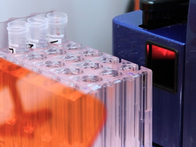
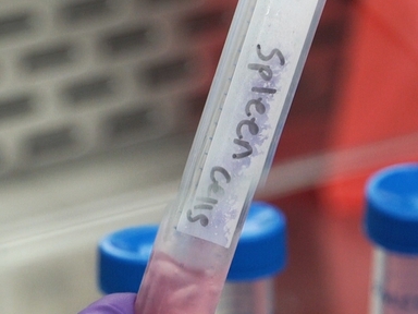
•We describe whole-animal imaging and flow cytometry-based techniques for monitoring expansion of antigen-specific CD8+ T cells in response to immunization with nanoparticles in a murine model of vaccination.
Tags
Vídeos Relacionados

Kupffer Cell Isolation for Nanoparticle Toxicity Testing

Isolation and Flow Cytometric Analysis of Immune Cells from the Ischemic Mouse Brain

Isolation and Flow Cytometric Analysis of Glioma-infiltrating Peripheral Blood Mononuclear Cells

Development of Stem Cell-derived Antigen-specific Regulatory T Cells Against Autoimmunity
Flow Cytometric Analysis of Natural Killer Cell Lytic Activity in Human Whole Blood

Isolation and Flow Cytometric Analysis of Human Endocervical Gamma Delta T Cells

Measurement of T Cell Alloreactivity Using Imaging Flow Cytometry

In Situ MHC-tetramer Staining and Quantitative Analysis to Determine the Location, Abundance, and Phenotype of Antigen-specific CD8 T Cells in Tissues
In Vivo Assay for Detection of Antigen-specific T-cell Cytolytic Function Using a Vaccination Model

Optimized Interferon-gamma ELISpot Assay to Measure T Cell Responses in the Guinea Pig Model after Vaccination
SOBRE A JoVE
Copyright © 2024 MyJoVE Corporation. Todos os direitos reservados