
Quantitative Fluorescence In Situ Hybridization (FISH) and Immunofluorescence (IF) of Specific Gene Products in KSHV-Infected Cells
August 27th, 2019
•We describe a protocol utilizing fluorescence in situ hybridization (FISH) to visualize multiple herpesviral RNAs within lytically infected human cells, either in suspension or adherent. This protocol includes quantification of fluorescence producing a nucleocytoplasmic ratio and can be extended for simultaneous visualization of host and viral proteins with immunofluorescence (IF).
Tags
Vídeos Relacionados
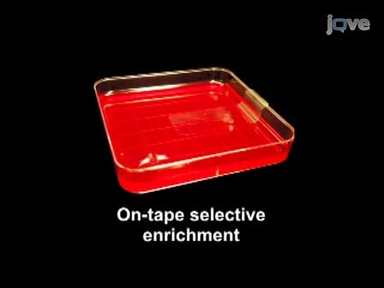
Combination of Adhesive-tape-based Sampling and Fluorescence in situ Hybridization for Rapid Detection of Salmonella on Fresh Produce
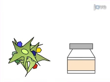
Generation of Multivirus-specific T Cells to Prevent/treat Viral Infections after Allogeneic Hematopoietic Stem Cell Transplant
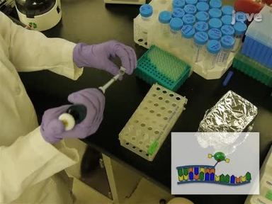
Locked Nucleic Acid Flow Cytometry-fluorescence in situ Hybridization (LNA flow-FISH): a Method for Bacterial Small RNA Detection

Hybridization in situ of Salivary Glands, Ovaries, and Embryos of Vector Mosquitoes

Fluorescent in situ Hybridization on Mitotic Chromosomes of Mosquitoes
piggyBac Transposon System Modification of Primary Human T Cells

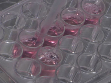
Quantitative In vitro Assay to Measure Neutrophil Adhesion to Activated Primary Human Microvascular Endothelial Cells under Static Conditions

Fluorescence in situ Hybridizations (FISH) for the Localization of Viruses and Endosymbiotic Bacteria in Plant and Insect Tissues

In Situ MHC-tetramer Staining and Quantitative Analysis to Determine the Location, Abundance, and Phenotype of Antigen-specific CD8 T Cells in Tissues
Visualization of Candida albicans in the Murine Gastrointestinal Tract Using Fluorescent In Situ Hybridization
SOBRE A JoVE
Copyright © 2024 MyJoVE Corporation. Todos os direitos reservados