
A subscription to JoVE is required to view this content. Sign in or start your free trial.
Method Article
Deciphering and Imaging Pathogenesis and Cording of Mycobacterium abscessus in Zebrafish Embryos
In This Article
Summary
Optically transparent zebrafish embryos are widely used to study and visualize in real time the interactions between pathogenic microorganisms and the innate immune cells. Micro-injection of Mycobacterium abscessus, combined with fluorescence imaging, is used to scrutinize essential pathogenic features such as cord formation in zebrafish embryos.
Abstract
Zebrafish (Danio rerio) embryos are increasingly used as an infection model to study the function of the vertebrate innate immune system in host-pathogen interactions. The ease of obtaining large numbers of embryos, their accessibility due to external development, their optical transparency as well as the availability of a wide panoply of genetic/immunological tools and transgenic reporter line collections, contribute to the versatility of this model. In this respect, the present manuscript describes the use of zebrafish as an in vivo model system to investigate the chronology of Mycobacterium abscessus infection. This human pathogen can exist either as smooth (S) or rough (R) variants, depending on cell wall composition, and their respective virulence can be imaged and compared in zebrafish embryos and larvae. Micro-injection of either S or R fluorescent variants directly in the blood circulation via the caudal vein, leads to chronic or acute/lethal infections, respectively. This biological system allows high resolution visualization and analysis of the role of mycobacterial cording in promoting abscess formation. In addition, the use of fluorescent bacteria along with transgenic zebrafish lines harbouring fluorescent macrophages produces a unique opportunity for multi-color imaging of the host-pathogen interactions. This article describes detailed protocols for the preparation of homogenous M. abscessus inoculum and for intravenous injection of zebrafish embryos for subsequent fluorescence imaging of the interaction with macrophages. These techniques open the avenue to future investigations involving mutants defective in cord formation and are dedicated to understand how this impacts on M. abscessus pathogenicity in a whole vertebrate.
Introduction
Mycobacterium abscessus is an emerging pathogen that causes a wide spectrum of clinical syndromes in humans. These include cutaneous infections as well as severe chronic pulmonary infections, mostly encountered in immunocompromised and in cystic fibrosis patients1,2,3,4. M. abscessus is also regarded as a major rapidly-growing mycobacterial species responsible for nosocomial and iatrogenic infections in humans. Moreover, several recent reports highlighted the possibility that M. abscessus could cross the blood-brain barrier and induce important lesions in the central nervous system (CNS)5,6. Despite being a rapid grower, M. abscessus exhibits also several pathogenic features that are related to those of Mycobacterium tuberculosis, including the capacity to remain silent for years within granulomatous structures and to generate caseous lesions in the lungs7. More alarming is the low sensitivity of M. abscessus to antibiotics, rendering these infections extremely difficult to treat leading to a significant therapeutic failure rate8,9. The important threat of this species is mainly its intrinsic resistance to antibiotics, which is of major concern in public health institutions10 and a contraindication to lung transplantation11.
M. abscessus displays smooth (S) or rough (R) colony morphotypes that lead to different clinical outcomes. In contrast to the S strain, R bacteria have a tendency to grow end to end, leading to a rope or cord-like structure12,13. Several independent studies based on either cellular or animal models revealed the hyper-virulence phenotype of the R morphotype14,15. From epidemiological studies, the most severe cases of M. abscessus pulmonary infections appear to be associated with R variants16 which are the only variant that has been seen to persist for years in an infected host3. The morphotype difference relies on the presence (in S) or loss (in R) of surface-associated glycopeptidolipids (GPL)12. However, due to the inherent limitations of the currently available cellular/animal models used to study M. abscessus infection, our knowledge regarding the pathophysiological events of the R or S variants remains obscure. Infection of immuno-competent mice via intravenous or aerosol routes leads to transient colonization, impeding the use of mice to study persistent infections and for in vivo drug susceptibility testing17. Therefore, developing animal models amenable to the manipulation of the host response is a major challenge. In this context, non-mammalian models of infection have been developed recently, including Drosophila melanogaster18 that offers several advantages such as cost, speed and ethical acceptability over the mouse model. The zebrafish (Danio rerio) model of infection has also been explored to visualize, by non-invasive imaging, the progression and chronology of M. abscessus infection in a live animal19. Importantly, a proof of concept was also established to demonstrate its suitability for in vivo antibiotic assessments against M. abscessus17,20.
The zebrafish have been widely used during the last two decades to study the interactions between various pathogens and the host immune system21. The increasing success of this alternative vertebrate model relies on major and unique opportunities that motivated and validated its use for a better understanding of numerous viral and bacterial infections19,22,23,24,25,26,27,28,29. As opposed to most other animal models, zebrafish embryos are optically transparent, allowing non-invasive fluorescence imaging30. This has led to study M. abscessus infected zebrafish embryos with unprecedented details, culminating with the description of extracellular cording, that represent an example of bacterial morphological plasticity. Cording represents a new mechanism of subversion of the immune system and a key mechanism promoting pathogenesis of acute M. abscessus infection19.
This report describes new tools and methods using the zebrafish embryo to decipher the pathophysiological traits of M. abscessus infection and to study the intimate interactions between the bacilli and the innate immune system. First, a detailed microinjection protocol that includes processing of the bacterial inoculum, embryo preparation, and infection per se, is presented. Methods specifically adapted to assess M. abscessus virulence by measuring various parameters, such as host survival and bacterial burden, are presented. Special focus is given on how to monitor, at a spatiotemporal level, the fate and progression of the infection and the host immune response to M. abscessus using video microscopy. Moreover, to investigate the contribution and role of macrophages during M. abscessus infection, methods to generate macrophages-depleted embryos (using either genetically- or chemically-based approaches) are described. Finally, protocols to visualize the specific interactions with macrophages or neutrophils using either fixed or living embryos are documented.
The aim of this report is to stimulate further studies to shed new light into M. abscessus virulence mechanisms and particularly the role of cording in the establishment of an acute and uncontrolled infection process.
Protocol
Zebrafish experimental procedures must comply with the relevant institutional and governmental regulations. For the present study, zebrafish experiments were done at the University Montpellier, according to European Union guidelines for handling of laboratory animals (http://ec.europa.eu/environment/chemicals/lab_animals/home_en.htm) and approved under the reference CEEA-LR-13007.
1. Preparation of Reagents and Microinjection Equipment
- Prepare fish water by dissolving 0.06 g Instant Ocean Sea salt in 1 L distilled water31, then autoclave to sterilize (120 °C for 20 min) and store at 28.5 °C for up to 1 month.
- Prepare methylene blue solution by dissolving 1 g of methylene blue in 1 L distilled water. Add 300 µl of methylene blue solution in 1 L fish water to obtain blue fish water, then autoclave to sterilize and store at 28.5 °C for up to 1 month.
NOTE: The addition of methylene blue in fish water prevents mold growth. - Preparation of Phosphate Buffered Saline (PBS)
- Prepare a 10 X PBS stock solution by dissolving 5.696 g Na2HPO4, 680 mg KH2PO4, 969 mg KCl and 78.894 g NaCl in 1 L distilled water and adjust the pH to 7.0 with HCl. To obtain 1 X PBS, dilute 100 ml of the 10 X PBS solution in 900 ml distilled water and autoclave during 20 min at 120 °C to sterilize.
- Add 0.05% Tween 80 to obtain 1 X PBST and store at RT for up to 1 year.
- Prepare oleic Acid Dextrose Catalase (OADC) enrichment by dissolving 8.5 g NaCl, 50 g Bovine Serum Albumin, 20 g D-Glucose, 40 mg Catalase from bovine liver and 0.5 g of oleic acid in 1 L distilled water then filter sterilize the solution and store it at 4 °C for up to 6 months.
- Prepare 100 ml of ethyl 3-aminobenzoate methanesulfonic acid (tricaine) stock solution, by dissolving 400 mg tricaine powder in 97.9 ml distilled water. Add 2.1 ml of 1 M Tris (pH 9) to adjust the pH at 7.0 and store the solution at -20 °C.
- Prepare the microcapillary injection needles by pulling borosilicate glass capillaries (1mm O.D. X 0.78 mm I.D.) using a micropipette puller device with the following settings : Air pressure 650; Heat 999; Pull 50; Velocity 80; Time 200.
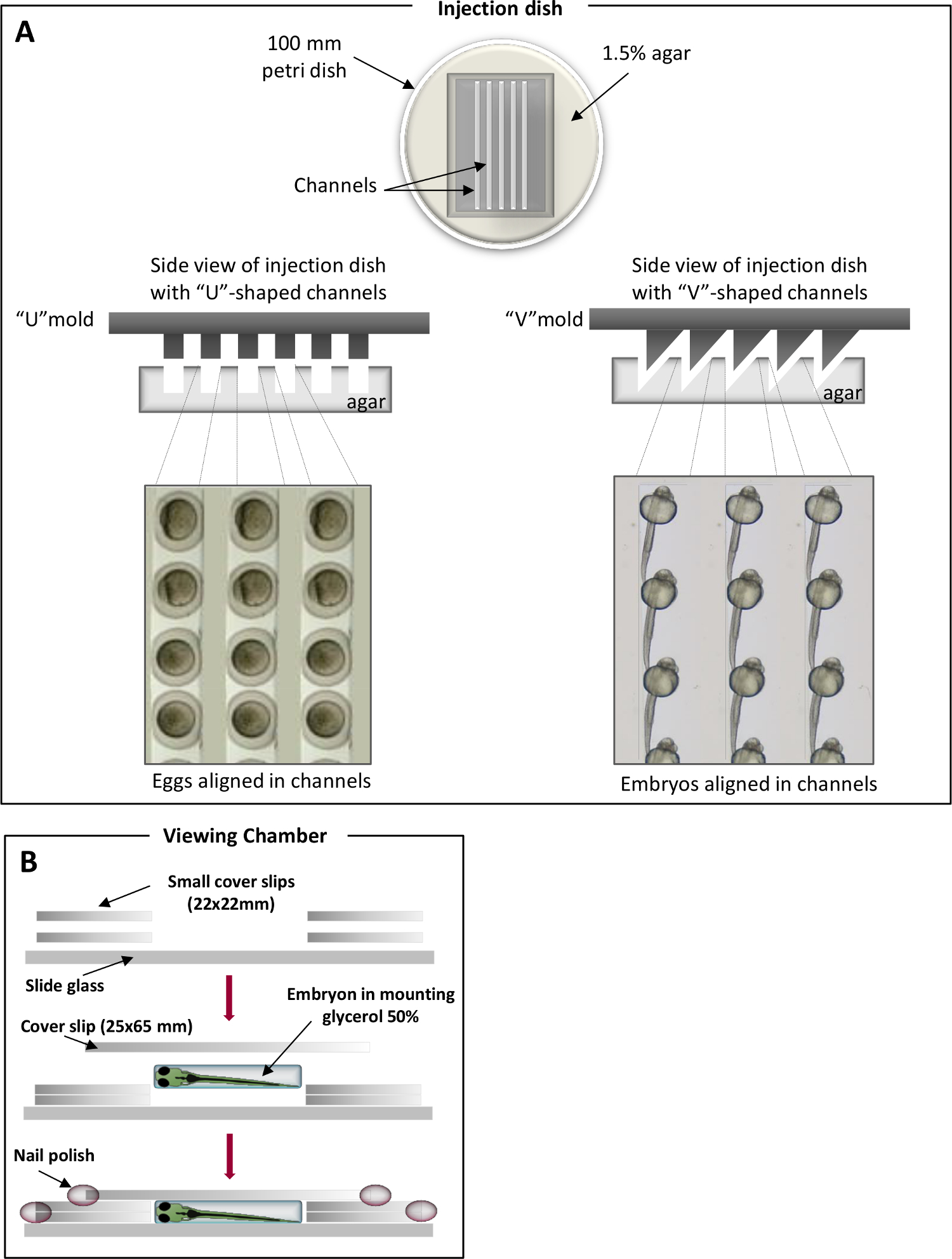
- Prepare a 1.5% agar solution in blue fish water and pour 25 ml of melted agar in 100 mm petri dishes. On the top of the melted agar, place homemade molds in order to imprint specific channels. “V”-shaped channels are used to position embryos while the “U”-shaped are used for eggs (Figure 1)31. Once solidified, remove carefully the imprint.
NOTE: Cover the agar with blue fish water to avoid dehydration and store at 4 °C for up to 2 months.

Figure 1. Positioning chambers for zebrafish injections. (A) U-shaped channels for eggs (left panel) and V-shaped channels for embryos (right panel). Zebrafish eggs/embryos are laid in the channels and aligned along the same axis. (B) Viewing chambers for microscopic observation. Please click here to view a larger version of this figure.
2. Preparation and Storage of the M. abscessus Inoculum
- M. abscessus growth conditions
- Plate out M. abscessus from a -80°C glycerol stock onto a Middlebrook 7H10 agar containing 10% of OADC and 0.5% glycerol (7H10OADC) and supplemented with the appropriate antibiotic depending on the resistance marker carried by the vector encoding the fluorescent protein. Incubate the plates at 30 °C for 4-5 days.
- Pick up a fluorescence-positive mycobacterial colony and inoculate 1 ml Middlebrook 7H9 broth supplemented with OADC, 0.2% glycerol, 0.05% Tween 80 (7H9OADC/T) with the required antibiotic in a 15 ml sterile plastic tube. Incubate at 30 °C for 1 week without shaking.
NOTE: Tween 80 restricts clumping and bacterial aggregation. - Resuspend the pre-culture obtained in 2.1.2 in 50 ml Middlebrook 7H9OADC/T in 150 cm2 tissue culture flasks to a final 0.1 OD600 (corresponding to around 5.107 bacteria/ml) and incubate further at 30 °C for 4 days to obtain an exponentially-growing culture (OD600 = 0.6 to 0.8).
NOTE: To minimize clumping, especially of the rough variant, incubation should not exceed 5 days. Both smooth and rough variants display a similar growth rate.
- Preparation of the M. abscessus inocula
NOTE: Due to high propensity of rough M. abscessus to form large aggregates and to produce cords, a specific treatment is necessary to generate homogeneous and quantitatively controlled inocula prior to microinjections in the embryos. This treatment is also applied to smooth bacteria that produce aggregates, albeit to a lesser extent than the rough strain.- Harvest exponentially-growing cultures from 150 cm2 tissue culture flasks by centrifugation at 4, 000 x g for 15 min at RT in a 50 ml sterile plastic tube and resuspend the bacterial pellet in 1 ml 7H9OADC/T. Aliquot 200 µl of bacterial suspensions into 1.5 ml microcentrifuge tubes.
NOTE: Working with small volumes in 1 ml syringes is necessary to obtain highly homogeneous suspensions. - Homogenize the bacterial suspensions with a 26-G needle (15 up-and-down sequences), sonicate three times for 10 sec (with 10 sec breaks between each sonication sequence) using a waterbath sonicator. Add 1 ml of 7H9OADC/T and vortex briefly. Centrifuge 3 min at 100 x g.
- Carefully collect the mycobacteria-containing supernatants to avoid the clumps and pool the homogenous suspensions in a 50 ml sterile plastic tube.
- Check visually for the presence of eventual remaining bacterial aggregates.
NOTE: If aggregates are still present, proceed to an additional centrifugation step at 100 x g for 2 min, and collect the supernatant. - Centrifuge the suspension at 4,000 x g for 5 min, resuspend the pellet in 200 µl 7H9OADC/Tand proceed to the injection.
- Assess the final bacterial concentration by plating serial dilutions (in 1 x PBST) on 7H10OADC agar and counting colony forming units (CFU) after 4 days incubation at 30 °C. CFUs should be determined for each new batch.
- Prepare frozen inocula stocks by storing 5 µl aliquots at -80 °C.
NOTE: The CFU assessment of the -80°C frozen aliquots is a pre-requisite to determine the exact number of viable bacteria as freeze/thawing may affect bacterial viability of the inoculum. These frozen inocula are ready to be used for subsequent injections. Since all aliquots contain the same number of CFU, they allow injecting bacteria in a reproducible manner from one experiment to another.
- Harvest exponentially-growing cultures from 150 cm2 tissue culture flasks by centrifugation at 4, 000 x g for 15 min at RT in a 50 ml sterile plastic tube and resuspend the bacterial pellet in 1 ml 7H9OADC/T. Aliquot 200 µl of bacterial suspensions into 1.5 ml microcentrifuge tubes.
- Inoculum quality control
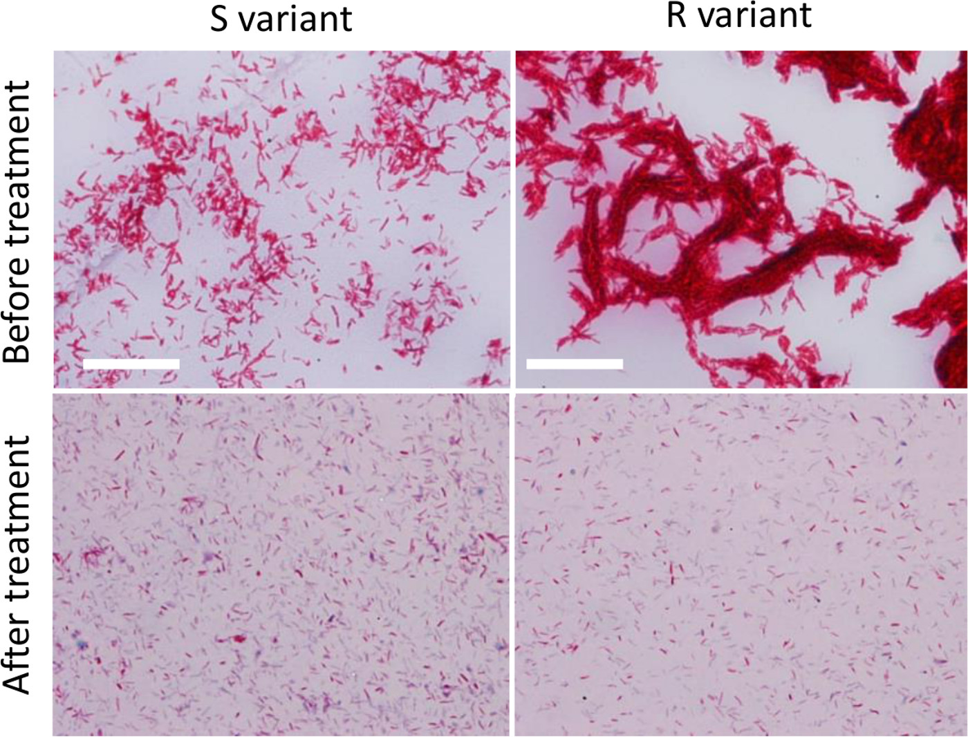
NOTE: Ziehl-Neelsen staining (specific to mycobacteria) can be used to compare the quality of the bacterial suspensions prior to and after the homogenization procedures described in 2.2.- Spot and spread out one droplet of the homogenized bacterial suspension on a glass slide and allow it to dry completely, fix the smear for at least 30 min on a hot plate set to 65-70 oC and stain using a Ziehl-Neelsen staining protocol.
- Observe the bacterial smear with a microscope using a 100 X objective and compare with a smear of a non-processed bacterial suspension (Figure 2).

Figure 2. Preparation of dispersed M. abscessus inocula. Ziehl-Neelsen staining of R or S variants grown on broth medium prior to any treatment (upper panels) or after treatment (lower panels) to reduce the size and number of mycobacterial aggregates (successive steps of syringing, sonication and decantation). Bacteria were observed using a microscope with 100 X APO oil 1.4 NA objective). Scale bars: 20 µm. Please click here to view a larger version of this figure.
3. Preparing Zebrafish Embryos
- Spawning and collecting zebrafish eggs
- The day before spawning, breed adult zebrafish by placing 2 males and 1 female (usually a 2:1 ratio allows optimal fertilization rate) into a breeding chamber, consisting of a separate fish tank with an egg collection basket31.
NOTE: Egg collection baskets are used to protect the eggs from being eaten by the parents and facilitate egg harvesting. - At 1 hpf, harvest eggs and only properly developed embryos in a 100 mm Petri dish filled with 25 ml blue fish water (maximum 100 embryos per dish) and keep them at 28.5 °C. Non-fertilized eggs are discarded.
- The day before spawning, breed adult zebrafish by placing 2 males and 1 female (usually a 2:1 ratio allows optimal fertilization rate) into a breeding chamber, consisting of a separate fish tank with an egg collection basket31.
- Preparation of zebrafish embryos for injections
- At about 24 hr post-fertilization (hpf), dechorionate the embryos with fine tweezers in a 100 mm Petri dish and keep them at 28.5 °C.
- Collect the 30-48 hpf embryos using a pipette and transfer them to a “V”-shaped positioning chamber filled with 25 ml of fish water containing 270 mg/L tricaine. Lay the embryos properly in the channels using a homemade microloader tip tool (homemade clipped micro loader tip) optimized for an easier micro-manipulation.
NOTE:Orientate the dorsal side downwards for intravenous injections in the caudal vein or the dorsal side upwards for tail muscle injections.
4. Micro-injection Procedure
NOTE: The micro-injection procedure for M. abscessus is similar to the one described previously for M. marinum injections32. To assess the chronology of M. abscessus infection (survival, bacterial loads, kinetic and characteristics of infection), injections in the caudal vein of 30 hpf embryos are preferred. To visualize the recruitment of immune cell, injections are done in localized sites such as is in tail muscles in 48 hpf embryo.
- Dilute the mycobacterial inoculum in 1 X PBST (depending on the number of CFU to be administered as determined in Step 2.2.6), containing 10% red phenol to check proper injection.
- Load a microcapillary injection needle with 5-10 µl of the bacterial inoculum using a microloader tip, connect the microinjection needle into the holder of the three-dimensional micromanipulator connected to the injector and break off the top of the needle with fine tweezers to obtain an opening diameter of 5-10 µm.
- Calibrate the injection volume by adjusting the microinjection pressure (20-50 hPa) and time (0.2 sec).
NOTE: The required injection volume is calibrated by visual determination of the diameter of a droplet expelled into the yolk of an embryo. - For caudal vein injection, position the embryo with the ventral side facing the needle (as described in section 3.2.2) and place the needle tip close to the urogenital opening, targeting the caudal vein, and gently push the tip of needle into the embryo until it just pierces the caudal vein region. Deliver the desired volume of bacterial preparation, usually 1-3 nl containing around 100 CFU/nl.
- For intramuscular injection, position the embryo with the dorsal side facing the needle (as in section 3.2.2), place the needle over one somite and inject a small volume (1 nl) of inoculum.
NOTE: As for caudal vein injections, intramuscular infections are also challenging to perform. Care must be taken to avoid an overloading that may injure the surrounding tissues. - At the end of the injection procedure, control the size of the inoculum by injecting the same volume of bacterial suspension in a drop of sterile PBST followed by plating on 7H10OADCand counting the CFU. Around 100 embryos can be injected with a needle.
NOTE: Because rough M. abscessus may continue to form aggregates in the needle, injections need to be done immediately after preparation of the inoculum. - Transfer the infected fish individually in 24-well plates containing 2 ml/well fish water and incubate at 28.5 °C. Embryos injected with 1-3 nl 1 X PBST can be used as controls.
- Proper infection of fluorescent bacteria in embryos is monitored using a fluorescence microscope.
5. Generation of Macrophage-Depleted Embryos
NOTE: Selective depletion of macrophages from tissues is used to investigate their contribution and role during infection. To visualize the proper depletion of macrophages, it is recommended to use a transgenic line with fluorescent macrophages, where mCherry is specifically expressed under the control of the macrophage specific mpeg1 promoter19.
- Lipo-clodronate-based procedure
NOTE: The lipo-clodronate procedure allows a selective depletion of macrophages from tissues (but no neutrophils)19. This compound does neither alter the survival of embryos nor affect the integrity of bacteria. A detailed protocol to prepare liposome-encapsulated clodronate has been reported previously33.- At 24 hpf, dechorionate the embryos and transfer them to a “V”-shaped injection dish filled with fish water supplemented with tricaine to maintain the embryos in an anesthetized state until the end of the injection procedure, as described in 3.2.2. Orientate the dorsal side downwards.
- Load a microcapillary injection needle with liposome-encapsulated clodronate (lipo-clodronate), connect the microinjection needle into the holder of the three-dimensional micromanipulator and break off the top of the needle with fine tweezers to obtain a tip opening diameter of 10 µm.
- To calibrate the injection volume, adjust the microinjection pressure to 20 hPa and the injection time to 0.2 sec. Place the needle tip close to the urogenital opening, targeting the caudal vein, and gently push the tip of needle into the embryo until it just pierces the caudal vein region and deliver the desired volume of solution (2-3 nl, 3 times).
NOTE: The lipo-clodronate suspension is very sticky and should be vortexed prior to injection to avoid blocking the vascular system and killing of the embryo. It is crucial to proceed to several successive injections of small volumes. - Transfer the embryos in 100 mm petri dishes containing fish water and keep them at 28.5 °C until infection.
- Control the proper depletion of macrophages by fluorescent microscopy.
NOTE: Complete macrophage depletion is effective for 4 days after lipo-clodoronate administration.
- Morpholino-based procedure
- The day before spawning, breed adult zebrafish by placing 2 males and 1 female separated by a plastic barrier in a breeding chamber.
- The next day, ≈ 30 min after that the light turns on in the zebrafish facility, remove the barrier separating males and females. Wait for about 20 min after beginning of the spawning to optimize the fertilization rate. Harvest the eggs and slow their development by transferring them in a 100 mm petri dish filled with cold blue fish water (4 °C).
NOTE: The control of the spawning is crucial for the morpholinos-based experiments. Morpholinos can only be injected into the eggs up to the four cells stage. - Collect the eggs using a pipette and deposit the eggs down in the “U”-shaped injection-chamber filled with cold blue fish water and block the eggs in the channels using a homemade microloader tip tool.
- Prepare the pu.1 morpholino solution containing 10% of red phenol, as previously described19. Load a microcapillary injection needle with the morpholino preparation and connect the microinjection needle into the holder of the three-dimensional micromanipulator, then break off the tip of the needle with fine tweezers to obtain a tip opening diameter of 5-10 µm.
- To calibrate the injection volume, adjust the microinjection pressure to 20-50 hPa and the injection time to 0.2 sec. Place the needle tip close to the egg and gently push the tip of needle through the chorion into the egg and deliver the desired morpholino solution volume, typically 3-5 nl.
- After micro-injection, transfer the eggs in a 100 mm petri dish in fish water and incubate at 28.5 °C till the infection procedure begins.
- Analyze the proper (complete) depletion of macrophages in pu.1 morphants by fluorescent microscopy at 30 to 48 hpf..
NOTE: Depending on the concentration injected, pu.1 morpholinos may also affect the number of early neutrophils. To confirm the proper depletion of macrophages without altering the number of neutrophils, it is recommended to use a double transgenic zebrafish line with fluorescent macrophages and neutrophils (with GFP expression specifically driven by the mpx promoter)19.
6. Bacterial Burden Quantification
- Determination by CFU enumeration
- At the desired time point, collect groups of infected embryo (5 per conditions) in 1.5 ml microcentrifuge tubes (1 embryo/tube), cryo-anesthetize the embryos by incubation on ice for 10 min and euthanize using an overdose of tricaine (300-500 mg/L)
- Wash the embryos with sterile water twice and dispense them in a new tube, lyse each embryo with 2% Triton X-100 diluted in 1 X PBST using a 26-G needle and homogenize the suspension until complete lysis. Centrifuge the suspension and resuspend the pellet in 1 X PBST with 0.05% Tween 80. Serial dilutions of the homogenates are plated on Middlebrook 7H10OADC and supplemented with BBL MGIT PANTA, as recommended by the supplier.
NOTE: M. abscessus being more susceptible to NaOH treatment than M. marinum, decontamination of fish lysates without affecting M. abscessus growth can be successfully achieved using the BBL MGIT PANTA antibiotic cocktail. - Incubate the plates for 4 days at 30 °C and count colonies.
- Determination by Fluorescence Pixel Count (FPC) measurements in live embryos
NOTE: To determine the bacterial loads via fluorescence emission, serial images of infected embryos are acquired and fluorescence intensity measured by FPC (identification of bacteria by particle analysis) using a homemade macro developed for the ImageJ freeware. A particle analysis requires a binary black and white image that relies on a threshold range that allows to discriminating the fluorescent signal of interest from the background.- At the desired time point, tricain-anesthetize infected embryos (5-10 per condition) in 35 mm petri dishes as described in 3.2.2.
- Remove water and submerge the embryos and the whole dish surface with 1% low melting point agarose in fish water, then align laterally the embryos. Cover the solidified agarose with fish water containing tricaine.
- Acquire fluorescence images of the entire embryo using an epifluorescence microscope with a 10 X objective.
NOTE: The fluorescence image acquisition is a critical step of the FPC measurements procedure. It is important to use a calibrated monitor with a colour calibration probe. Adjusting the optimal time of exposure to obtain a minimal background during acquisition prevents saturation of the signal. Images must be in an 8-bit tiff format. Identical settings are kept during the whole experiment for quantitative purposes. - Remove carefully the agarose from the embryos with a homemade microloader tip tool. These embryos can be used for subsequent analyses if required. Prior to the quantification of the bacterial load, each picture shall be quality controlled. To analyze the images and convert the fluorescence intensity into FPC per fish, open the ImageJ freeware.
NOTE: The FPC value reflects the bacterial burden as shown previously using M. marinum34. - Determine the required threshold for image analysis using an image of an uninfected embryo (several control embryos can be used to obtain an average threshold). Go to Image → Adjust → Threshold and then move the bottom slider in the threshold window to the right until the background becomes completely black (i.e., to obtain a background should be at zero). Record the corresponding value.
- Within Image J, open the FPC macro (supplementary file) and follow the prompts: locate the folder of images to be measured and then enter the threshold as previously determined and click on “OK”.
NOTE: The macro automatically subtracts the background. A threshold is then applied. Florescent signals are identified following the particle analysis. - Copy and paste the data from the summary window to the desired software for data analysis.
NOTE: In contrast to the CFU determination method, the FPC method is non-invasive and allows re-use the embryos for subsequent analyses and, importantly, monitoring the dynamics/kinetics of the bacterial burden on an individual basis. It, therefore, allows to significantly reducing the number of animals, in agreement with ethical rules.
7. Imaging M. abscessus- infected Embryos
- Live imaging of M. abscessus infection
- Mount tricaine-anesthetized embryos (see 3.2.2) in 1% low melting point agarose in a 35 mm petri dish prior to epifluorescence microscopy observations. Use a glass bottom dish for inverted confocal microscopy or in a single cavity depression slide for upright confocal microscopy. Orientate the embryo to the desired position and cover the solidified agarose with fish water containing tricaine.
- Use an epifluorescence microscope with a 10 X objective for sequential fluorescence acquisition and transmission images of the entire embryo. Alternatively, use a confocal microscope with 40X or 63X objectives to visualize the activity of immune cells after an infection.
- Imaging fixed M. abscessus infected embryos
- Euthanize the embryos as described in 6.1.1 and transfer them to 1.5 ml micro-centrifuge tubes and fix the embryos in 4% paraformaldehyde in 1 X PBST for 2 hr at RT or O/N at 4 °C. Remove paraformaldehyde by washing the embryos twice with 1 X PBST for 10 min.
NOTE: To preserve fluorescence, fixed embryos must be protected from light. - To preserve the integrity of the tissues and fluorescence, embryos are incubated successively in increasing glycerol concentration solutions (10, 20, 30, 40 and 50% diluted in 1 X PBST), for 10 min per condition.
NOTE: Fixed embryos can be stored for several months in 50% glycerol at 4 °C. - Lay the embryo embedded in 50% glycerol in a viewing chamber (Figure 1B)31.
- Image using 40X or 63X objectives on a confocal microscope.
- Euthanize the embryos as described in 6.1.1 and transfer them to 1.5 ml micro-centrifuge tubes and fix the embryos in 4% paraformaldehyde in 1 X PBST for 2 hr at RT or O/N at 4 °C. Remove paraformaldehyde by washing the embryos twice with 1 X PBST for 10 min.
Results
Although various anatomical sites can be injected32, caudal vein injections are often used to generate systemic infection for subsequent analyses including survival experiments, bacterial burden determination, phagocytosis activity or cord formation. Injections in the tail muscles are used to assess the recruitment of macrophages at the site of injection (Figure 3A). To investigate and compare the virulence of R and S variants of M. abscessus, fluorescent bacterial suspensions are inj...
Discussion
The zebrafish has recently emerged as an excellent vertebrate model system for studying the dynamics of bacterial infection using wide field and confocal imaging in real-time36. The combination of dispersed mycobacterial suspensions (protocol 2.2) together with micro-injection methods (protocol 4) allows reproducible systemic infections, and subsequent monitoring and visualization of the progression of the infection with a special focus on the bacterial interactions with host macrophages. Virulence of M. a...
Disclosures
The authors have nothing to disclose.
Acknowledgements
The authors are grateful to K. Kissa for helpful discussions and for providing lipo-clodronate and L. Ramakrishnan for the generous gift of pTEC27 and pTEC15 that allow expression of tdTomato and Wasabi, respectively. This work forms part of the projects of the French National Research Agency (ZebraFlam ANR-10-MIDI-009 and DIMYVIR ANR-13-BSV3-007-01) and the European Community’s Seventh Framework Program (FP7-PEOPLE-2011-ITN) under grant agreement no. PITN-GA-2011-289209 for the Marie-Curie Initial Training Network FishForPharma. We wish also to thank the Association Gregory Lemarchal and Vaincre La Mucoviscidose (RF20130500835) for funding C.M. Dupont.
Materials
| Name | Company | Catalog Number | Comments |
| BBL MGIT PANTA | BD Biosciences | 245114 | |
| Bovine Serum Albumin | Euromedex | 04-100-811-E | |
| Catalase from Bovine Liver | Sigma-Aldrich | C40 | |
| Difco Middlebrook 7H10 Agar | BD Biosciences | 262710 | |
| Difco Middlebrook 7H9 Broth | BD Biosciences | 271310 | |
| Ethyl 3-aminobenzoate methanesulfonate salt (Tricaine) | Sigma-Aldrich | A5040 | |
| Oleic Acid | Sigma-Aldrich | O1008 | |
| Paraformaldehyde | Delta Microscopie | 15710 | |
| Phenol Red | Sigma-Aldrich | 319244 | |
| Tween 80 | Sigma-Aldrich | P4780 | |
| Agar | Gibco Life Technologie | 30391-023 | |
| Low melting agarose | Sigma-Aldrich | ||
| Instant Ocean Sea Salts | Aquarium Systems Inc | ||
| Borosilicate glass capillaries | Sutter instrument Inc | BF100-78-10 | 1 mm O.D. X 0.78 mm I.D. |
| Micropipette puller device | Sutter Instrument Inc | Flamming/Brown Micropipette Puller p-87 | |
| Microinjector | Tritech Research | Digital microINJECTOR, MINJ-D | |
| Tweezers | Sciences Tools inc | Dumont # M5S | |
| Microloader Tips | Eppendorf |
References
- Brown-Elliott, B. A., Wallace, R. J. Clinical and taxonomic status of pathogenic nonpigmented or late-pigmenting rapidly growing mycobacteria. Clinical Microbiology Reviews. 15 (4), 716-746 (2002).
- Aitken, M. L., Limaye, A., et al. Respiratory outbreak of Mycobacterium abscessus subspecies massiliense in a lung transplant and cystic fibrosis center. American Journal of Respiratory and Critical Care Medicine. 185 (2), 231-232 (2012).
- Gilljam, M., Lindblad, A., Ridell, M., Wold, A. E., Welinder-Olsson, C. Molecular epidemiology of Mycobacterium abscessus, with focus on cystic fibrosis. Journal of Clinical Microbiology. 45 (5), 1497-1504 (2007).
- Roux, A. -. L., Catherinot, E., et al. Multicenter study of prevalence of nontuberculous mycobacteria in patients with cystic fibrosis in France. Journal of Clinical Microbiology. 47 (12), 4124-4128 (2009).
- Lee, M. -. R., Cheng, A., et al. CNS infections caused by Mycobacterium abscessus complex: clinical features and antimicrobial susceptibilities of isolates. The Journal of Antimicrobial Chemotherapy. 67 (1), 222-225 (2012).
- Talati, N. J., Rouphael, N., Kuppalli, K., Franco-Paredes, C. Spectrum of CNS disease caused by rapidly growing mycobacteria. The Lancet Infectious Diseases. 8 (6), 390-398 (2008).
- Medjahed, H., Gaillard, J. -. L., Reyrat, J. -. M. Mycobacterium abscessus: a new player in the mycobacterial field. Trends in Microbiology. 18 (3), 117-123 (2010).
- Griffith, D. E., Girard, W. M., Wallace, R. J. Clinical features of pulmonary disease caused by rapidly growing mycobacteria. An analysis of 154 patients. The American Review of Respiratory Disease. 147 (5), 1271-1278 (1993).
- Nessar, R., Cambau, E., Reyrat, J. M., Murray, A., Gicquel, B. Mycobacterium abscessus: a new antibiotic nightmare. The Journal of Antimicrobial Chemotherapy. 67 (4), 810-818 (2012).
- Sanguinetti, M., Ardito, F., et al. Fatal pulmonary infection due to multidrug-resistant Mycobacterium abscessus a patient with cystic fibrosis. Journal of Clinical Microbiology. 39 (2), 816-819 (2001).
- Griffith, D. E., Aksamit, T., et al. An official ATS/IDSA statement: diagnosis, treatment, and prevention of nontuberculous mycobacterial diseases. American Journal of Respiratory and Critical Care Medicine. 175 (4), 367-416 (2007).
- Howard, S. T., Rhoades, E., et al. Spontaneous reversion of Mycobacterium abscessus a smooth to a rough morphotype is associated with reduced expression of glycopeptidolipid and reacquisition of an invasive phenotype. Microbiology (Reading, England). 152 (Pt 6), 1581-1590 (2006).
- Chardi, A., Olivares, F., Byrd, T. F., Julián, E., Brambilla, C., Luquin, M. Demonstration of cord formation by rough Mycobacterium abscessus variants: implications for the clinical microbiology laboratory. Journal of Clinical Microbiology. 49 (6), 2293-2295 (2011).
- Byrd, T. F., Lyons, C. R. Preliminary characterization of a Mycobacterium abscessus mutant in human and murine models of infection. Infection and Immunity. 67 (9), 4700-4707 (1999).
- Catherinot, E., Clarissou, J., et al. Hypervariance of a rough variant of the Mycobacterium abscessus type strain. Infection and Immunity. 75 (2), 1055-1058 (2007).
- Catherinot, E., Roux, A. -. L., et al. Acute respiratory failure involving an R variant of Mycobacterium abscessus. Journal of Clinical Microbiology. 47 (1), 271-274 (2009).
- Bernut, A., Le Moigne, V., Lesne, T., Lutfalla, G., Herrmann, J. -. L., Kremer, L. In vivo assessment of drug efficacy against Mycobacterium abscessus using the embryonic zebrafish test system. Antimicrobial Agents and Chemotherapy. 58 (7), 4054-4063 (2014).
- Oh, C. -. T., Moon, C., Jeong, M. S., Kwon, S. -. H., Jang, J. Drosophila melanogaster for Mycobacterium abscessus infection. Microbes and Infection / Institut Pasteur. 15 (12), 788-795 (2013).
- Bernut, A., Herrmann, J. -. L., et al. Mycobacterium abscessus cording prevents phagocytosis and promotes abscess formation. Proceedings of the National Academy of Sciences of the United States of America. 111 (10), E943-E952 (2014).
- Dubée, V., Bernut, A., et al. β-Lactamase inhibition by avibactam in Mycobacterium abscessus. Journal of Antimicrobial Chemotherapy. 70 (4), 1051-1058 (2015).
- Torraca, V., Masud, S., Spaink, H. P., Meijer, A. H. Macrophage-pathogen interactions in infectious diseases: new therapeutic insights from the zebrafish host model. Disease Models Mechanisms. 7 (7), 785-797 (2014).
- Alibaud, L., Rombouts, Y., et al. A Mycobacterium marinum TesA mutant defective for major cell wall-associated lipids is highly attenuated in Dictyostelium discoideum and zebrafish embryos. Molecular Microbiology. 80 (4), 919-934 (2011).
- Clay, H., Volkman, H. E., Ramakrishnan, L. Tumor necrosis factor signaling mediates resistance to mycobacteria by inhibiting bacterial growth and macrophage death. Immunity. 29 (2), 283-294 (2008).
- Palha, N., Guivel-Benhassine, F., et al. Real-time whole-body visualization of Chikungunya Virus infection and host interferon response in zebrafish. PLoS pathogens. 9 (9), e1003619 (2013).
- Mostowy, S., Boucontet, L., et al. The zebrafish as a new model for the in vivo study of Shigella flexneri with phagocytes and bacterial autophagy. PLoS pathogens. 9 (9), e1003588 (2013).
- Prajsnar, T. K., Cunliffe, V. T., Foster, S. J., Renshaw, S. A. A novel vertebrate model of Staphylococcus aureus reveals phagocyte-dependent resistance of zebrafish to non-host specialized pathogens. Cellular Microbiology. 10 (11), 2312-2325 (2008).
- Van der Sar, A. M., Appelmelk, B. J., Vandenbroucke-Grauls, C. M. J. E., Bitter, W. A star with stripes: zebrafish as an infection model. Trends in Microbiology. 12 (10), 451-457 (2004).
- Vergunst, A. C., Meijer, A. H., Renshaw, S. A., O’Callaghan, D. Burkholderia cenocepacia creates an intramacrophage replication niche in zebrafish embryos, followed by bacterial dissemination and establishment of systemic infection. Infection and Immunity. 78 (4), 1495-1508 (2010).
- Levraud, J. -. P., Disson, O., et al. Real-time observation of Listeria monocytogenes-phagocyte interactions in living zebrafish larvae. Infection and Immunity. 77 (9), 3651-3660 (2009).
- Meijer, A. H., Spaink, H. P. Host-pathogen interactions made transparent with the zebrafish model. Current Drug Targets. 12 (7), 1000-1017 (2011).
- Westerfield, M. . The Zebrafish Book: A Guide for the Laboratory Use of Zebrafish (Danio Rerio). , (2007).
- Benard, E. L., van der Sar, A. M., Ellett, F., Lieschke, G. J., Spaink, H. P., Meijer, A. H. Infection of zebrafish embryos with intracellular bacterial pathogens. Journal of Visualized Experiments. (61), e3781 (2012).
- Van Rooijen, N., Sanders, A. Liposome mediated depletion of macrophages: mechanism of action, preparation of liposomes and applications. Journal of Immunological Methods. 174 (1-2), 83-93 (1994).
- Adams, K. N., Takaki, K., et al. Drug tolerance in replicating mycobacteria mediated by a macrophage-induced efflux mechanism. Cell. 145 (1), 39-53 (2011).
- Ramakrishnan, L. Looking within the zebrafish to understand the tuberculous granuloma. Advances in Experimental Medicine and Biology. 783, 251-266 (2013).
- Davis, J. M., Clay, H., Lewis, J. L., Ghori, N., Herbomel, P., Ramakrishnan, L. Real-time visualization of Mycobacterium-macrophage interactions leading to initiation of granuloma formation in zebrafish embryos. Immunity. 17 (6), 693-702 (2002).
- Lamason, R. L., Mohideen, M. -. A. P. K., et al. SLC24A5, a putative cation exchanger, affects pigmentation in zebrafish and humans. Science (New York, NY). 310 (5755), 1782-1786 (2005).
- Renshaw, S. A., Loynes, C. A., Trushell, D. M. I., Elworthy, S., Ingham, P. W., Whyte, M. K. B. A transgenic zebrafish model of neutrophilic inflammation. Blood. 108 (13), 3976-3978 (2006).
- Hall, C., Flores, M. V., Storm, T., Crosier, K., Crosier, P. The zebrafish lysozyme C promoter drives myeloid-specific expression in transgenic fish. BMC developmental biology. 7, 42 (2007).
- Takaki, K., Davis, J. M., Winglee, K., Ramakrishnan, L. Evaluation of the pathogenesis and treatment of Mycobacterium marinum in zebrafish. Nature Protocols. 8 (6), 1114-1124 (2013).
- Stoop, E. J. M., Schipper, T., et al. Zebrafish embryo screen for mycobacterial genes involved in the initiation of granuloma formation reveals a newly identified ESX-1 component. Disease Models Mechanisms. 4 (4), 526-536 (2011).
- Carvalho, R., de Sonneville, J., et al. A high-throughput screen for tuberculosis progression. PloS One. 6 (2), e16779 (2011).
Reprints and Permissions
Request permission to reuse the text or figures of this JoVE article
Request PermissionThis article has been published
Video Coming Soon
Copyright © 2025 MyJoVE Corporation. All rights reserved