A subscription to JoVE is required to view this content. Sign in or start your free trial.
Method Article
Shrinky-Dink Hanging Drops: A Simple Way to Form and Culture Embryoid Bodies
In This Article
Summary
We show a simple and rapid method to load pre-defined numbers of cells into microfabricated wells and maintain them for embryoid body development.
Abstract
Embryoid bodies (EB) are aggregates of embryonic stem cells. The most common way of creating these aggregates is the hanging drop method, a laborious approach of pipetting an arbitrary number of cells into well plates. The interactions between the stem cells forced into close proximity of one another promotes the generation of the EBs. Because the media in each of the wells has to be manually exchanged every day, this approach is manually intensive.
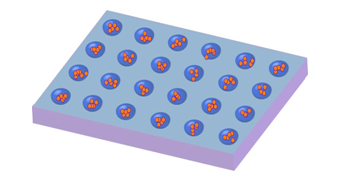
Moreover, because environmental parameters including cell-cell, cell-soluble factor interactions, pH, and oxygen availability can be functions of EB size, cell populations obtained from traditional hanging drops can vary dramatically even when cultured under identical conditions. Recent studies have indeed shown that the initial number of cells forming the aggregate can have significant effects on stem cell differentiation. We have developed a simple, rapid, and scalable culture method to load pre-defined numbers of cells into microfabricated wells and maintain them for embryoid body development. Finally, these cells are easily accessible for further analysis and experimentation. This method is amenable to any lab and requires no dedicated equipment. We demonstrate this method by creating embryoid bodies using a red fluorescent mouse cell line (129S6B6-F1).
Protocol
1. Making Shrinky-Dink Mold
- Print the desired pattern on shrinky-dink sheet using a good definition printer.
- Bake shrinky-dink sheet at 163° C for about 10 minutes, or until fully shrunk and having aquired a regular shape.
- After shrinky-dink mold has cooled down, submerge it in an isopropanol bath until the complete surface is barely covered.
- Carefully, spray some acetone over the mold and shake container a few times. Add more isopropanol to wash out acetone excess and repeat this step a few times until shrinky-mold looks clean.
- Immerse mold in distilled water for 10 minutes to wash off any remaining organic solvent.
- Air clean shrinky-mold. Re-heat it for about 5 minutes at 163° C. This will compact ink and evaporate any remaining solvent.
2. Making PDMS microwells
- Prepare a 10:1 PDMS/curing agent mixture, and agitate vigorously for few minutes.
- Place shrinky-dink mold in a small petri dish. Pour PDMS mixture until it reaches about 1/2 cm over the mold surface.
- Place dish under vacuum bell to eliminate all bubbles from PDMS mixture.
- Place dish in oven at 70°C, overnight.
- Cut off solid PDMS from mold and bind it to a glass slide just by applying pressure.
- Discard first microwell-chip, since it has ink residues incrusted between PDMS.
- Repeat this procedure to produce a second chip that is ink-free and has a more defined shape.
- Clean microwell-chip using 70% ethanol solution. Place it under UV light source for 10 minutes to sterilize it.
3. Trapping cells in microwells
- Count cells and dilute them in culture media to desired concentration (depending on how many initial cells in wells you would like). For example, to get approximately 10-15 cells per well (average = 11, SD = 5.4, loading rate= 93%), we used a concentration of 8×10⁴ cells/ml. For a concentration of 17×104 cells/ml, we could reliably get between 25 and 35 cells per well (average = 27.17857 SD = 7.7, loading rate = 100%).
- Carefully place microwell chip in a 50 ml centrifuge tube containing a solidified PDMS base.
- Add about 2JDP ml of the cell solution.
- Centrifuge for 5 minutes at 760 rpm and 4°C.
- Pipet out excess solution and carefully wash microwell with PBS 1X solution.
- Place microwell in a small petry dish, being careful while taking the chip out of the centrifuge tube.
- Wash cell excess using 1 X PBS solution.
- Place microwell chip under an inverted microscope to verify intended number of cells per well.
- Incubate microwell containing cells at standard conditions.

4. Cell incubation
- Follow the normal EB protocol in the lab.
- Change the medium slowly from the side of the chamber; avoid disturbing the cell in the microwell.
Discussion
We have developed a simple, rapid, and scalable culture method to load pre-defined numbers of cells into microfabricated wells (molded from Shrinky-Dinks) and maintain them for embryoid body development. Finally, these cells are easily accessible for further analysis and experimentation. This method is amenable to any lab and requires no dedicated equipment because we obviate the need for photolithography. We can vary the size of the microwells as well as the concentration of cells/ wells to change the number and size of...
Acknowledgements
We would like to thank CIRM for support of this work. The cell line was generously donated from Dr. Andras Nagy at Mount Sinai Hospital, Toronto, Ontario.
Materials
| Name | Company | Catalog Number | Comments |
| Material Name | Type | Company | Catalogue Number |
| Shrink-Dink Film | Material | K&B Innovations | D300-10A |
| PDMS | Material | Dow Corning | Sylgard 184 |
| Acetone | Reagent | Fisher Scientific | A16P-4 |
| Ethanol | Reagent | Fisher Scientific | A405P-4 |
| PBS | Reagent | Sigma | P4417 |
| BMP-4 | Reagent | R and D systems | 314-BP-010 |
| Knock-Out DMEM (KO DMEM) | Reagent | Invitrogen | 10829 |
| KnockOut Sirum Replacement (KSR) | Reagent | Invitrogen | 10828 |
| Penn-Strep | Reagent | Invitrogen | 15070-063 |
| L-glutamine | Reagent | Invitrogen | 25030-081 |
| Non-essential Amino Acids (NEAA) | Reagent | Invitrogen | 11140 |
| D-mercaptoethanol (BME) | Reagent | Calbiochem | 444203 |
| Leukemia Inhibitory Factor (LIF) {ESGRO 106 units} | Reagent | Chemicon | ESG1106 |
| Printer | Tool | HP | Laser Jet 2420d |
| Oven | Tool | Yamato Scientific | DP-22 |
| For the mESC media(McCloskey lab protocol): (for total media prepared: 50ml; 100ml) KO DMEM: 40.8ml; 81.6ml 15% KSR: 7.5ml; 15ml 1x Penn-Strep: .5ml; 1ml 2mM L-glutamine: .5ml; 1ml NEAA : .5ml; 1ml LIF: 100ul; 200ul BMP-4 (10ng/ml): 50ul; 100ul Diluted BME: 50ul; 100ul (Add 35ul of sterile filtered BME to 5ml of PBS and syringe filter sterilize. Discard after 2 weeks. Final concentration in the solution is .1mM) | |||
References
- Keller, G. M. In vitro differentiation of embryonic stem cells. Curr Opin Cell Biol. 7, 862-869 (1995).
- Doetschman, T. C., Eistetter, H., Katz, M., Schmidt, W., Kemler, R. The in vitro development of blastocyst-derived embryonic stem cell lines: Formation of visceral yolk sac, blood islands and myocardium. Journal of Embryology and Experimental Morphology. 87, 27-45 (1985).
- Park, J., Cho, C. H., Parashurama, N., Li, Y., Berthiaume, F., Toner, M., Tilles, A. W., Yarmush, M. L. Microfabrication-based modulation of embryonic stem cell differentiation. Lab Chip. 7, 1018-1028 (2007).
- Koike, M., Sakaki, S., Amano, Y., Kurosawa, H. Characterization of embryoid bodies of mouse embryonic stem cells formed under various culture conditions and estimation of differentiation status of such bodies. J Biosci Bioeng. 104, 294-299 (2007).
- Hwang, N. S., Varghese, S., Elisseeff, J. Controlled differentiation of stem cells. Advanced Drug Delivery Reviews. 60, 199-204 (2008).
- Adelman, C. A., Chattopadhyay, S., Bieker, J. J. The BMP/BMPR/Smad pathway directs expression of the erythroid-specific EKLF and GATA1 transcription factors during embryoid body differentiation in serum-free media. Development. 129, 539-549 (2002).
- Tanaka, N., Takeuchi, T., Neri, O. V., Sills, E. S. Laser-assisted blastocyst dissection and subsequent cultivation of embryonic stem cells in a serum/cell free culture system: applications and preliminary results in a murine model. J Transl Med. 4, (2006).
Reprints and Permissions
Request permission to reuse the text or figures of this JoVE article
Request PermissionThis article has been published
Video Coming Soon
Copyright © 2025 MyJoVE Corporation. All rights reserved