
Visualizing Soil Microorganisms via the Contact Slide Assay and Microscopy
Overview
Source: Laboratories of Dr. Ian Pepper and Dr. Charles Gerba - The University of Arizona
Demonstrating Author: Bradley Schmitz
Soil comprises the thin layer at the earth’s surface, containing biotic and abiotic factors that contribute to life. The abiotic portion includes inorganic particles ranging in size and shape that determine the soil’s texture. The biotic portion incorporates plant residues, roots, organic matter, and microorganisms. Soil microbe abundance and diversity is expansive, as one gram of soil contains 107-8 bacteria, 106-8 actinomycetes, 105-6 fungi, 103 yeast, 104-6 protozoa, 103-4 algae, and 53 nematodes. Together, the biotic and abiotic factors form architectures around plant roots, known as the rhizosphere, that provide favorable conditions for soil microorganisms.
Biotic and abiotic factors promote life in soils. However, they also contribute stressful dynamics that limit microbes. Biotic stress involves competition amongst life to adapt and survive in environmental conditions. For example, microbes can secrete inhibitory or toxic substances to harm neighboring microorganisms. Penicillium notatum is a notorious fungus, as it reduces competition for nutrients by producing an antimicrobial, which humans harvest to create the pharmaceutical penicillin. Abiotic stresses arise from physical or chemical properties limiting microbial survival, such as light, moisture, temperature, pH, nutrients, and texture.
Procedure
1. Soil Slide Microcosm Preparation
- Collect garden soil from the surface (0-6” depth), and weigh 150 g soil into two separate cups.
- If soil has high density of organic matter, weigh 100 g.
- Label one cup “Treatment” and the other “Control.”
- Calculate amount of water needed to alter moisture content.
- Moisture content is often close to field capacity.

- Moisture content is often close to field capacity.
Results
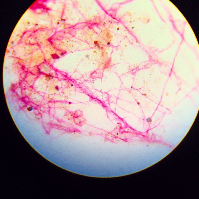
Fungi display thick, filamentous hyphae (Figure 2). Actinomycetes display thin, filamentous hyphae. Bacteria display small cocci or rod shapes. They’re often found in clumps, on soil particles, or lining fungal hyphae. Soil particles display irregular, dark shapes (Figure 3).

Figure 2. Contact slide image using 100X objective lens.
Photo courtesy W.H. Ful
Application and Summary
The contact slide assay, also referred to as the buried-slide, is a simple technique utilized to qualitatively observe soil biota. This assay qualitatively shows the spatial interactions between fungal hyphae, actinomycete filaments, bacteria, and soil particles. Individuals or industry can employ this assay to gather knowledge on a particular soil’s health in regards to agriculture, gardening, composting, teaching, and studying. However, this technique does not quantify soil micro biota, as it only encompasses a s
References
- Pepper, I. L., & Gerba, C P. 'Contact Slide Assay.' Environmental Microbiology A Laboratory Manual. 2nd ed. Elsevier 19-25 (2004).
- Pepper, I. L., Gerba, C. P., & Gentry, T. J. 'Earth Environments.' Environmental Microbiology. 3rd ed. Elsevier 59-88 (2014).
- Rossi, G., Ricardo, S., Gesue, G., Stanganelli, M., and Want, T.K. Direct Microscopic and bacteriological investigations of the soil. Soil Science. 41, 52 – 66 (1936).
Skip to...
Videos from this collection:

Now Playing
Visualizing Soil Microorganisms via the Contact Slide Assay and Microscopy
Environmental Microbiology
42.2K Views

Determination of Moisture Content in Soil
Environmental Microbiology
359.3K Views

Aseptic Technique in Environmental Science
Environmental Microbiology
126.4K Views

Gram Staining of Bacteria from Environmental Sources
Environmental Microbiology
100.2K Views

Filamentous Fungi
Environmental Microbiology
57.3K Views

Community DNA Extraction from Bacterial Colonies
Environmental Microbiology
28.8K Views

Detecting Environmental Microorganisms with the Polymerase Chain Reaction and Gel Electrophoresis
Environmental Microbiology
44.5K Views

RNA Analysis of Environmental Samples Using RT-PCR
Environmental Microbiology
40.4K Views

Quantifying Environmental Microorganisms and Viruses Using qPCR
Environmental Microbiology
47.8K Views

Water Quality Analysis via Indicator Organisms
Environmental Microbiology
29.5K Views

Isolation of Fecal Bacteria from Water Samples by Filtration
Environmental Microbiology
39.3K Views

Detection of Bacteriophages in Environmental Samples
Environmental Microbiology
40.7K Views

Culturing and Enumerating Bacteria from Soil Samples
Environmental Microbiology
184.3K Views

Bacterial Growth Curve Analysis and its Environmental Applications
Environmental Microbiology
295.9K Views

Algae Enumeration via Culturable Methodology
Environmental Microbiology
13.8K Views
Copyright © 2025 MyJoVE Corporation. All rights reserved