
Quantitative Strain Mapping of an Abdominal Aortic Aneurysm
Overview
Source: Hannah L. Cebull1, Arvin H. Soepriatna1, John J. Boyle2 and Craig J. Goergen1
1Weldon School of Biomedical Engineering, Purdue University, West Lafayette, Indiana
2Mechanical Engineering & Materials Science, Washington University in St. Louis, St Louis, Missouri
The mechanical behavior of soft tissues, such as blood vessels, skin, tendons, and other organs, are strongly influenced by their composition of elastin and collagen, which provide elasticity and strength. The fiber orientation of these proteins depends on the type of soft tissue and can range from a single preferred direction to intricate meshed networks, which can become altered in diseased tissue. Therefore, soft tissues often behave anisotropically on the cellular and organ level, creating a need for three-dimensional characterization. Developing a method for reliably estimating strain fields within complex biological tissues or structures is important to mechanically characterize and understande disease. Strain represents how soft tissue relatively deforms over time, and it can be described mathematically through various estimations.
Acquiring image data over time allows deformation and strain to be estimated. However, all medical imaging modalities contain some amount of noise, which increases the difficulty of accurately estimating in vivo strain. The technique described here successfully overcomes these issues by using a direct deformation estimation (DDE) method to calculate spatially varying 3D strain fields from volumetric image data.
Current strain estimation methods include digital image correlation (DIC) and digital volume correlation. Unfortunately, DIC can only accurately estimate strain from a 2D plane, severely limiting the application of this method. While useful, 2D methods such as DIC have difficulty quantifying strain in regions that undergo 3D deformation. This is because out-of-plane motion creates deformation errors. Digital volume correlation is a more applicable method that divides the initial volume data into regions and finds the most similar region of the deformed volume, thereby reducing out-of-plane error. However, this method proves to be sensitive to noise and requires assumptions about the mechanical properties of the material.
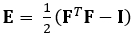
The technique demonstrated here eliminates these issues by using a DDE method, thus making it very useful in the analysis of medical imaging data. Furthermore, it is robust to high or localized strain. Here we describe the acquisition of gated, volumetric 4D ultrasound data, its conversion into an analyzable format, and the use of a custom Matlab code to estimate 3D deformation and corresponding Green-Lagrange strains, a parameter that better describes large deformations. The Green-Lagrange strain tensor is implemented in many 3D strain estimation methods because it allows for F to be calculated from a Least Squares Fit (LSF) of the displacements. The equation below represents the Green-Lagrange strain tensor, E, where F and I represent the deformation gradient and second order identity tensor, respectively.
 (1)
(1)
Procedure
1. 4D Ultrasound Set-up
- When using the imaging software, use a laptop capable of running mathematical computing software to automate the 4D acquisition process. Connect the laptop with this custom code to the ultrasound system via the USB port. Note that the imaging software has a 4D ultrasound feature integrated into the software.
- After turning on the ultrasound system, set up the physiological monitoring unit while also ensuring that the heart rate and temperature buttons are on. Initialize the 3D
Results
Using the procedure described above, 4D ultrasound of an angiotensin II-induced suprarenal dissecting abdominal aortic aneurysm (AAA) of a mouse was acquired. Multiple short-axis EKV video loops were acquired along the aorta and combined to create 4D data, as shown in Figure 1. This data was then converted into a MAT file using a custom code, which was then analyzed in a 3D strain calculation code using a warping function. After optimizing the parameters of the code for a specific data set,
Application and Summary
Localized in vivo mechanical characterization is an important part of understanding the growth and remodeling of biological tissues. Compared to existing approaches, the strain quantification procedure described here uses an improved method of accurately calculating 3D strain through optimally warping the undeformed image before cross-correlation. This method does not use any material assumptions in determining strains within tissue volumes. Unfortunately, the strain estimation is reliable only down to a kernel
Skip to...
Videos from this collection:

Now Playing
Quantitative Strain Mapping of an Abdominal Aortic Aneurysm
Biomedical Engineering
4.6K Views

Imaging Biological Samples with Optical and Confocal Microscopy
Biomedical Engineering
35.8K Views

SEM Imaging of Biological Samples
Biomedical Engineering
23.8K Views

Biodistribution of Nano-drug Carriers: Applications of SEM
Biomedical Engineering
9.3K Views

High-frequency Ultrasound Imaging of the Abdominal Aorta
Biomedical Engineering
14.5K Views

Photoacoustic Tomography to Image Blood and Lipids in the Infrarenal Aorta
Biomedical Engineering
5.7K Views

Cardiac Magnetic Resonance Imaging
Biomedical Engineering
14.8K Views

Computational Fluid Dynamics Simulations of Blood Flow in a Cerebral Aneurysm
Biomedical Engineering
11.8K Views

Near-infrared Fluorescence Imaging of Abdominal Aortic Aneurysms
Biomedical Engineering
8.3K Views

Noninvasive Blood Pressure Measurement Techniques
Biomedical Engineering
11.9K Views

Acquisition and Analysis of an ECG (electrocardiography) Signal
Biomedical Engineering
105.9K Views

Tensile Strength of Resorbable Biomaterials
Biomedical Engineering
7.5K Views

Micro-CT Imaging of a Mouse Spinal Cord
Biomedical Engineering
8.0K Views

Visualization of Knee Joint Degeneration after Non-invasive ACL Injury in Rats
Biomedical Engineering
8.2K Views

Combined SPECT and CT Imaging to Visualize Cardiac Functionality
Biomedical Engineering
11.0K Views
Copyright © 2025 MyJoVE Corporation. All rights reserved