
In Vivo Two-Color 2-Photon Imaging of Genetically-Tagged Reporter Cells in the Skin
July 11th, 2019
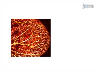
•Morphological changes occur in immune responsive fibroblast cells following activation and promote alterations in cellular recruitment. Utilizing 2-photon imaging in conjunction with a genetically engineered Fibroblast-specific protein 1 (FSP1)-cre; tdTomato floxed-stop-floxed (TB/TB) mouse line and green fluorescently tagged lipopolysaccharide-FITC, we can illustrate highly specific uptake of lipopolysaccharide in dermal fibroblasts and morphological changes in vivo.
Tags
Related Videos
Three-dimensional Optical-resolution Photoacoustic Microscopy

Multiparametric Optical Mapping of the Langendorff-perfused Rabbit Heart

Optical Mapping of Action Potentials and Calcium Transients in the Mouse Heart

Viral Nanoparticles for In vivo Tumor Imaging

A Full Skin Defect Model to Evaluate Vascularization of Biomaterials In Vivo

Quantification of Global Diastolic Function by Kinematic Modeling-based Analysis of Transmitral Flow via the Parametrized Diastolic Filling Formalism

Generation of a Three-dimensional Full Thickness Skin Equivalent and Automated Wounding

Electrotaxis Studies of Lung Cancer Cells using a Multichannel Dual-electric-field Microfluidic Chip

An In Vitro Organ Culture Model of the Murine Intervertebral Disc
Rigid Embedding of Fixed and Stained, Whole, Millimeter-Scale Specimens for Section-free 3D Histology by Micro-Computed Tomography
ABOUT JoVE
Copyright © 2024 MyJoVE Corporation. All rights reserved