
Time-lapse Imaging of Mouse Macrophage Chemotaxis
April 2nd, 2020
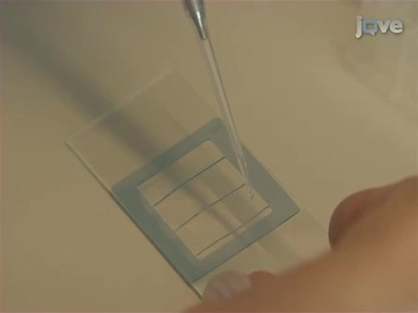
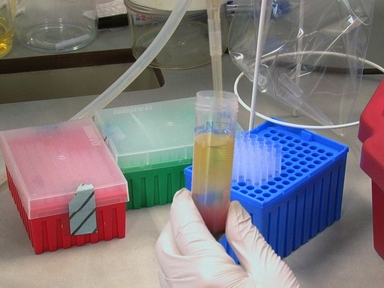
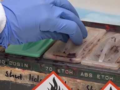
•Here we describe methods using time-lapse, phase-contrast microscopy to image mouse resident peritoneal macrophages in a chemotactic complement C5a gradient. The protocols can be extended to other immune cells.
Related Videos
Live Cell Imaging of Bacillus subtilis and Streptococcus pneumoniae using Automated Time-lapse Microscopy

Intravital Imaging of the Mouse Popliteal Lymph Node

Non-invasive Optical Imaging of the Lymphatic Vasculature of a Mouse
Studying Interactions of Staphylococcus aureus with Neutrophils by Flow Cytometry and Time Lapse Microscopy

Bone Marrow-derived Macrophage Production

Fluorescence Time-lapse Imaging of the Complete S. venezuelae Life Cycle Using a Microfluidic Device

An All-on-chip Method for Rapid Neutrophil Chemotaxis Analysis Directly from a Drop of Blood
Visualizing Macrophage Extracellular Traps Using Confocal Microscopy

Fabricating Optical-quality Glass Surfaces to Study Macrophage Fusion

Time-lapse 3D Imaging of Phagocytosis by Mouse Macrophages
ABOUT JoVE
Copyright © 2024 MyJoVE Corporation. All rights reserved