
Histological-Based Stainings Using Free-Floating Tissue Sections
August 25th, 2020
•The free-floating technique allows researchers to perform histological-based stainings including immunohistochemistry on fixed tissue sections to visualize biological structures, cell type, and protein expression and localization. This is an efficient and reliable histochemical technique that can be useful for investigating a multitude of tissues, such as brain, heart, and liver.
Tags
Related Videos

Immunohistochemistry: Paraffin Sections Using the Vectastain ABC Kit from Vector Labs

Immunohistochemistry on Paraffin Sections of Mouse Epidermis Using Fluorescent Antibodies

Rapid Genotyping of Mouse Tissue Using Sigma's Extract-N-Amp Tissue PCR Kit

Actin Co-Sedimentation Assay; for the Analysis of Protein Binding to F-Actin
Paraffin-Embedded and Frozen Sections of Drosophila Adult Muscles

Micro-Mechanical Characterization of Lung Tissue Using Atomic Force Microscopy

Chromatin Immunoprecipitation (ChIP) using Drosophila tissue
A Rapid Automated Protocol for Muscle Fiber Population Analysis in Rat Muscle Cross Sections Using Myosin Heavy Chain Immunohistochemistry

Use of Anti-phospho-girdin Antibodies to Visualize Intestinal Tuft Cells in Free-Floating Mouse Jejunum Cryosections
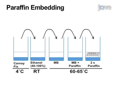
Pancreatic Islet Embedding for Paraffin Sections
ABOUT JoVE
Copyright © 2024 MyJoVE Corporation. All rights reserved