
Method Article
Endoscopic Balloon Dilatation of the Eustachian Tube via the Soft Palate Approach in Miniature Pigs
* These authors contributed equally
In This Article
Summary
In this study, we present a protocol to explore an eustachian tube surgery approach via the soft palate in miniature pigs. The surgical procedure is simple, with short surgical time, and wound healing is rapid; it is thus a good choice for procedures such as balloon dilation of the eustachian tube.
Abstract
The eustachian tube (ET) is one of the most complex organs in the human body, and its dysfunction may lead to a variety of diseases. In recent years, an increasing number of scholars have opted to conduct ET-related studies using large experimental animals such as miniature pigs or sheep, yielding promising results. Typically, conventional endoscopic procedures are performed through the nasal approach for large experimental animals. However, due to the elongated and narrow nasal cavity in these animals, transnasal surgeries are challenging.
To address this issue, we explored an ET surgery approach via the soft palate. The animal was placed in a supine position. After endotracheal intubation under general anesthesia, a mouth opener was used to fully expose the upper palate. Local infiltration with diluted adrenal fluid was performed for anesthesia of the area. A sickle knife was then used to make a longitudinal soft palate incision at the junction of the soft and hard palates. After hemostasis, an endoscope was inserted into the nasopharynx cavity, allowing the visualization of the pharyngeal opening of the ET on the posterior lateral wall of the nasal cavity.
Subsequently, a specialized pusher was used to insert a balloon into ET. The balloon was inflated, maintained at 10 bar for 2 min, and then removed. The incision in the soft palate was then sutured to ensure proper alignment. The soft palate healed well after the operation. This surgical approach is suitable for ET-related procedures in large experimental animals (e.g., miniature pigs, sheep, and dogs). The surgical procedure is simple, with a short surgical time, and wound healing is rapid. Under endoscopy, the pharyngeal opening of the ET is visible, and it is thus a good choice for procedures such as balloon dilation of the ET.
Introduction
The Eustachian tube (ET), one of the most complex organs in the human body, is a vital component of the middle ear and plays a crucial role in maintaining its physiological function1. The physiological functions of the ET are as follows2,3: first, it balances the pressure in the middle ear. When negative pressure forms in the middle ear due to gas exchange with the mucosa, the ET opens intermittently to equalize the pressure with the atmospheric pressure. This is essential for maintaining the lowest impedance conditions in the middle ear sound transmission system, ensuring the most effective transmission of sound to the inner ear. Second, it facilitates the drainage of mucus from the middle ear to the nasopharynx, preventing the accumulation of fluids and the development of secretory otitis media. Third, it has a protective role. Under normal conditions, the ET remains closed most of the time, preventing the entry of nasopharyngeal secretions or pathogens into the middle ear. The surfactant secreted by the mucosa of the ET also inhibits microbial infection.
Bluestone and Beery4 believe that the ET also has a thin neck effect similar to a flask, which is also a mechanism showing the protective effect of the ET. Fourth, it has a sound protection function. In the closed state, the ET prevents respiratory and swallowing sounds in the respiratory tract from entering the middle ear, avoiding interference with hearing. The physiological functions mentioned above are intricately connected to the complex anatomical structure of the ET. Currently, it is known that the total length of the human ET is approximately 31-40 mm, with an average length of 36 mm. Anatomically, it is divided into the bony part, the isthmus, and the cartilaginous part. In adults, the ET is inclined at an angle of 30-40° in the horizontal position and has an angle of approximately 45° in the sagittal position. The bony and cartilaginous parts do not form a straight line but rather have an angle of ~160°. The ET serves as a conduit between the nasopharynx and the middle ear. Its structure includes ET cartilage attached to the skull base, the tensor veli palatini muscle, the levator veli palatini muscle, the salpingopharyngeus muscle, the glands around the eustachian tube, and various epithelial components.
ET dysfunction may lead to various diseases, such as abnormal ET opening, ET blockage5,6, and ET pressure injury. Abnormal ET opening may result in symptoms such as tinnitus and autophony. ET blockage may affect the ventilation and drainage functions of the middle ear, leading to diseases such as secretory otitis media or cholesteatoma. ET pressure injury may cause aerotitis media.
Small-sized animals are often used in studies on ET, including mice7,8,9, rats10,11, guinea pigs12,13,14, chipmunks15,16,17,18, and rabbits19,20. These animals are cost-effective, reproduce quickly, and are easy to manage. After many years of research, substantial progress has been made in the study of ET structure, physiological functions, and pathological models using these animals. In the future, it is expected that they remain critical choices for ET research.
However, the fact that the ET has a very small volume within the animal's body and that it is even smaller in small-sized experimental animals, is a big obstacle faced by researchers. The structure and some functions of ET in these animals are quite different from those of humans, thus limiting their application in the field of ET research. In recent years, an increasing number of scholars have been utilizing large experimental animals such as miniature pigs or sheep for research related to the ET21,22,23,24,25. They have found that these large experimental animal models are more suitable for ET studies.
Pracy et al.24 discovered that ET and middle ear structures of miniature pigs are very similar to those of humans, with nearly identical lengths and similar courses. This finding aligns with the preliminary results of our research group23. Some scholars have also utilized non-human primates, such as monkeys26 and rhesus monkeys27,28,29. Their eustachian tubes are anatomically, physiologically, and genetically closer to humans. However, it is undeniable that these animals are expensive, challenging to obtain, and, in some cases, may have smaller body sizes, making surgical procedures more difficult. These characteristics contribute to the limited application of these animals in medical research.
The use of large experimental animals for ET research has recently become a hotspot in this field. In the past, the conventional endoscopic transnasal approach was used for ET research by which the eustachian tube was exposed after reaching the nasopharynx, followed by essential procedures. This process closely mimics human ET surgery. However, the anatomical characteristics of the nasal cavity and skull in large animals differ from those in humans. This may lead to challenges such as prolonged surgical duration, noticeable collateral damage, and fatigue for the surgeon during the transnasal surgery. These issues may potentially affect ET research. To address this problem, our research group has explored ET surgery via the soft palate approach and has refined the surgical process, which is introduced below.
Protocol
This study was conducted in accordance with the guidelines for the care and use of laboratory animals of the National Institutes of Health and was approved by the Animal Experiment Ethics Committee of the PLA General Hospital.
1. Preparation of surgical instruments
- Mouth opener (Figure 1)
- Make the opening device out of a stainless-steel rod with a diameter of 2 mm and weld the connection.
- Ensure that the external frame is rectangular in shape with a length of 30 cm and a width of 20 cm. Weld two stainless steel metal rings of 1 cm diameter at the midpoint of the short side of the frame.
- Connect two equilateral triangular stainless steel metal frames with a height of 7 cm to the metal rings through springs with a length of ~3 cm. Pass the upper and lower jaws of the miniature pig through the two triangles and fix the jaws to them. The triangular metal rings, under the action of spring force, separate the upper and lower jaws of the miniature pig, exposing the surgical area on the soft palate.
- Retractor (Figure 2)
- Modify the retractor on the base of the original one shown in the Table of Materials. Adjust the angle of the two edges of the original retractor to the one shown in Figure 2, to facilitate the opening of the incision.
- Balloon pusher (Figure 3)
- Modify the pusher used in this study on the base of the original one. Cut off a part of the top of the pusher, keep it ~3 mm, and adjust the end to 140° to facilitate pushing the balloon into the eustachian tube.
NOTE: Other surgical instruments are listed in the Table of Materials.
- Modify the pusher used in this study on the base of the original one. Cut off a part of the top of the pusher, keep it ~3 mm, and adjust the end to 140° to facilitate pushing the balloon into the eustachian tube.
2. Experimental animals and anesthesia
- Select healthy, 10-month-old, female Bama pigs, weighing 25 kg, and fast them for 1 day before the procedure.
- Administer tiletamine and zolazepam via intramuscular injection at a dosage of 10-15 mg/kg.
- Place the pig in a supine position on the surgical table, intubate the trachea, connect the pig to a ventilator, and maintain anesthesia with isoflurane at the flow rate of 2 L/min. Monitor blood oxygen levels (>90%), respiratory rate (15-20/min), and heart rate (60-120 beats/min)30. Check for the disappearance of corneal reflex to confirm successful anesthesia, and use medical tape to keep the eyes closed to prevent dryness.
3. Surgical procedures (Figure 4)
- Use a mouth opener (Figure 1) to fix the upper jaw downward at the lower edge of the mouth opener. Bind the tongue along with the lower jaw, suspending it upward at the upper edge of the mouth opener. Secure the tip of the tongue with sterile gauze to the lower jaw. Completely open the pig's mouth, and expose the upper palate up to 3 cm behind the junction of the soft and hard palate.
- Disinfect the oral cavity and surrounding skin with iodine, especially the junction of the soft and hard palate. Place a sterile drape and connect a 0° endoscope of 14 cm length and 3 mm diameter to a high-resolution camera system.
- Under the illumination and guidance of the endoscope, use the forefinger to reach the junction of the soft and hard palates, and find the position of the incision in the soft palate (1 cm behind the junction, 2-3 mm to the surgical side of the median suture of the upper palate). Use 1 mL of adrenaline injection with a concentration of 0.02% for local infiltration, covering the entire layers of the incised soft palate.
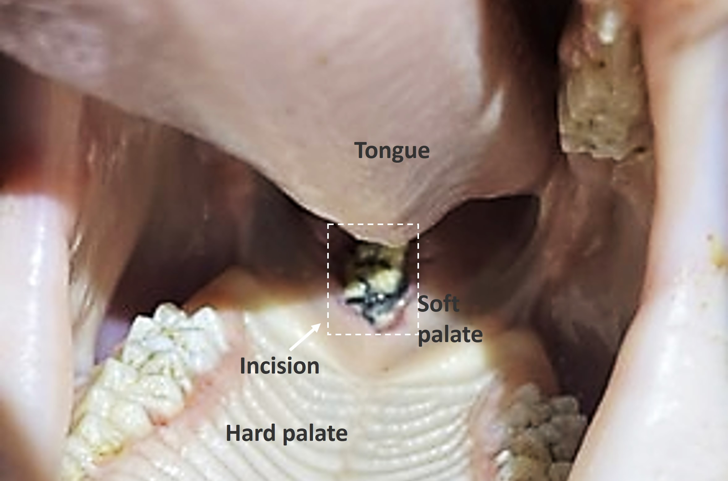
- Use a sickle knife to make a vertical incision downward at the above-mentioned location. A sense of breakthrough indicates that the soft palate has been cut through and the nasal cavity is reached. The incision length is approximately 5-7 mm.
NOTE: The downward incision should be vertical because the soft palate is thick, and if the direction is changed, it may cut inward into the submucosa of the nasal septum or outward into the submucosa of the lateral wall of the nasal cavity. Whether deviating inward or outward, it may be challenging to navigate once in the submucosa, when observed under an endoscope. - After the incision, use a self-made retractor (Figure 2) to pull it open, soak a small gauze strip in adrenaline injection with a concentration of 0.1%, and immediately insert it into the incision. Observe the incision for 2-3 min. Generally, there is no obvious bleeding but if bleeding is heavy and affects endoscopic observation, use adrenaline-soaked gauze strips until bleeding stops.
- Use the self-made retractor to fully open the incision and expose the deep structures of the soft palate.
NOTE: If it is completely opened, the opening of the nasal proximal wall can be seen through endoscopy. If the opening is not visible, the incision may be too shallow or has cut into the submucosa of the lateral wall of the nasal cavity or the submucosa of the nasal septum, which can be further adjusted according to the depth of the incision. - Insert the endoscope into the incision to visualize the structures at the back of the nasal cavity and nasopharynx, and the pharyngeal opening of ET was examined.
NOTE: The pharyngeal opening of ET is generally located at the posterior part of the lateral wall of the nasal cavity, appearing as a slightly crescent-shaped small fissure. The morphology of the cartilage of the posterior wall of the pharyngeal opening can also be observed. - Use a specially designed miniature pig balloon pusher (self-made) (Figure 3) or a bent suction device for exploring the pharyngeal opening of ET, which is a deeper fissure with a downward and outward pathway.
- Install a balloon catheter with an applied length of 35.5 cm, a balloon length of 2 cm, and a dilation diameter of 3 mm on the pusher while tightening the screws. After inserting the specially designed miniature pig balloon pusher into the ET, release the fixation screw, gently push the balloon in, pressurize it to 10 bar, and maintain it for 2 min. After that, release the pressure of the balloon, and slowly pull out the balloon together with the pusher.
- Under endoscopic examination of the pharyngeal opening and nasopharynx, perform subsequent procedures if there is no heavy bleeding. After disinfecting the soft palate incision, perform the suturing to bring it back into alignment with the 4# nylon suture.
NOTE: Full-thickness or partial-thickness sutures can be used. Generally, 1-2 stitches are sufficient. The mucosa of the incision is adjusted for proper alignment to facilitate wound healing.
4. Postoperative care
- Remove the mouth opener and bandage gauze. Place the pig in a lateral position with the tongue pulled out of the mouth to prevent the tongue from falling backward and blocking the respiratory tract. Monitor the animal closely until it is fully awake to prevent injuries due to unconscious movements.
- Place the miniature pig back into an individual cage.
- Administer one dose of antibiotics postoperatively, fast the animal for 1 day, and administer oral antibiotics for 6 days.
Results
This study performed balloon dilation of the ET via the soft palate approach on 53 miniature pigs, which were used to explore the effect of balloon dilation on ET mucosa. Balloons were inserted into the ET lumen successfully for all experimental animals and dilated smoothly, and the expected surgical objectives were achieved. Among the experimental animals, 50 pigs recovered well after surgery, with complete wound healing and successful surgery. The healing rate of the incision was 94.34%. One week postoperatively, the sutures remained intact and the incision closed well (Figure 5). Four weeks after surgery, during sample collection, we found that sutures had naturally fallen off, and healing recovered well. Almost no incision traces were visible (Figure 6). The soft palate incisions of the other three animals showed varying degrees of unhealing 1 week after surgery, leading to the formation of soft palate fistulas (Figure 7).
The soft palate approach for ET surgery proved to be convenient, and a good view of the pharyngeal opening of ET was achieved. It resulted in minimal local tissue damage, controllable bleeding, smooth balloon dilation procedures, short surgical time (an average of 15-20 min), and fast postoperative healing. No complications were indicated.

Figure 1: Diagram of self-made frame-type mouth opener. The dimensions are presented in the figure. Please click here to view a larger version of this figure.

Figure 2: Retractor. When the dilator is inserted into the incision, the elasticity of the two long sides will stretch the tissue on both sides, allowing better exposure of the surgical area below the incision. Please click here to view a larger version of this figure.

Figure 3: Balloon pusher. When the metal ring at the rear is pushed, the metal block in the red area pushes the balloon out of the tube of the pusher and into the eustachian tube. Please click here to view a larger version of this figure.

Figure 4: Surgical process of balloon dilation of the eustachian tube via the soft palate approach. (A) Surgical area and incision position; the dashed line indicates the position of the incision. (B) Local infiltration injection of adrenaline. (C) Incision with a sickle knife. (D) Post incision morphology. (E) The incision is pulled by a distractor and adrenaline-soaked gauze is used for hemostasis. (F) Observing the position of the pharyngeal orifice of ET through the soft palate incision; the dashed outlines indicate the pharyngeal orifice of ET. (G) Endoscope insertion into the nasopharynx and exposure of the pharyngeal orifice of the eustachian tube; the dashed outlines indicate the pharyngeal orifice of ET; (H) Balloon pusher at the pharyngeal orifice of ET; balloon insertion into the lumen of ET; the white arrow indicates the pharyngeal orifice of ET. (I) Pressurization of the balloon for Eustachian tube dilation; the white arrow indicates the dilation of ET. (J) Suture of the incision and apposition of the mucosa. Please click here to view a larger version of this figure.

Figure 5: One week after surgery in animals showing healing. The suture remains intact, and the incision has healed well. The dashed outlines indicate the healed incision. Please click here to view a larger version of this figure.

Figure 6: Four weeks after surgery. During sample collection, the incision is examined. Sutures have naturally fallen off, and the healing is excellent. The dashed outlines indicate the healed incision. Please click here to view a larger version of this figure.

Figure 7: One week after surgery in animals not showing healing. The incision was unclosed and an irregular pseudomembrane and fistula formed on the surface, with some sutures falling off. The dashed outlines indicated the soft palate fistula. Please click here to view a larger version of this figure.

Figure 8: Miniature pig head CT scan (sagittal section). (A) Curved nasal cavity. The yellow vertical line on the right side of the image is the scale bar, and the yellow horizontal line below is 5 cm, marking the length of a part of the soft palate, which indicates that the length of the soft palate in miniature pigs exceeds 5 cm. (B) Diagram of endoscopy insertion into the nasal cavity. Friction between endoscopy and nasal mucosa when inserting into the nasal cavity. Please click here to view a larger version of this figure.

Figure 9: Miniature pig head CT scan (Transverse section). (A-C) Normal miniature pig head CT, no high-density shadows found in bilateral middle ear and mastoid cavities (red arrow). (D) The left hamulus of the pterygoid process of miniature pig A was severed, and a CT scan was performed 3 months after surgery. No high-density shadow was found in the middle ear mastoid cavity (red arrow). (E) Miniature pig B underwent left eustachian tube balloon dilation via the soft palate pathway. A CT scan was performed 3 months after surgery, and no high-density shadow was observed in the middle ear mastoid cavity (red arrow). (F) Miniature pig C was subjected to left eustachian tube cauterization, and a CT scan was performed one month after surgery. An increased density shadow (yellow arrow) was visible in the left middle ear mastoid cavity. Please click here to view a larger version of this figure.
Discussion
ET, connecting the nasopharynx and the middle ear, plays a crucial role in regulating middle ear pressure and is implicated in the pathogenesis of otitis media25. To comprehensively study the function of ET, researchers have expanded beyond small-sized animal models to other models such as rodents or rhesus monkeys. Currently, multiple studies have used sheep or miniature pigs. Through CT scans and three-dimensional structures, Miller et al. confirmed that the anatomy of the eustachian tube in Blackfaced Sheep is very similar to that in humans22. Pohl et al. verified the feasibility of treating dysfunction of ET by implanting a stent into the sheep's ET21.
Miniature pigs, characterized by small size, early sexual maturity, rapid reproduction, and ease of management, have become increasingly popular as large animal models in medical research24. Previous studies from our team indicated that miniature pigs serve as excellent models for the study of the middle ear and inner ear30,31,32, given their morphological and functional similarities to humans. Remarkable achievements have been made in investigating the morphology, electrophysiological characteristics, development of the inner ear33,34, gene expression in the auditory system35, gene regulation36,37,38, and gene transformation39,40. Simultaneously, in-depth research has been conducted in the realm of ET function. Therefore, to investigate ET structure and function more effectively, we explored the novel surgical approach using miniature pigs.
In the investigation of surgical approaches, we initially employed the transnasal approach23, which is a conventional approach for all large animal surgeries involving ET25. Subsequently, we attempted the hard palate and soft palate approaches, and the soft palate approach was finally selected. The advantages and disadvantages of each approach are as follows:
Transnasal approach
The transnasal approach is a conventional approach for clinical examinations and surgeries involving the ET. Due to the relatively short yet spacious human nasal cavity and the large space, adrenaline-soaked cotton can be used to constrict the nasal mucosa, facilitating the passage of an endoscope to the nasopharynx. The ample space allows for comfortable manipulation, making the surgery relatively simple. Naturally, when performing surgery on large experimental animals, this approach is preferred.
The advantages of this routine approach are that the anatomical structure is relatively simple, and the use of nasal endoscopy allows for clear visualization, making it a familiar choice for operators. The disadvantages of this approach are first that the nasal cavity in miniature pigs is elongated and narrow, which can be seen from the CT results of our previous research (Figure 8A)23, restricting the mobility of instruments after deep insertion. Maneuvering becomes challenging, particularly when a rotation or angulated movement is required, making it notably difficult when attempting to reach outward and upward into the pharyngeal opening of ET.
Second, bleeding from the nasal mucosa may occur, leading to endoscopic lens contamination and an unclear view of the surgical field (Figure 8B). This issue significantly affects the surgery, and much time is spent during the process of inserting the endoscope into the nasal cavity, cleaning the lens after lens contamination, and then reinserting the endoscope. Additionally, prolonged surgery leads to congestion and swelling of the nasopharyngeal mucosa, exacerbating nasal cavity narrowing and increasing blood loss. Transnasal surgeries often take 3 h or longer; during this period, the nasal cavity is in a state of continuous bleeding, resulting in low surgical efficiency. Prolonged procedures may lead to operator fatigue and increased damage to the nasal cavity, nasopharynx, and pharyngeal opening of ET. In ET experiments, changes in pharyngeal opening morphology and function are critical points for observation. Excessive surgical damage to this area can impact observation results and, consequently, affect experimental conclusions, which should be avoided as far as possible during the operation.
Hard palate approach
The advantage of this approach is that the incision is positioned closer to the front during surgical procedures, allowing for a larger oral space. Instrument deployment is convenient, rendering the surgery relatively simple. The palatal mucosa is thin, and no instruments are needed.
The disadvantages of this approach are that the surgical steps are complex, and equipment such as a powered system drill is required to open the hard palate bone. This complexity hinders the widespread application of the approach. Most importantly, postoperative healing is often poor, leading to the formation of a hard palatal fistula, which may adversely affect ET function and even the nutritional status of the pigs. Due to these complications, this approach was discarded.
Soft palate approach
The advantages of this approach include clear anatomical layers with recognizable landmarks. The surgical incision is small, only 5-7 mm, and can be fully exposed for sufficient hemostasis so that blood loss is limited. The surgical procedures are simple, and after opening the soft palate, the nasal cavity from the level of the junction of the soft and hard palate to the level of the throat can be observed using a 0° endoscope, the pharyngeal opening of ET is directly exposed. The surgery takes approximately 20 min, and skilled personnel can complete it in just over 10 min, increasing surgical efficiency. Postoperative recovery is fast, and the wound heals within 1 week.
The disadvantages of this approach are that the incision position during surgery is posterior, resulting in a smaller oral space and making the operation more challenging. However, these challenges can be overcome. This approach involves cutting a portion of the levator veli palatini (LVP) muscle of the soft palate, which may affect the function of the ET.
To exclude the impact of muscle injury on the function of the eustachian tube, we conducted related studies. First, we severed the hamulus of the pterygoid process to weaken the contractile ability of the tensor veli palatini (TVP). Postoperative CT scans were conducted monthly, and no otitis media was formed at the end of 3 months, confirming that the eustachian tube function was not affected (Figure 9A and Figure 9D). Second, we performed CT scans on the miniature pigs 3 months after making the incision on the soft palate, and the results also showed no formation of otitis media, confirming that the eustachian tube function was not affected (Figure 9B and Figure 9E). However, eustachian tube cauterization can damage the function of the eustachian tube, leading to the formation of otitis media (Figure 9C and Figure 9F). The comparison of postoperative CT scans of the three pigs showed that the soft palate surgical pathway did not lead to the formation of otitis media in miniature pigs, indicating that this surgical method did not affect the function of the eustachian tube. Moreover, the length of the incision is only 5-7 mm, while the length of the soft palate exceeds 5 cm, providing an anatomical basis for not affecting the function of the ET (Figure 8A). We also sutured the incisions promptly, with the incision healing 1 week postoperatively. These findings, both from the anatomical structure and postoperative CT scans, confirm that the surgical incision did not affect the function of the ET. This may be due to the different effects of the pig's TVP and LVP on the ET compared to humans. Additionally, the miniature pigs were raised for four weeks after the surgery to observe the recovery of the eustachian tube. During this period, no complications such as difficulty in eating or breathing occurred. Therefore, the soft palate approach is safe and effective.
Key points of the surgical procedure for the soft palate approach include infection prevention and critical anatomical landmarks. For infection prevention, the primary focus is preventing the formation of a soft palate fistula. Once it occurs, it may severely affect the pig's water intake and nutritional status, and compromise ET function. It is imperative to follow strict aseptic procedures, ensuring thorough disinfection of the surgical area, which includes the entire upper palate mucosa, both sides of the gums and dental arches, portions of the tongue surface, and the skin surrounding the oral cavity. Additionally, antibiotics are prescribed during and after surgery, with daily oral antibiotics intake for one week postoperatively.
Critical anatomical landmarks are the posterior edge of the hard palate and the median suture of the soft palate. The posterior edge of the hard palate is located at the junction of the soft and hard palate and is palpable with the forefinger. Identification of this landmark is crucial to avoid cutting forward and risking damage to the hard palate bone, making it difficult to incise, or making it impossible to gain access to the nasopharynx after incision. Cutting backward may cause excessive damage to the soft palate, influencing the localization of the pharyngeal opening of ET. For the median suture of the soft palate, the incision should be approximately 2-3 mm to the surgical side of the median suture of the soft palate. Cutting too close to the median suture may inadvertently enter the nasal septum submucosa, hindering access to the pharyngeal opening of ET. If the incision is too far from the median suture, there is a risk of proximity to the lateral aspect of the nasopharynx, potentially damaging the pharyngeal opening of ET. In extreme cases, it may even enter the submucosa of the lateral wall, making it difficult to locate the pharyngeal opening of ET and causing potential damage to the surrounding tissues, leading to deep tissue infection postoperatively.
Fifty-three miniature pigs were involved in this research dedicated to the study of eustachian tube function. In addition to the soft palate pathway as a surgical method, we also studied the impact of balloon dilation on the structure and function of the eustachian tube and the mechanism of injury and repair process of the eustachian tube mucosa. Subsequent studies included dilation with balloons of different diameters and the histological characteristics of the eustachian tube mucosa at various time points after dilation. Fifty miniature pigs were successfully operated on for the experimental research described above.
Our surgical approach failed in 3 cases. These results could be caused by multiple factors, including some of the following. First, the location of the incision was not accurate enough, leaning towards the nasal septum side so that the position of the pharyngeal opening of the eustachian tube could not be observed effectively through the original incision. To complete the surgery, the incision was expanded, resulting in an unhealed wound. Second, the artery was injured during the cutting of the soft palate, surgical time was prolonged due to repeated hemostasis, and the residual blood stains adhered to the nasal cavity, serving as a culture medium for accelerating bacterial growth, which led to wound infection and affected healing. Third, the sutured depth was insufficient, and food friction during eating caused the suture to fall off in an untimely manner, resulting in the wound not healing. Although non-healing was presented in particular cases, the surgical success rate was still at a high level.
The limitation of this study is that we have not performed a statistical analysis on blood loss and surgical time between the transnasal and soft palatal pathways. We initially used the transnasal approach but found the operation quite challenging. The endoscope needed to be repeatedly removed to clean the blood, further aggravating the damage to the nasal mucosa and making the operation difficult. It took over 3 h before the balloon dilation was completed. Despite being able to complete the procedure, we did not continue to use this surgical method owing to the animal experiment ethics. Due to the limited number of early transnasal procedures, we just made preliminary calculations of the surgical time. However, it was precisely the disadvantages of long surgical time and excessive bleeding that inspired us to consider other pathways to avoid these issues and achieve the goal of Eustachian tube balloon dilation.
In conclusion, the surgical approach described in this study is highly suitable for performing ET procedures in large animals such as miniature pigs, sheep, and dogs. The technique is characterized by its simplicity, easy learning, short surgical time, high efficiency, minimal tissue damage, and rapid healing. Direct visualization of the pharyngeal opening of ET is possible during surgery, and the procedure has no adverse effects on the normal function of ET. This approach facilitates various interventions at the pharyngeal opening, including the balloon dilation mentioned in this study, and is equally applicable to other procedures such as ET stent placement in other literature.
Disclosures
The authors have no conflicts of interest to declare.
Acknowledgements
This study was funded by grants from the National Key Research and Development Program of China (2023YFC2508400), Beijing Municipal Natural Science Foundation (7212096), and Beijing Nova Program (Z201100006820133)
Materials
| Name | Company | Catalog Number | Comments |
| Amoxicillin Soluble Powder | Hefei dragon god Animal Pharmaceutical Co., Ltd. | Veterinary Drug License No. 120.51199 | Amoxicillin Soluble Powder is an antibiotic used in veterinary medicine. It is effective against a wide range of bacterial infections in animals, especially respiratory and gastrointestinal infections. Easily dissolved in water, it is convenient for oral administration to livestock and poultry.To prevent postoperative infections following Eustachian tube balloon dilation surgery. |
| Anesthesia Circuit | INSPIRED MEDICAL | YZB/Guangdong 0016-2009 | The anesthesia circuit is a system of tubes and components in an anesthesia machine, which delivers anesthetic gases to the patient and removes exhaled gases. It ensures a continuous flow of gases, allowing precise control of anesthesia levels during surgical procedures. |
| Anesthesia Machine | Dräger | ARHB-0015 | An anesthesia machine is a medical device used to administer and maintain anesthesia during surgical procedures. It precisely mixes gases and vapors, delivering a controlled flow of anesthesia to the patient while monitoring vital signs to ensure safety |
| Balloon Dilatation Catheter | BIOVAS | ETB30200 | An Eustachian tube balloon dilatation catheter is a specialized medical device designed to treat Eustachian tube dysfunction. It features a small balloon at its tip, which, when inflated, gently opens and widens the Eustachian tube, aiding in restoring normal function and relieving symptoms. |
| Balloon Pusher | BIOVAS | DYQ42 | Used in conjunction with a balloon dilation catheter, this device is employed during Eustachian tube balloon dilation surgery. It is inserted through the nasal cavity to the opening of the Eustachian tube, establishing a working channel. This guides the balloon catheter into the canal to complete the balloon dilation. After the procedure, it is removed from the canal along with the balloon dilation catheter. In this experiment, it is inserted through the soft palate to the opening of the Eustachian tube." |
| Cefuroxime Sodium for Injection | SHANDONG RUNZE PHARMACEUTICAL CO., LTD | National Medicine Approval Number H20066112 | Cefuroxime sodium for injection is a broad-spectrum antibiotic used to treat various bacterial infections. It's effective against respiratory, urinary, skin, and soft tissue infections, working by interfering with bacterial cell wall synthesis, thereby eliminating the infection-causing bacteria.To prevent postoperative infections following Eustachian tube balloon dilation surgery. |
| Disposable Balloon Inflation Pressure Pump | FERVID | FXB-20-30 | In Eustachian tube balloon dilation surgery, a single-use balloon inflation pressure pump is employed. This device precisely controls the balloon's expansion, ensuring safe and optimal pressure during the procedure. It's crucial for achieving effective dilation while minimizing the risk of tissue damage. |
| Endoscope | Karl Storz | 7220AA | In Eustachian tube balloon dilation surgery, an endoscope is used for direct visualization of the Eustachian tube. This allows for precise guidance of the balloon catheter, ensuring accurate placement and dilation, crucial for the success of the procedure and reducing the risk of complications.Used in conjunction with an endoscopic imaging system. |
| Endoscopic Imaging System | DELON | HD3808 | In Eustachian tube balloon dilation surgery, an endoscopic imaging system provides high-resolution visuals of the nasal and Eustachian tube areas. This facilitates precise balloon placement and expansion, ensuring the procedure's effectiveness while minimizing the risk of tissue damage and complications. |
| Epinephrine Hydrochloride Injection | HENGTONG | Veterinary Drug License No. (2013) 220381220 | Epinephrine hydrochloride injection is widely used for its vasoconstrictive properties, effectively constricting blood vessels to reduce bleeding. This makes it a crucial agent in controlling hemorrhage, particularly during surgical procedures and in certain bleeding disorders. |
| Tiletamine Hydrochloride and Zolazepam Hydrochloride Injection | Virbac | Zoletil 50 | Tilidine hydrochloride and naloxone hydrochloride injection is a medication combining an opioid analgesic and an opioid antagonist. It's used for severe pain management, where tilidine provides pain relief while naloxone reduces the risk of opioid-induced side effects, particularly respiratory depression.Used for the anesthesia of experimental animals. |
| Trauma Continuous Drainage Suction Device | Shanghai S.MANF | YZB/Shanghai 5094-54-2014 | Trauma continuous drainage aspirator is a medical device used for prolonged suction of fluids from surgical or traumatic wounds. It helps to remove exudate and blood, reducing infection risk and promoting faster healing by maintaining a clean and dry wound environment. |
| Xylazine Hydrochloride Injection | Chang shabest biological technology institute CO., LTD. | Veterinary Drug License Number180121777 | Cetirizine hydrochloride injection is used in experimental animal anesthesia for its sedative properties. It aids in the induction of mild sedation, reducing stress and discomfort in animals during procedures. Its application is crucial in ensuring the welfare and minimizing distress in various animal studies. |
References
- Smith, M. E., Scoffings, D. J., Tysome, J. R. Imaging of the eustachian tube and its function: A systematic review. Neuroradiology. 58 (6), 543-556 (2016).
- Di Martino, E. F. eustachian tube function tests: An update. Hno. 61 (6), 467-476 (2013).
- Bluestone, C. D. Impact of evolution on the eustachian tube. Laryngoscope. 118 (3), 522-527 (2008).
- Bluestone, C. D., Beery, Q. C. Concepts on the pathogenesis of middle ear effusions. Ann Otol Rhinol Laryngol. 85, 2 Suppl 25 Pt 2 182-186 (1976).
- Derkay, C. S., Bluestone, C. D., Thompson, A. E., Kardatske, D. Otitis media in the pediatric intensive care unit: A prospective study. Otolaryngol Head Neck Surg. 100 (4), 292-299 (1989).
- Bluestone, C. D., Doyle, W. J. Anatomy and physiology of eustachian tube and middle ear related to otitis media. J Allergy Clin Immunol. 81 (5), Pt 2 997-1003 (1988).
- Li, G., et al. Surfactant protein a can affect the surface tension of the eustachian tube and macrophage migration. Laryngoscope. 133 (7), 1726-1733 (2023).
- Kouhi, A., Xia, A., Khomtchouk, K., Santa Maria, P. L. Minimally invasive trans-tympanic eustachian tube occlusion animal model. Int J Pediatr Otorhinolaryngol. 156, 111070(2022).
- Nicholas, B. D., et al. Changes in eustachian tube mucosa in mice after short-term tobacco and e-cigarette smoke exposure. Laryngoscope. 132 (3), 648-654 (2022).
- Kim, Y., et al. Serial histological changes in the cartilaginous eustachian tube in the rat following balloon dilation. PLoS One. 17 (5), e0268763(2022).
- Yang, J., Zhao, C., Chen, P., Zhao, S. Morphological and pathological changes of eustachian tube mucosa in an animal model of eosinophilic otitis media. Braz J Otorhinolaryngol. 88 (5), 701-707 (2022).
- Yu, C., et al. Effect of glucocorticoids on aquaporin-1 in guinea pigs with otitis media with effusion. Exp Ther Med. 5 (6), 1589-1592 (2013).
- Choi, H. S., et al. Functional study of mucus secretion of the eustachian tube in guinea pigs. Otol Neurotol. 31 (5), 817-822 (2010).
- Jang, C. H., Park, H., Choi, C. H., Cho, Y. B., Park, I. Y. Efficacy of transnasal nebulized surfactant on experimental otitis media with effusion in guinea pig. Int J Pediatr Otorhinolaryngol. 74 (1), 71-74 (2010).
- Murrah, K. A., et al. Replication of type 5 adenovirus promotes middle ear infection by streptococcus pneumoniae in the chinchilla model of otitis media. Pathog Dis. 73 (2), 1-8 (2015).
- Novotny, L. A., Mason, K. M., Bakaletz, L. O. Development of a chinchilla model to allow direct, continuous, biophotonic imaging of bioluminescent nontypeable haemophilus influenzae during experimental otitis media. Infect Immun. 73 (1), 609-611 (2005).
- Tong, H. H., Grants, I., Liu, X., Demaria, T. F. Comparison of alteration of cell surface carbohydrates of the chinchilla tubotympanum and colonial opacity phenotype of streptococcus pneumoniae during experimental pneumococcal otitis media with or without an antecedent influenza a virus infection. Infect Immun. 70 (8), 4292-4301 (2002).
- Giebink, G. S. Otitis media: The chinchilla model. Microb Drug Resist. 5 (1), 57-72 (1999).
- Wang, B., Xu, X., Lin, J., Jin, Z. Dynamic rabbit model of ear barotrauma. Aerosp Med Hum Perform. 90 (8), 696-702 (2019).
- Koten, M., et al. Nebulized surfactant as a treatment choice for otitis media with effusion: An experimental study in the rabbit. J Laryngol Otol. 115 (5), 363-368 (2001).
- Pohl, F., et al. Stenting the eustachian tube to treat chronic otitis media - a feasibility study in sheep. Head Face Med. 14 (1), 8(2018).
- Miller, F., et al. Treatment of middle ear ventilation disorders: Sheep as animal model for stenting the human eustachian tube--a cadaver study. PLoS One. 9 (11), e113906(2014).
- An, F. W., et al. Establishment of a large animal model for eustachian tube functional study in miniature pigs. Anat Rec (Hoboken). 302 (6), 1024-1038 (2019).
- Pracy, J. P., White, A., Mustafa, Y., Smith, D., Perry, M. E. The comparative anatomy of the pig middle ear cavity: A model for middle ear inflammation in the human. J Anat. 192, Pt 3 359-368 (1998).
- Oppel, N., et al. Development of an in vivo model for eustachian tube dysfunction). Bioengineering (Basel). 9 (7), (2022).
- Alper, C. M., Swarts, J. D., Doyle, W. J. Prevention of otitis media with effusion by repeated air inflation in a monkey model). Arch Otolaryngol Head Neck Surg. 126 (5), 609-614 (2000).
- Van Cauwenberge, P. animal experiment concerning the relationship between tubal function and nasal resistance. Acta Otorhinolaryngol Belg. 43 (5), 499-513 (1989).
- Casselbrant, M. L., Cantekin, E. I., Dirkmaat, D. C., Doyle, W. J., Bluestone, C. D. Experimental paralysis of tensor veli palatini muscle. Acta Otolaryngol. 106 (3-4), 178-185 (1988).
- Doyle, W. J., Ingraham, A. S., Fireman, P. The effects of intranasal histamine challenge on eustachian tube function. J Allergy Clin Immunol. 76 (4), 551-556 (1985).
- Ji, X., et al. The miniature pig: A large animal model for cochlear implant research. J Vis Exp. (185), e64174(2022).
- Yi, H. J., et al. The temporal bone microdissection of miniature pigs as a useful large animal model for otologic research. Acta Otolaryngol. 134 (1), 26-33 (2014).
- Yi, H., et al. Miniature pigs: A large animal model of cochlear implantation. Am J Transl Res. 8 (12), 5494-5502 (2016).
- Guo, W., et al. The morphology and electrophysiology of the cochlea of the miniature pig. Anat Rec (Hoboken). 298 (3), 494-500 (2015).
- Guo, W., et al. The morphological and functional development of the stria vascularis in miniature pigs. Reprod Fertil Dev. 29 (3), 585-593 (2017).
- Wang, Q., et al. Transcription analysis of cochlear development in minipigs. Acta Otolaryngol. 137 (11), 1166-1173 (2017).
- Chen, L., et al. A de novo silencer causes elimination of mitf-m expression and profound hearing loss in pigs. BMC Biol. 14, 52(2016).
- Hai, T., et al. Pilot study of large-scale production of mutant pigs by enu mutagenesis. Elife. 6, (2017).
- Hai, T., et al. Creation of miniature pig model of human waardenburg syndrome type 2a by enu mutagenesis. Hum Genet. 136 (11-12), 1463-1475 (2017).
- Ma, Y., et al. Transplantation of human umbilical cord mesenchymal stem cells in cochlea to repair sensorineural hearing. Am J Transl Res. 8 (12), 5235-5245 (2016).
- Shi, X., et al. Adeno-associated virus transformation into the normal miniature pig and the normal guinea pigs cochlea via scala tympani. Acta Otolaryngol. 137 (9), 910-916 (2017).
Reprints and Permissions
Request permission to reuse the text or figures of this JoVE article
Request PermissionExplore More Articles
This article has been published
Video Coming Soon
Copyright © 2025 MyJoVE Corporation. All rights reserved