A subscription to JoVE is required to view this content. Sign in or start your free trial.
Method Article
الذهب Nanostar التجميعي مع أسلوب بذور فضة النمو وساطة
In This Article
Summary
نحن nanostars توليفها على شكل نجمة ذهبية باستخدام بوساطة البذور الفضة طريقة النمو. القطر من nanostars يتراوح 200-300 نانومتر ، وعدد من النصائح تختلف من 7 إلى 10. النانوية يكون لها صدى واسع وضع سطح مأكل تركزت في الأشعة تحت الحمراء القريب.
Abstract
الخصائص الفيزيائية والكيميائية والضوئية للنانو النطاق الغرويات تعتمد على حجمها المادي ، وتكوين وشكل 1-5. هناك اهتمام كبير في استخدام نانو الغرويات عن الصورة الحرارية الاجتثاث ، لتقديم الأدوية والعديد من التطبيقات الطبية الحيوية الأخرى 6. ويستخدم الذهب لا سيما بسبب سميتها منخفضة 7-9. خاصية من الغرويات المعدنية نانو هو أنها يمكن أن يكون لها صدى مأكل السطح قوي 10. ذروة وضع سطح صدى مأكل يعتمد على بنية وتركيبة المعدن نانو الغرويات. منذ يتم تحفيز وضع سطح صدى مأكل بالضوء هناك حاجة لجعل قمة في امتصاص الأشعة تحت الحمراء القريبة حيث transmissivity الأنسجة البيولوجية القصوى 11 و 12.
نقدم طريقة لتجميع نجمة ذهبية على شكل الغروية ، والمعروف أيضا باسم نجم على شكل جزيئات أو nanostars 13-15 16. ويستند هذا الأسلوب على النحوolution تحتوي على بذور الفضة التي يتم استخدامها كعامل nucleating للنمو متباين من الغرويات الذهب 17-22. وأظهر المسح المجهري الإلكتروني (SEM) تحليل غرواني الذهب مما أدى إلى أن 70 ٪ من النانو وnanostars. والآخر 30 ٪ من كتل الجسيمات غير متبلور وdecahedra rhomboids. تم الكشف عن ذروة الامتصاصية للnanostars ليكون في الأشعة تحت الحمراء بالقرب من (840 نانومتر). وهكذا ، ينتج لدينا وسيلة مناسبة لnanostars الذهب التطبيقات الطبية الحيوية ، وخاصة للصورة الحرارية الاجتثاث.
Protocol
1. الفضة البذور إعداد
- إعداد محلول المخزون من نترات الفضة (آنيو 3) عن طريق اتخاذ كتلة التعسفي وخلطها مع 10 مل من الماء (DI) منزوع الأيونات. حساب molarity من الحل. يبقي الحل في مكان مظلم لعزلها عن الضوء.
- إضافة 14.7 ملغ من الصوديوم سترات ثلاثي القواعد (نا 3 C 6 H 5 O 7) إلى 10 مل من الماء DI لجعل الحل 5 ملم. هزة القارورة حتى يذوب المسحوق.
- إضافة 15.1 ملغ من الصوديوم بوروهيدريد (NABH 4) إلى قارورة أخرى مع 10 مليلتر من الماء لجعل DI حل 40 ملم. إغلاق القارورة فورا. يهز بلطف حل باليد ووضعه في كوب مع الجليد. ضع الكأس في الثلاجة وبدء الموقت (T1 = 0). وسيتم استخدام الحل الطازجة في 15 دقيقة وهو وقت كاف لتبريده.
- من محلول المخزون من نترات الفضة ، 1.1) ، وإعداد 10 مل عند 0.25 ملم. وضع مغناطيس التحريك في القارورة وشالتحريك الفن.
- إضافة 0.25 مل من محلول سترات الصوديوم ثلاثي القواعد 1.2) إلى 1.4).
- في ر 1 = 15 دقيقة إزالة حل بوروهيدريد الصوديوم ، 1.3) ، من الثلاجة. باستخدام ماصة تتخذ 0.4 مل من هذا الحل وإضافته إلى 1.5). ملاحظة : الحل في إضافة السكتة سريعة واحدة. وسوف تتحول إلى اللون الأصفر. تحريك حل لمدة 5 دقائق.
- في ر 1 = 20 دقيقة توقف التحريك ، وإزالة المغناطيس من القارورة والحفاظ على قنينة في مكان مظلم. لا تغلق القارورة.
- يبقي الحل في الظلام في درجة حرارة الغرفة لمدة لا تقل عن 2 ساعة قبل استخدامها. يفضل استخدام البذور في غضون أسبوع واحد من التحضير.
2. نمو اعداد حل
- إعداد 80 ملم من حمض الاسكوربيك (C 6 H 8 O 6) بإضافة 140 ملغ إلى 10 مليلتر من الماء DI.
- تحضير 10 مل من محلول كلوريد الذهب (HAuCl 4). حساب molarity من الحل. الحفاظ على محلولن معزولة بعيدا عن الضوء.
- تحضير 20 مل من 50 ملم من بروميد cetyltrimethylammonium (CTAB -- C 19 H 42 BRN) بإضافة 364 ملغ إلى قارورة مع 20 مل من الماء DI. المكان على الفور المغناطيس التحريك في قارورة وبدء التحريك على طبق دافئ في 30 درجة مئوية. بعد حله تماما CTAB مسحوق والحل يصبح بدوره قبالة سخان شفافة من لوحة ولكن يبقي التحريك من خلال الخطوة 2.7).
- إضافة حل 1.1) إلى حل 2.3) للحصول على molarity النهائي 4.9x10 ملي -2. بدء الموقت (ر 2 = 0).
- في ر 2 = 1 دقيقة إضافة حل 2.2) إلى 2.4) للحصول على molarity النهائي من 0.25 ملم.
- في ر 2 = 2 دقيقة إضافة 0.1 مل من 2.1) إلى 2.5). سيكون الحل بدوره اللون.
- في ر 2 = 2 دقائق و 20 ثانية إضافة 0.05 مل من 1.8) (البذور الفضة) إلى 2.6). تحريك تعليق لمدة 15 دقيقة. وسوف توقف بدوره في البداية ثم الأزرق والبني.
- في ر 2 = 17 دقيقة توقف التحريك ، وإزالة مagnet والحفاظ على نظام التعليق في درجة حرارة الغرفة لمدة 24 ساعة.
3. فصل الذهب من nanostars CTAB التصوير ، وتوصيف أو التجريب
ملاحظة : CTAB قد تتبلور في درجة حرارة الغرفة. حل لحرارة تصل إلى بلورات غرواني الذهب إلى 30 درجة مئوية أو تزج القارورة في الاستفادة من الماء الساخن حتى تذوب البلورات.
- يصوتن للتعليق لمدة 2 دقيقة.
- تعليق الطرد المركزي لمدة 5 دقائق على 730 إطار التعاون الإقليمي. سوف Nanostars تتراكم على جدار الأنبوب.
- إزالة أكبر قدر من وقف مع ماصة مع الحرص على عدم إزالة nanostars.
- DI إضافة إلى أنبوب المياه ويصوتن لمدة 2 دقيقة.
- تعليق الطرد المركزي لمدة 3 دقائق على 460 إطار التعاون الإقليمي. تعليق يحتوي على أقل CTAB ، هناك حاجة أقل قوة الطرد المركزي لفصل بالتالي nanostars.
- كرر الخطوات من 3.3) و 3.4).
- إضافة DI المياه إلى التعليق والطرد المركزي لمدة 3 دقائق على 380 إطار التعاون الإقليمي.
- Repea ر خطوات 3.3) و 3.4). وnanostars مستعدون التصوير ، والتحليل الطيفي ، أو التجريب.
4. ممثل النتائج :
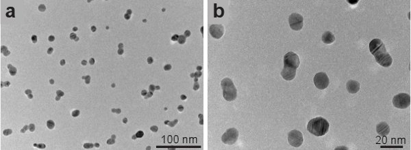
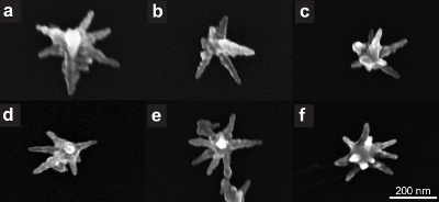
الشكل 1 يبين انتقال الإلكترون المجهر (تيم) وصور من البذور الفضة تصوير باستخدام JEOL 2010 - F TEM. بذور لها شكل كروي ، ومتوسط حجم 15 نانومتر. يتم تصوير nanostars الذهب باستخدام هيتاشي S - 5500 في المسح المجهر الإلكتروني (SEM) واسطة. ويبين الشكل 2 تكبير المتزايد للnanostars توليفها مع أسلوبنا. الجسيمات على شكل نجمة ما يقرب من 70 ٪ من الجسيمات في جميع الغروانية. غير شكلت النجوم تبدو وكأنها كتل غير متبلور وdecahedra rhomboids (لا يظهر). الشكل 3 يبين عدة nanostars الذهب واحد. حجم يتراوح من 200 نانومتر nanostars إلى 300 نانومتر ، وعدد من النصائح تختلف من 7 إلى 10. إذا كان الذهب النانوية توليفها بهذه الطريقة يتم ترك في CTAB أنها تحتفظ بشكلها لمدة لا تقل عن 1 في الشهر بعد التوليف.
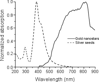
e_content "> قياس ونحن الأطياف امتصاص بذور الفضة وnanostars باستخدام كاري 14 OLIS معمل ، وذروة الامتصاص من البذور وكان في 400 نانومتر ، في حين امتصاص ذروة nanostars كان ما بين 800 نانومتر و 850 نانومتر (الشكل 4 ).
الشكل 1. نقل الصور من المجهر الالكتروني بذور الفضة.

الشكل 2. مسح الصور من المجهر الالكتروني nanostars الذهب.

الشكل 3. مسح الصور من المجهر الالكتروني nanostars ذهبية واحدة.

الشكل 4. تطبيع الأطياف امتصاص بذور الفضية (خط متقطع) والذهبnanostars (خط الصلبة).
Access restricted. Please log in or start a trial to view this content.
Discussion
في هذا العمل قدمنا طريقة لتجميع الذهب nanostars باستخدام بذور الفضة. وجدنا أن بذور الفضة أسفرت عن محصول إنتاج 70 ٪ من nanostars. وnanostars لها بالقرب من ذروة امتصاص الأشعة تحت الحمراء ، الموافق طريقتهم سطح صدى مأكل ، تركز بين 800 نانومتر و 850 نانومتر 7 و 23. هذه الخصائص خصائص ت?...
Access restricted. Please log in or start a trial to view this content.
Disclosures
الإعلان عن أي تضارب في المصالح.
Acknowledgements
وأيد هذا البحث من قبل مؤسسة العلوم الوطنية شراكات للبحوث والتعليم في المواد (PREM) منحة رقم DMR - 0934218. كان مدعوما أيضا من حيث عدد 2G12RR013646 - 11 جائزة من المركز الوطني لبحوث الموارد. المحتوى هو فقط من مسؤولية الكتاب ولا تمثل بالضرورة وجهة النظر الرسمية للمركز القومي للبحوث الموارد أو المعاهد الوطنية للصحة.
Access restricted. Please log in or start a trial to view this content.
Materials
| Name | Company | Catalog Number | Comments |
| اسم الكاشف | شركة | فهرس العدد | نقاء |
| يذوى سيترات الصوديوم ثلاثي القواعد | سيغما | S4641 | 99.0 ٪ |
| نترات الفضة | الدريتش | 204390 | 99.9999 ٪ |
| بوروهيدريد الصوديوم | الدريتش | 213462 | 99 ٪ |
| L - حمض الأسكوربيك | سيغما الدريخ | 255564 | 99 + ٪ |
| هيدرات كلوريد الذهب | الدريتش | 520918 | 99.9 ٪ + |
| بروميد Hexadecyltrimethylammonium (CTAB) | سيغما | H6269 |
| اسم المعدات | شركة | تعليقات |
| JEOL 2010 - F | JEOL | انتقال الإلكترون المجهر |
| هيتاشي S - 5500 | هيتاشي | المسح الضوئي المستخدمة في وضع المجهر الإلكترون |
| OLIS كاري 14 معمل | OLIS | معمل |
References
- Irimpan, L., Nampoori, V. P. N., Radhakrishnan, P., Krishnan, B., Deepthy, A. Size-dependent enhancement of nonlinear optical properties in nanocolloids of ZnO. Journal of Applied Physics. 103, (2008).
- Sharma, V., Park, K., Srinivasarao, M. Colloidal dispersion of gold nanorods: Historical background, optical properties, seed-mediated synthesis, shape separation and self-assembly. Materials Science and Engineering: R: Reports. 65, 1-38 (2009).
- El-Sayed, M. A. Some interesting properties of metals confined in time and nanometer space of different shapes. Accounts of Chemical Research. 34, 257-2564 (2001).
- Daniel, M. C., Astruc, D. Gold nanoparticles: assembly, supramolecular chemistry, quantum-size-related properties, and applications toward biology, catalysis, and nanotechnology. Chemical reviews. 104, 293-346 (2004).
- Burda, C., Chen, X., Narayanan, R., El-Sayed, M. A. Chemistry and Properties of Nanocrystals of Different Shapes. Chemical reviews. 105, 1025-1102 (2005).
- Hu, M., Chen, J. Y. X., Li, J. Y., Au, L., Hartland, G. V., Li, X. D., Marquez, M., Xia, Y. N. Gold nanostructures: engineering their plasmonic properties for biomedical applications. Chemical Society Reviews. 35, 1084-1094 (2006).
- Seo, J. T., Yang, Q., Kim, W. J., Heo, J., Ma, S. M., Austin, J., Yun, W. S., Jung, S. S., Han, S. W., Tabibi, B., Temple, D. Optical nonlinearities of Au nanoparticles and Au/Ag coreshells. Opt. Lett. 34, 307-309 (2009).
- Jeong, S., Choi, S. Y., Park, J., Seo, J. -H., Park, J., Cho, K., Joo, S. -W., Lee, S. Y. Low-toxicity chitosan gold nanoparticles for small hairpin RNA delivery in human lung adenocarcinoma cells. Journal of Materials Chemistry. 21, 13853-13859 (2011).
- Huang, X., Jain, P. K., El-Sayed, I. H., El-Sayed, M. A. Gold nanoparticles: interesting optical properties and recent applications in cancer diagnostics and therapy. Nanomedicine. 2, 681-693 (2007).
- Link, S., El-Sayed, M. A. Shape and size dependence of radiative, non-radiative and photothermal properties of gold nanocrystals. International Reviews in Physical Chemistry. 19, 409-453 (2000).
- El-Sayed, I. H., Huang, X. H., El-Sayed, M. A. Selective laser photo-thermal therapy of epithelial carcinoma using anti-EGFR antibody conjugated gold nanoparticles. Cancer Letters. 239, 129-135 (2006).
- O'Neal, D. P., Hirsch, L. R., Halas, N. J., Payne, J. D., West, J. L. Photo-thermal tumor ablation in mice using near infrared-absorbing nanoparticles. Cancer Letters. 209, 171-176 (2004).
- Nehl, C. L., Liao, H. W., Hafner, J. H. Optical properties of star-shaped gold nanoparticles. Nano Letters. 6, 683-688 (2006).
- Pazos-Perez, N., Rodriguez-Gonzalez, B., Hilgendorff, M., Giersig, M., Liz-Marzan, L. M. Gold encapsulation of star-shaped FePt nanoparticles. Journal of Materials Chemistry. 20, 61-64 (2010).
- Sahoo, G. P., Bar, H., Bhui, D. K., Sarkar, P., Samanta, S., Pyne, S., Ash, S., Misra, A. Synthesis and photo physical properties of star shaped gold nanoparticles. Colloids and Surfaces A: Physicochemical and Engineering Aspects. , 375-371 (2011).
- Senthil Kumar, P., Pastoriza-Santos, I., Rodriguez-Gonzalez, B., Garcia de Abajo, F. J., Liz-Marzan, L. M. High-yield synthesis and optical response of gold nanostars. Nanotechnology. 19, (2008).
- Goodrich, G. P., Bao, L. L., Gill-Sharp, K., Sang, K. L., Wang, J., Payne, J. D. Photothermal therapy in a murine colon cancer model using near-infrared absorbing gold nanorods. Journal of Biomedical Optics. 15, (2010).
- Zhang, D., Neumann, O., Wang, H., Yuwono, V. M., Barhoumi, A., Perham, M., Hartgerink, J. D., Wittung-Stafshede, P., Halas, N. J. Gold Nanoparticles Can Induce the Formation of Protein-based Aggregates at Physiological pH. Nano Lett. 9, 666-671 (2009).
- Alkilany, A. M., Nagaria, P. K., Hexel, C. R., Shaw, T. J., Murphy, C. J., Wyatt, M. D. Cellular uptake and cytotoxicity of gold nanorods: molecular origin of cytotoxicity and surface effects. Small. 5, 701-708 (2009).
- Sun, L., Liu, D., Wang, Z. Functional gold nanoparticle-peptide complexes as cell-targeting agents. Langmuir. 24, 10293-10297 (2008).
- Park, J., Estrada, A., Sharp, K., Sang, K., Schwartz, J. A., Smith, D. K., Coleman, C., Payne, J. D., Korgel, B. A., Dunn, A. K., Tunnell, J. W. Two-photon-induced photoluminescence imaging of tumors using near-infrared excited gold nanoshells. Opt. Express. 16, 1590-1599 (2008).
- Nikoobakht, B., El-Sayed, M. A. Preparation and growth mechanism of gold nanorods (NRs) using seed-mediated growth method. Chemistry of Materials. 15, 1957-1962 (2003).
- Hao, F., Nehl, C. L., Hafner, J. H., Nordlander, P. Plasmon resonances of a gold nanostar. Nano Letters. 7, 729-732 (2007).
- Hao, F., Nordlander, P., Sonnefraud, Y., Dorpe, P. V. an, Maier, S. A. Tunability of Subradiant Dipolar and Fano-Type Plasmon Resonances in Metallic Ring/Disk Cavities: Implications for Nanoscale Optical Sensing. ACS Nano. 3, 643-652 (2009).
- Sweeney, C. M., Hasan, W., Nehl, C. L., Odom, T. W. Optical Properties of Anisotropic Core-Shell Pyramidal Particles. Journal of Physical Chemistry A. 113, 4265-4268 (2009).
- Dickerson, E. B., Dreaden, E. C., Huang, X. H., El-Sayed, I. H., Chu, H. H., Pushpanketh, S., McDonald, J. F., El-Sayed, M. A. Gold nanorod assisted near-infrared plasmonic photothermal therapy (PPTT) of squamous cell carcinoma in mice. Cancer Letters. 269, 57-66 (2008).
- Jana, N. R., Gearheart, L., Murphy, C. J. Wet chemical synthesis of high aspect ratio cylindrical gold nanorods. Journal of Physical Chemistry B. 105, 4065-4067 (2001).
- Jana, N. R., Gearheart, L., Murphy, C. J. Seed-mediated growth approach for shape-controlled synthesis of spheroidal and rod-like gold nanoparticles using a surfactant template. Advanced Materials. 13, 1389-1393 (2001).
- Xiao, J., Qi, L. Surfactant-assisted, shape-controlled synthesis of gold nanocrystals. Nanoscale. 3, 1383-1396 (2011).
- Tao, A. R., Habas, S., Yang, P. Shape control of colloidal metal nanocrystals. Small. 4, 310-325 (2008).
- Cole, J. R., Mirin, N. A., Knight, M. W., Goodrich, G. P., Halas, N. J. Photothermal Efficiencies of Nanoshells and Nanorods for Clinical Therapeutic Applications. Journal of Physical Chemistry C. 113, 12090-12094 (2009).
- Choi, J. S., Park, J. C., Nah, H., Woo, S., Oh, J., Kim, K. M., Cheon, G. J., Chang, Y., Yoo, J., Cheon, J. A hybrid nanoparticle probe for dual-modality positron emission tomography and magnetic resonance imaging. Angew. Chem. Int. Ed. Engl. 47, 6259-6262 (2008).
- Chithrani, B. D., Ghazani, A. A., Chan, W. C. W. Determining the Size and Shape Dependence of Gold Nanoparticle Uptake into Mammalian Cells. Nano Letters. 6, 662-668 (2006).
Access restricted. Please log in or start a trial to view this content.
Reprints and Permissions
Request permission to reuse the text or figures of this JoVE article
Request PermissionExplore More Articles
This article has been published
Video Coming Soon
Copyright © 2025 MyJoVE Corporation. All rights reserved