JoVE 비디오를 활용하시려면 도서관을 통한 기관 구독이 필요합니다. 전체 비디오를 보시려면 로그인하거나 무료 트라이얼을 시작하세요.
Method Article
실버 씨앗 중재 성장 방법과 골드 Nanostar 합성
요약
우리는 은색 씨앗 중재의 성장 방법을 사용하여 별 모양의 골드 nanostars을 합성. nanostars의 직경이 200-300 nm의 범위에서 및 도움말의 숫자 7에서 10에 따라 다릅니다. nanoparticles는 가까운 적외선 중심으로 광범위한 표면 plasmon 공진 모드를있다.
초록
나노 스케일 colloids의 물리적, 화학적 및 광학 성질들은 재료 성분, 크기와 모양 1-5에 따라 달라집니다. 사진 열 절제, 약물 전달 및 기타 여러 생명 의학 애플 리케이션 6 나노 colloids를 사용하여 큰 관심이 있습니다. 골드는 특히 때문에 낮은 독성 7-9 중 사용됩니다. 금속 나노 colloids의 속성들은 강력한 표면 plasmon 공명 10 가질 수 있습니다. 표면 plasmon 공명 모드의 절정은 금속 나노 colloids의 구조와 구성에 따라 달라집니다. 표면 plasmon 공명 모드가 빛을 자극되기 때문에 생물 학적 조직 투과율이 11, 12 최대한의 어디에 가까운 적외선의 최대 흡광도를 가질 필요가있다.
우리는 또한 별 모양 nanoparticles 13-15 또는 nanostars 16 알려져 별 모양의 금 콜로이드를 합성하는 방법을 제시한다. 이 방법은로 기반황금 colloids 17-22의 비등 방성 성장을위한 nucleating 에이전트로 사용되는 은색 씨앗을 포함 olution. 결과 금 콜로이드의 스캐닝 전자 현미경 (SEM) 분석은 nanostructures의 70 %가 nanostars 것을 보여주었다. 입자의 다른 30 % decahedra 및 rhomboids의 비정질 클러스터했다. nanostars의 흡광도 피크는 가까운 적외선 (840 NM)에 있어야 발견했습니다. 따라서, 우리의 방법은 특히 사진 열 절제를 위해, 생명 의학 애플 리케이션에 적합한 골드 nanostars을 생산하고 있습니다.
프로토콜
1. 실버 종자 준비
- 임의의 질량을 감수하면서 deionized (DI) 물 10 ML로 혼합하여 실버 질산 (AgNO 3) 재고 솔루션을 준비합니다. 솔루션의 molarity을 계산합니다. 라이트에서 분리 어두운 장소에 솔루션을 보관하십시오.
- 5 MM 솔루션을 만들기 위해 DI 물 10 ML에 나트륨 구연산 tribasic의 14.7 MG (나 3 C 6 H 5 O 7) 추가합니다. 파우더가 해산 때까지 병을 흔들어.
- 40 MM 솔루션을 만들기 위해 DI 물 10 ML 다른 유리병에 나트륨 borohydride (NaBH 4) 15.1 밀리그램을 추가합니다. 즉시 병을를 닫습니다. 부드럽게 손으로 솔루션을 흔들 얼음 비커에 넣습니다. 냉장고에 비커를 놓고 (T1 = 0) 타이머를 시작합니다. 갓 만들어진 솔루션은 충분히 그것을 아래로 냉각 시간 15 분 사용됩니다.
- 실버 질산, 1.1)의 주식 솔루션에서, 0.25 MM 10 ML을 준비합니다. 유리병와 ST의 감동 자석 장소예술 감동.
- 1.4 나트륨 구연산 tribasic 솔루션 1.2))의 0.25 ML을 추가합니다.
- t 1 = 15 분 냉장고에서 나트륨 borohydride 솔루션, 1.3)를 제거하십시오. 피펫이 솔루션 0.4 ML을 가지고) 1.5에 추가 사용. 참고 : 단일 빠른 스트로크에 솔루션을 추가합니다. 색깔이 노란색으로 바뀝니다. 5 분 솔루션을 저어.
- t 1 = 20 분 중지 감동, 유리병에서 자석을 제거하고 어두운 장소에 보관. 약병을 닫지 마십시오.
- 사용하기 전에 최소한 2 시간 동안 실온에서 어둠의 솔루션을 유지. 가급적 준비 한 주일 이내에 씨앗을 사용합니다.
2. 성장 솔루션 준비
- DI 워터 10 ML에 140 밀리그램을 추가하여 아스코르비 산 (C 6 H 8 O 6)의 80 MM을 준비합니다.
- 골드 염화 (HAuCl 4) 농축 용액 10 ML을 준비합니다. 솔루션의 molarity을 계산합니다. 솔루션을 유지n은 빛으로부터 격리.
- DI 워터 20 ML과 유리병에 364 MG를 추가하여 - cetyltrimethylammonium의 브로마이드 50 MM (C 19 H 42 BrN CTAB)의 20 ML을 준비합니다. 즉시 유리병에 감동 자석을 배치하고 30 따뜻한 접시에 감동을 시작 ° C. CTAB 분말이 완전히 해산 및 솔루션은 투명 판의 히터 해제하지만, 단계 2.7을 통해 감동 유지)되는 후.
- 4.9x10 -2 mm의 최종 molarity를 구하는 해결책 2.3)에 솔루션 1.1) 추가합니다. (T 2 = 0) 타이머를 시작합니다.
- t 2 = 1 분, 0.25 mm의 최종 molarity를 얻을 2.4)에 솔루션 2.2) 추가합니다.
- t 2 = 2 분) 2.5-2.1의 0.1 ML)를 추가합니다. 이 솔루션은 무색으로 바뀔 것이다.
- t 2 = 2 분 20 초)이 2.6-1.8의 0.05 ML) (실버 씨앗)을 추가합니다. 15 분에 대한 정지를 저어. 정지는 처음에 파란색 후 갈색으로 바뀔 것이다.
- t 2 = 17 분 정지의 감동, m을 제거agnet 24 시간 동안 실온에서 정학을 유지.
3. 이미징, 특성 또는 실험에 대해 CTAB에서 골드 nanostars 분리
참고 : CTAB는 실온에서 구체화 수 있습니다. 30 ° C로 골드 콜로이드 최대 크리스탈 열을 분해 또는 수정이 용해 때까지 탭 물을 고온에 병을 담가합니다.
- 2 분 정지를 Sonicate.
- 730 rcf에서 5 분 현탁액을 원심. Nanostars은 튜브의 벽에 축적됩니다.
- nanostars을 제거하지 않도록 관리하는 피펫와 마찬가지로 많은 정지 제거합니다.
- 튜브에 DI 물을 추가 2 분 sonicate.
- 460 rcf에서 3 분 현탁액을 원심. 정지 덜 CTAB를 포함, 따라서 낮은 원심력은 nanostars을 구분이 필요합니다.
- ) 단계 3.3) 및 3.4를 반복합니다.
- 380 rcf에서 3 분 서스펜션과 원심에 DI 워터를 추가합니다.
- 반복 하오 t 3.3) 및 3.4) 단계를 반복합니다. nanostars는 이미징, 분광, 또는 실험을위한 준비가되어 있습니다.
4. 대표 결과 :
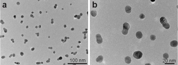
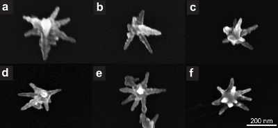
그림 1은 실버 씨앗 JEOL 2010 - F TEM을 사용하여 몇 군데의 전송 전자 현미경 (TEM) 이미지를 보여줍니다. 씨앗은 구형 모양과 15 nm의의 평균 크기를했습니다. 골드 nanostars은 히타치를 사용하여 몇 군데 아르 S - 5500 전자 현미경 (SEM) 모드를 스캔 인치 그림 2는 우리의 방법과 합성 nanostars의 증가 배율을 보여줍니다. 스타 모양의 입자 콜로이드의 모든 입자의 약 70 %입니다. 비 형성 스타 decahedra 및 rhomboids의 비정질 클러스터 (표시되지 않음)처럼 나타납니다. 그림 3은 여러 싱글 골드 nanostars을 보여줍니다. 200 NM에서 300 NM 및 도움말의 개수에 nanostars 범위의 크기는 7에서 10에 따라 다릅니다. 황금 그들이 합성 후 최소 1 개월 그 모양을 그대로 유지이 방법 CTAB에 남아 의해 합성 nanoparticles 경우.
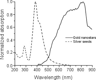
e_content "> 우리는 Olis 캐리 - 14 분광 광도계를 사용하여 실버 종자와 nanostars의 흡수 스펙트럼을 측정. 종자의 최대 흡수가 400 NM에서 되었음 nanostars의 최대 흡수 800 NM 850 NM (그림 4의 사이 동안 ).
그림 1. 은빛 씨앗의 전송 전자 현미경 이미지.

그림 2. 금을 nanostars의 전자 현미경 이미지를 검사합니다.

그림 3. 단일 골드 nanostars의 전자 현미경 이미지를 검사합니다.

그림 4. 표준화 흡수 실버 종자의 스펙트럼 (점선)과 금nanostars (실선).
Access restricted. Please log in or start a trial to view this content.
토론
이 작품에서 우리는 실버 종자를 사용하여 골드 nanostars를 종합하는 방법을 제시합니다. 우리는 실버 씨앗이 nanostars의 70 %를 생산 수율 결과 것으로 나타났습니다. nanostars 800 NM 850 NM 7, 23 사이를 중심으로 그들의 표면 plasmon 공진 모드에 해당하는 가까운 적외선 흡수 피크를,있다. 이러한 속성 속성은 우리의 골드 nanostars 이러한 사진 열 절제로 24-26 생물 의학 응용 프로그램에 사용...
Access restricted. Please log in or start a trial to view this content.
공개
관심 없음 충돌 선언하지 않습니다.
감사의 말
이 연구는 재료의 연구와 교육에 대한 국립 과학 재단 (National Science Foundation) 협력 (PREM) 부여 번호 DMR - 0934218에 의해 지원되었다. 또한 연구 자원에 대한 국립 센터에서 보너스 번호 2G12RR013646 - 11에 의해 지원되었다. 내용은 전적으로 저자의 책임이며 반드시 연구 자원이나 국립 보건원에 대한 국립 센터의 공식 견해를 대변하지 않습니다.
Access restricted. Please log in or start a trial to view this content.
자료
| Name | Company | Catalog Number | Comments |
| 시약의 이름 | 회사 | 카탈로그 번호 | 순도 |
| 나트륨 구연산 tribasic의 탈수 | 시그마 | S4641 | 99.0 % |
| 실버 질산염 | 올드 리치 | 204,390 | 99.9999 % |
| 나트륨 borohydride | 올드 리치 | 213,462 | 99% |
| L - 아스코르비 산 | 시그마 - 올드 리치 | 255,564 | 99 이상 % |
| 골드 염화물 trihydrate | 올드 리치 | 520,918 | 99.9 이상 % |
| Hexadecyltrimethylammonium의 브로마이드 (CTAB) | 시그마 | H6269 |
| 장비의 명칭 | 회사 | 댓글 |
| JEOL 2010 - F | JEOL | 전송 전자 현미경 |
| 히타치 S - 5500 | 히타치 | 전자 현미경 모드를 스캔에 사용 |
| Olis 캐리 - 14 분광 광도계 | Olis | 분광 광도계 |
참고문헌
- Irimpan, L., Nampoori, V. P. N., Radhakrishnan, P., Krishnan, B., Deepthy, A. Size-dependent enhancement of nonlinear optical properties in nanocolloids of ZnO. Journal of Applied Physics. 103, (2008).
- Sharma, V., Park, K., Srinivasarao, M. Colloidal dispersion of gold nanorods: Historical background, optical properties, seed-mediated synthesis, shape separation and self-assembly. Materials Science and Engineering: R: Reports. 65, 1-38 (2009).
- El-Sayed, M. A. Some interesting properties of metals confined in time and nanometer space of different shapes. Accounts of Chemical Research. 34, 257-2564 (2001).
- Daniel, M. C., Astruc, D. Gold nanoparticles: assembly, supramolecular chemistry, quantum-size-related properties, and applications toward biology, catalysis, and nanotechnology. Chemical reviews. 104, 293-346 (2004).
- Burda, C., Chen, X., Narayanan, R., El-Sayed, M. A. Chemistry and Properties of Nanocrystals of Different Shapes. Chemical reviews. 105, 1025-1102 (2005).
- Hu, M., Chen, J. Y. X., Li, J. Y., Au, L., Hartland, G. V., Li, X. D., Marquez, M., Xia, Y. N. Gold nanostructures: engineering their plasmonic properties for biomedical applications. Chemical Society Reviews. 35, 1084-1094 (2006).
- Seo, J. T., Yang, Q., Kim, W. J., Heo, J., Ma, S. M., Austin, J., Yun, W. S., Jung, S. S., Han, S. W., Tabibi, B., Temple, D. Optical nonlinearities of Au nanoparticles and Au/Ag coreshells. Opt. Lett. 34, 307-309 (2009).
- Jeong, S., Choi, S. Y., Park, J., Seo, J. -H., Park, J., Cho, K., Joo, S. -W., Lee, S. Y. Low-toxicity chitosan gold nanoparticles for small hairpin RNA delivery in human lung adenocarcinoma cells. Journal of Materials Chemistry. 21, 13853-13859 (2011).
- Huang, X., Jain, P. K., El-Sayed, I. H., El-Sayed, M. A. Gold nanoparticles: interesting optical properties and recent applications in cancer diagnostics and therapy. Nanomedicine. 2, 681-693 (2007).
- Link, S., El-Sayed, M. A. Shape and size dependence of radiative, non-radiative and photothermal properties of gold nanocrystals. International Reviews in Physical Chemistry. 19, 409-453 (2000).
- El-Sayed, I. H., Huang, X. H., El-Sayed, M. A. Selective laser photo-thermal therapy of epithelial carcinoma using anti-EGFR antibody conjugated gold nanoparticles. Cancer Letters. 239, 129-135 (2006).
- O'Neal, D. P., Hirsch, L. R., Halas, N. J., Payne, J. D., West, J. L. Photo-thermal tumor ablation in mice using near infrared-absorbing nanoparticles. Cancer Letters. 209, 171-176 (2004).
- Nehl, C. L., Liao, H. W., Hafner, J. H. Optical properties of star-shaped gold nanoparticles. Nano Letters. 6, 683-688 (2006).
- Pazos-Perez, N., Rodriguez-Gonzalez, B., Hilgendorff, M., Giersig, M., Liz-Marzan, L. M. Gold encapsulation of star-shaped FePt nanoparticles. Journal of Materials Chemistry. 20, 61-64 (2010).
- Sahoo, G. P., Bar, H., Bhui, D. K., Sarkar, P., Samanta, S., Pyne, S., Ash, S., Misra, A. Synthesis and photo physical properties of star shaped gold nanoparticles. Colloids and Surfaces A: Physicochemical and Engineering Aspects. , 375-371 (2011).
- Senthil Kumar, P., Pastoriza-Santos, I., Rodriguez-Gonzalez, B., Garcia de Abajo, F. J., Liz-Marzan, L. M. High-yield synthesis and optical response of gold nanostars. Nanotechnology. 19, (2008).
- Goodrich, G. P., Bao, L. L., Gill-Sharp, K., Sang, K. L., Wang, J., Payne, J. D. Photothermal therapy in a murine colon cancer model using near-infrared absorbing gold nanorods. Journal of Biomedical Optics. 15, (2010).
- Zhang, D., Neumann, O., Wang, H., Yuwono, V. M., Barhoumi, A., Perham, M., Hartgerink, J. D., Wittung-Stafshede, P., Halas, N. J. Gold Nanoparticles Can Induce the Formation of Protein-based Aggregates at Physiological pH. Nano Lett. 9, 666-671 (2009).
- Alkilany, A. M., Nagaria, P. K., Hexel, C. R., Shaw, T. J., Murphy, C. J., Wyatt, M. D. Cellular uptake and cytotoxicity of gold nanorods: molecular origin of cytotoxicity and surface effects. Small. 5, 701-708 (2009).
- Sun, L., Liu, D., Wang, Z. Functional gold nanoparticle-peptide complexes as cell-targeting agents. Langmuir. 24, 10293-10297 (2008).
- Park, J., Estrada, A., Sharp, K., Sang, K., Schwartz, J. A., Smith, D. K., Coleman, C., Payne, J. D., Korgel, B. A., Dunn, A. K., Tunnell, J. W. Two-photon-induced photoluminescence imaging of tumors using near-infrared excited gold nanoshells. Opt. Express. 16, 1590-1599 (2008).
- Nikoobakht, B., El-Sayed, M. A. Preparation and growth mechanism of gold nanorods (NRs) using seed-mediated growth method. Chemistry of Materials. 15, 1957-1962 (2003).
- Hao, F., Nehl, C. L., Hafner, J. H., Nordlander, P. Plasmon resonances of a gold nanostar. Nano Letters. 7, 729-732 (2007).
- Hao, F., Nordlander, P., Sonnefraud, Y., Dorpe, P. V. an, Maier, S. A. Tunability of Subradiant Dipolar and Fano-Type Plasmon Resonances in Metallic Ring/Disk Cavities: Implications for Nanoscale Optical Sensing. ACS Nano. 3, 643-652 (2009).
- Sweeney, C. M., Hasan, W., Nehl, C. L., Odom, T. W. Optical Properties of Anisotropic Core-Shell Pyramidal Particles. Journal of Physical Chemistry A. 113, 4265-4268 (2009).
- Dickerson, E. B., Dreaden, E. C., Huang, X. H., El-Sayed, I. H., Chu, H. H., Pushpanketh, S., McDonald, J. F., El-Sayed, M. A. Gold nanorod assisted near-infrared plasmonic photothermal therapy (PPTT) of squamous cell carcinoma in mice. Cancer Letters. 269, 57-66 (2008).
- Jana, N. R., Gearheart, L., Murphy, C. J. Wet chemical synthesis of high aspect ratio cylindrical gold nanorods. Journal of Physical Chemistry B. 105, 4065-4067 (2001).
- Jana, N. R., Gearheart, L., Murphy, C. J. Seed-mediated growth approach for shape-controlled synthesis of spheroidal and rod-like gold nanoparticles using a surfactant template. Advanced Materials. 13, 1389-1393 (2001).
- Xiao, J., Qi, L. Surfactant-assisted, shape-controlled synthesis of gold nanocrystals. Nanoscale. 3, 1383-1396 (2011).
- Tao, A. R., Habas, S., Yang, P. Shape control of colloidal metal nanocrystals. Small. 4, 310-325 (2008).
- Cole, J. R., Mirin, N. A., Knight, M. W., Goodrich, G. P., Halas, N. J. Photothermal Efficiencies of Nanoshells and Nanorods for Clinical Therapeutic Applications. Journal of Physical Chemistry C. 113, 12090-12094 (2009).
- Choi, J. S., Park, J. C., Nah, H., Woo, S., Oh, J., Kim, K. M., Cheon, G. J., Chang, Y., Yoo, J., Cheon, J. A hybrid nanoparticle probe for dual-modality positron emission tomography and magnetic resonance imaging. Angew. Chem. Int. Ed. Engl. 47, 6259-6262 (2008).
- Chithrani, B. D., Ghazani, A. A., Chan, W. C. W. Determining the Size and Shape Dependence of Gold Nanoparticle Uptake into Mammalian Cells. Nano Letters. 6, 662-668 (2006).
Access restricted. Please log in or start a trial to view this content.
재인쇄 및 허가
JoVE'article의 텍스트 или 그림을 다시 사용하시려면 허가 살펴보기
허가 살펴보기더 많은 기사 탐색
This article has been published
Video Coming Soon
Copyright © 2025 MyJoVE Corporation. 판권 소유