
Method Article
捕获组织修复的斑马鱼幼虫与时间推移明视立体显微镜
摘要
We present a protocol for capturing the dynamics of zebrafish larval tail fin regeneration on a whole-tissue scale using brightfield-based stereomicroscopy. This technique enables capturing the regeneration dynamics with single cell resolution. This methodology can be adapted to any stereomicroscope equipped with a CCD camera and time-lapse software.
摘要
The zebrafish larval tail fin is ideal for studying tissue regeneration due to the simple architecture of the larval fin-fold, which comprises of two layers of skin that enclose undifferentiated mesenchyme, and because the larval tail fin regenerates rapidly within 2-3 days. Using this system, we demonstrate a method for capturing the repair dynamics of the amputated tail fin with time-lapse video brightfield stereomicroscopy. We demonstrate that fin amputation triggers a contraction of the amputation wound and extrusion of cells around the wound margin, leading to their subsequent clearance. Fin regeneration proceeds from proximal to distal direction after a short delay. In addition, developmental growth of the larva can be observed during all stages. The presented method provides an opportunity for observing and analyzing whole tissue-scale behaviors such as fin development and growth in a simple microscope setting, which is easily adaptable to any stereomicroscope with time-lapse capabilities.
引言
The ability of an organism to orchestrate tissue repair processes after injury is crucial for its survival 1. While all animals have the capacity to heal their wounds, the extent to which tissues regenerate differs greatly among species. Vertebrate species such as zebrafish, salamanders and frog tadpoles have the remarkable ability to regenerate lost tissues, including their appendages, portions of their eyes, heart, and central nervous system 2-4. Mammalian species, such as the African spiny mouse and rabbits, are capable of regenerating holes in their pinnae 5-7, and humans and mice regenerate portions of their liver as well as their digit tips during fetal and juvenile stages 8-12. Although it is not well understood yet why and how certain species regenerate tissues more effectively than others, the presence of similar genetic pathways suggests that these mechanisms may lie dormant in species without great regeneration potential 13,14. Thus elucidating tissue repair and regeneration mechanisms in species with satisfactory regeneration outcomes will benefit regeneration in humans.
We have chosen the larval zebrafish tail fin as a paradigm to demonstrate its regeneration with time-lapse brightfield stereomicroscopy. The zebrafish larval tail fin is anatomically simple as compared to the more complex adult structures, consisting of a two-layered, infolded epithelium with somatosensory axons innervating the skin that surrounds medially located mesenchymal cells 15. Despite the anatomical differences, larval tail fin regeneration is somewhat comparable to adult fin regeneration in terms of the molecular signatures and the outgrowth responses 16,17. As compared to the adult fin, imaging larval tail fin regeneration has however several advantages: 1) larval fin regeneration is completed within just 2-3 days 16, 2) larvae can be mounted in low-melt agarose, and 3) larvae do not require feeding until ~ 5 days post fertilization (dpf) due to the presence of the yolk sac. This makes zebrafish larvae ideal for observing tissue repair dynamics in vivo.
The presented method enables the capture of detailed dynamics underlying the early processes of fin regeneration. Many studies have utilized fluorescence-based confocal microscopy to study cellular and subcellular biological processes in embryonic and larval zebrafish. Sophisticated confocal imaging setups are however often not accessible to everyone and highly expensive as compared to other imaging techniques. In contrast, the presented methodology utilizes a Discovery V12 stereomicroscope equipped with Axiovision software and a time-lapse module, thus providing a more affordable alternative to expensive imaging equipment to examine tissue behaviors. We demonstrate that this method can be utilized for imaging tissue regeneration with high temporal resolution at a minimal cost. The implications for this method could extend beyond basic biology to advance mammalian regeneration studies using organ cultures, for therapeutic development through pharmacological and genetic screens, and it can serve as a teaching tool in a classroom setting.
研究方案
斑马鱼( 珍珠层株)根据既定协议孕育和提高。所作的所有努力,尽量减少痛苦,使用0.4毫米的三卡因麻醉和1 mM的三卡因安乐死。斑马鱼胚胎和幼虫严格按照良好的动物的做法被处理为经适当委员会(MDI生物实验室动物的核心IACUC数13-20)。这项研究是根据协议,#14-09经国家人类基因组研究所动物护理和使用委员会,MDIBL制度保障#A-3562-01。
注意:成像程序,捕捉在斑马鱼幼体总结在下面的步骤鳍再生:
1.提高斑马鱼幼虫阶段
- 收集鸡蛋,并放置约50个卵到含0.03%的即时海洋盐在补充有0.00004%亚甲蓝去离子水的100×25毫米的培养皿。孵育O / N在28.5℃的孵化器。
- 第二天早晨取出死胚用玻璃吸管和冲洗鸡蛋与去离子水(称为胚胎培养基)0.03%即时海洋盐过滤。
注:中等如林格18,汉克斯18,E2 19 E3 20和Danieau 21可优选的。 - SUP>添加新鲜胚胎中期的菜。如果使用含颜料的菌株,任选添加0.2mM的1-苯基-2-硫脲(PTU),如PTU将阻止黑色素生成,因而色素沉着的幼虫。让胚胎进一步发展在孵化器直到后2天受精或任何其它期望的幼虫阶段。
2.准备成像室的
- 方法1:由PVC或聚四氟乙烯管制成成像腔室( 图1)
注意:此方法类似于孔查和Adams(1998)22。- 获得饮用水级塑料或聚四氟乙烯管从五金商店有25毫米的外哒20毫米内径。切管,使大约10毫米厚的环带的平滑表面在每一侧。使用> 200砂纸平滑边缘。
- 清洁用温水和70%乙醇的戒指,让他们风干。
- 用移液管尖,适用硅润滑脂的一半的环的和环附加到75毫米×25毫米的玻璃盖玻片。可替代地,使用3毫升注射器填充有硅润滑脂,而不是一个枪头。
注意:因为它是难以插入硅润滑脂分成3毫升注射器的,硅润滑脂首先添加到30ml的注射器中,并使用这个填充3毫升注射器。
- 方法2:培养皿作为成像室的制备。
- 获得35或60毫米直径的培养皿与附连到所述盖( 图2A)的玻璃盖玻片。可替代地,如图迪斯泰尔&斯科斯特(2007)23的图3中 ,足够钻的开口小至h旧盖玻片成小培养皿的盖子,并应用硅润滑脂向外部用3毫升注射器。使用干净的移液管尖,小心地附加一个圆形的或期望的厚度的平方盖玻片( 图2B)的外部。
- 以确保被用于安装所述成像程序期间牢固地附着撑琼脂糖,附加环内一个细塑料网。首先,切断了从五金店获得成使用细剪刀与一角度的内环直径大小窗口屏面的网格。再切一个小矩形>幼虫的两倍大小成网状的中间( 图1,2)。
- 适用的无毒硅润滑脂4小点到盖玻片和腔环( 图1A)之间的接口。
- 使用镊子牢牢附着网格到玻璃盖玻片的底部。
3.安装和预注射的成像置的幼虫(这一步是可选)
注意:此步骤是适合的截肢和再生翅片长度之间的比较,如鳍再生后的截肢平面不识别在斑马鱼的幼虫。
- 准备在胚胎培养基中0.5-1.2%低熔点琼脂糖溶液为试样的固定。
- 加热在微波琼脂糖和液体琼脂糖转移到1.5毫升管中的加热块被预加热至42℃。
- 让热琼脂糖冷却至42℃,其可保持在几个星期在该温度下。尽量避免吹打幼虫进入琼脂糖上述42°C,因为这将不利于动物。
- 麻醉几个幼虫用在一个直径60毫米的培养皿中加入10ml 0.4毫三卡因(pH值为7)中的胚胎培养基。根据斑马鱼书食谱18准备三卡因。
- 另外,使用110毫升:1000稀释99%2-苯氧乙醇在60毫米直径的培养皿。捅幼虫有封顶微加载枪头评估,然后再继续他们的触摸响应。只能使用无响应的幼虫。
- 传送一个幼虫入用玻璃巴斯德吸管将42℃的琼脂糖溶液。不要过多的液体转移到琼脂糖,否则琼脂糖也将被摊薄,而不是固化。
- 丢弃从吸管剩余的液体,并与一滴琼脂糖到一个小培养皿(35或60毫米直径)转移幼虫。放置在其一侧幼虫的尾鳍的成像。
- 允许琼脂糖固化。评估用塑料移液管尖的琼脂糖;尖端将浸入琼脂糖如果过液体。一旦琼脂糖已经凝固,一个小的压痕会在与一个枪头触摸可见。以下凝固,添加三卡因溶液并继续到稍后将用于延时成像的立体显微镜。 注:此步骤也可通过将麻醉的幼虫上的培养皿涂覆在胚胎培养基1.5%琼脂糖进行的。
- 使用随时间推移软件立体显微镜。选择一个合适的目标和放大,这将在稍后用于延时成像。在这里,使用3.5×16毫米的工作距离物镜上的立体显微镜。根据需要,利用替代显微镜物镜,但选择合适的放大倍率来解释潜在的XY-漂移和鳍生长的成像过程中。
- 选择在显微镜的照相机检测模式。
- 在软件中,选择"活"模式,以查看屏幕上的幼虫。
- 打开"属性"窗口,自动检测亮度。
- 手动调整在显微镜跨照明基地的对比度。
- 移动幼虫出视场的选择和遮蔽校正功能,以减少背景无伊势。
- 幼虫位置返回进入视场,并采取快照。保存图像。
- 先刮琼脂糖落头取下幼虫琼脂糖。这样,幼虫可通过轻轻拉动头部远离其余琼脂糖了封顶微加载枪头或昆虫引脚溜出琼脂糖。
- 用玻璃巴斯德吸管的幼虫转移到新鲜的三卡因液。
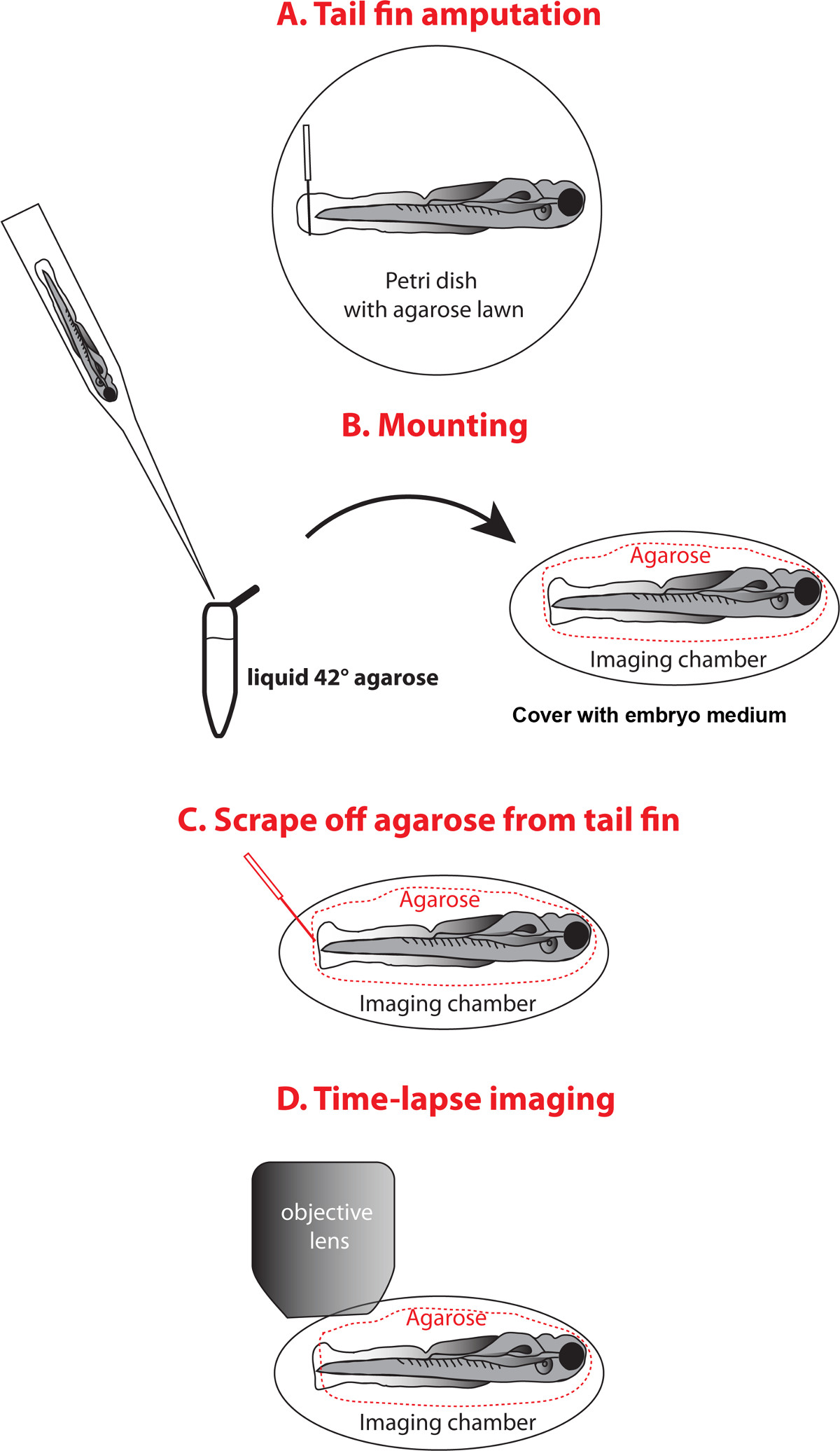
4.截肢分析
- 准备使用胚胎培养基中的1.5%琼脂糖溶液倒入一薄层到培养皿。让琼脂糖巩固。
- 在立体显微镜下,将幼虫向侧上固化琼脂糖和截肢的尾鳍用23政注射器针头用轻微的压力( 图3A)。
5.安装幼虫的时间推移成像
- 继续按照步骤3.1中所述- 3.5( 图3B)。
- 转移下跌0.5-1.2%琼脂糖液在42℃,含有幼虫进入成像室环(步骤2.1),东方幼虫,让琼脂糖巩固。填补了环三卡因液。替代地,如果在培养皿室(步骤2.2)的利用,装入幼虫到盖子盖玻片并填写三卡因溶液盖子。
- 要允许适当的伤口愈合或组织再生的发生,小心刮去使用上限微加载枪头或昆虫针周围远端尾鳍琼脂糖。尽量不要伤害鳍反复( 图3C)。
- 倒出含有去除琼脂糖的三卡因溶液,填补了腔环食三卡因液。
- 适用的硅润滑脂腔环的顶部和附加75毫米×25毫米的玻璃载玻片上。尽量避免气泡在室内,因为它们会干扰明成像和变干幼虫随着时间的推移。
- 如果使用的培养皿作为成像室中,适用的硅润滑脂的底部腔室的顶部边缘和填充底腔与三卡因溶液。小心倒出在盖的三卡因溶液,并打开盖过沉浸幼虫进入底部腔室的一个很小的角度的三卡因溶液,以避免气穴。该室会由于硅润滑脂进行密封。
6.时间推移成像
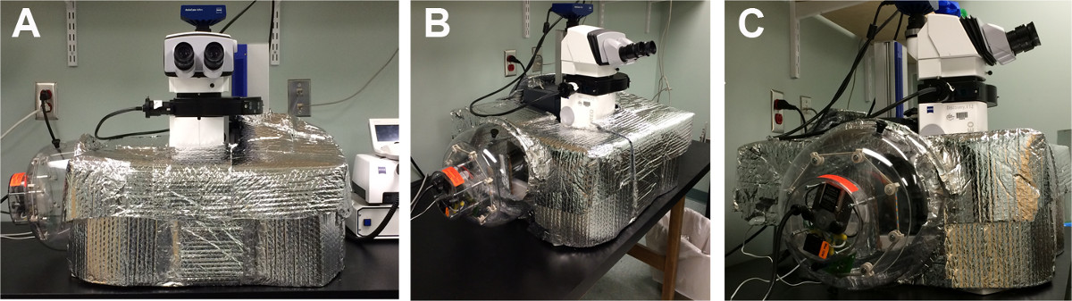
- 组装加热孵化室在23,24( 图4).Place显微镜周围的孵化室,打开热描述。调节温度到28℃,进行约10 - 20分钟或直到温度稳定。
- 打开加热孵育室的前部,并放置在成像室到显微镜支架朝上朝向目标盖玻片。
- 定位幼虫鳍的方式,视场的2/3仍然未被占用。这确保的捕获增长和鳍在成像过程的过程中再生,而无需重新定位幼虫。
- 调整安装幼虫28℃〜30分钟开始时间推移录音,以避免改变在明强度或琼脂糖的潜在变化之前。另外,利用预热的缓冲后较短的调整时间开始成像。
- 要设置延时录制中,打开的AxioVision软件6D多维采集窗口,选择z-堆栈和延时选项。
- 可选)在Z堆栈选项卡,然后切片模式,选择层厚,然后选择启动/停止模式。
- 定义的堆栈的上和下位置。
- 在延时选项卡,选择间隔和电影的时间,然后按下启动按钮开始播放电影。我们发现,30分钟的时间间隔是足够和该区间不产生过大的数据;然而shorteR间都可以使用。
- 在第一个小时期间,检查位置和Z堆叠的尺寸,如果幼虫未预调整。如果有必要,一天之后,再次重新定位幼虫,因为幼虫可能已经转移。
- 将文件保存在延时录制结束并继续进行后期处理和利用现有的图像分析软件,如25了Imaris或开源软件包的Image J 26和斐济27的量化值。
7.数据分析
- 确定鳍的长度。
- 打开在成像软件的定时短片和保存文件的专有文件格式,以提高软件性能。
- 选择正交视图来显示各个部分的预计栈。对于漂移校正,在斐济的菜单中选择插件,然后注册,和"正确的3D漂移"。这将打开一个窗口,斐济,进行漂移校正。正确旋转漂移在了Imaris景点的功能。此外,安装StackReg和TurboReg插件在图像J和导入到斐济。选择在StackReg插件所需的变换算法。
- 测量距离( 例如 ,伤口或直径翅片长度),选择在左上角工具栏上的"添加新测量点"选项。
- 在底部左侧菜单"可见价值统计列表配置",选择要显示的统计值。
- 在设置选项卡下的"行模式"选择"对(AB,CD ......)"。
- 在"标签属性"中选择"姓名"和"距离",显示A和B点旁边的测量线之间的距离。
- 切换到"编辑"模式,按住shift键选择在脊索结束的第一点。然后使用相同的配置来选择的第二点处的前端缘翅片。
- 根据"统计信息"选项卡,在右下角显示和出口的距离图像中选择磁盘按钮(导出所有)。
- 通过移动图像下方的滑块向右重复在选定的时间进行测量。而不是创建新的测量点,以前的可以先选择点用鼠标左键,然后同时按下Shift和鼠标左键在新的位置重新定位。
- 可替代地,以测量点的选择,利用切片观众测量距离。在片浏览模式,滚动到所需的位置,然后单击用鼠标左键第一和第二的位置。的距离将被显示。此选项但不允许进行数据导出。
- 确定鳍的长度和面积ImageJ的。
- 使用一个插件,识别.zvi文件格式打开ImageJ的时间推移电影。另外,加载在曲ickTime文件或TIFF序列。
注意:如果使用未压缩的TIFF文件格式,图像尺寸不需要被指定。 - 如果没有文件信息打开不同的文件格式,选择"分析"菜单下的"设置规模"首先定义图像距离和单位。在"设置缩放菜单',式中'的距离中的像素",下面的像素值的"已知距离"型(这可通过测量在被添加到图像比例尺的像素数获得的后点击测量,得到的结果),和"单位长度"(通常"微米")。然后单击确定。
- 对于鳍片面积测量选择工具栏中的"手绘选择"工具,按住鼠标左键,同时沿着散热片的外形图勾勒出鳍片面积。对于鳍片长度测量,选择'直'线工具,绘制所需点之间的线路是测量。
- 点击下的"分析"的"测量"选项,显示散热片的面积和长度。根据需要重复对电影的多个时间点这一步骤的次数。
- 使用一个插件,识别.zvi文件格式打开ImageJ的时间推移电影。另外,加载在曲ickTime文件或TIFF序列。
- 使用统计软件进行数据可以图形方式显示。
结果
所提出的技术是适用于阐明组织修复的动态响应截肢。电影表明,翅片的截肢最初触发荷包的效果,通过肌动蛋白-肌球蛋白电缆中存在的特征在于收缩翅片折28( 图5的A,B)。伴随地,将细胞从伤口(见电影)挤出。因而收缩可以是驱逐细胞很可能注定要经历细胞死亡的方法。我们的研究结果进一步表明,幼虫的生长发育出现独立再生(电影),而鳍再生不会启动,直到大约14小时后截肢由鳍片长度和面积超过36小时的时间过程以下截肢( 图5C测,D)。 1.5天后的总再生鳍生长为约60%的原始翼片长度( 图5E)的。两者合计,这些结果表明,截肢TR iggers翅片收缩,从伤口,并且在时间上延迟的再生反应的细胞挤出。而挤出的细胞可能注定要经历细胞死亡,需要这些细胞的性质,以进一步澄清。

图1.成像室环组件
(A)中显示的是一个连接到硅润滑脂盖玻片一个塑料环。的塑料网格连接于所述腔室的与硅润滑脂四个小点的内部。 (B)中含有的安装幼虫的腔室填充有三卡因溶液和载玻片被附连到顶部。 (C)在安装2天岁的幼虫(箭头)显示在更高的放大倍率来描绘它的尺寸相对于网格。"_blank">点击此处查看该图的放大版本。

图2.成像室组件从培养皿中进行 。 (A)中显示的是一个商用玻璃顶玻璃底培养皿与附连到玻璃盖玻片一塑料网。 (B)中显示的是一个自建培养皿室钻入盖和连接的从硅润滑脂外侧的盖玻片的孔。网格和幼虫安装包含三卡因溶液室内。密封室,硅脂适用于底部室和连接顶盖的上方,外缘。 请点击此处查看该图的放大版本。
图截肢和安装幼虫的成像3.计划 。 (A)中对于截肢,放置一个麻醉幼虫于琼脂糖被覆培养皿和截肢的尾鳍用注射器针头。 (B)的安装时,用移液管转移幼虫到1.5毫升管填充有42℃的液体琼脂糖和吸取含有幼虫到成像室中的下降,定位鱼和覆盖胚胎培养基中的固化琼脂糖。 (C)的刮去从尾鳍琼脂糖使用封微加载枪头或类似的工具,并用新鲜培养基替换培养基的胚胎。 (D)图像尾鳍在立体显微镜下。 请点击此处查看该图的放大版本。

图4.自建加热孵化室 。 (AC)显示的是一个温水孵化室用纸板,泡沫包装和魔术贴。有线圆顶加热器(最初设计用于鸡卵孵化)连接到使用铝带室。 请点击此处查看该图的放大版本。

图5.鳍再生动力 。所利用的尾鳍截肢测定和定量方法(A) 的计划,以确定该翼片长度(红色箭头)和面积(翅片的红色轮廓)。 (B)的尾鳍截肢最初触发收缩翅片,接着雷杰nerative组织生长。散热片也经历了发展和成长,就证明了横向尺寸增加。 (c)中所示的翼片长度为时间的函数,露出一个线性再生开始生长在〜14 HPA。翅片面积(D)的定量揭示一个最初减小尺寸,这可以归因于该鳍的收缩。后〜14小时,以线性速率翅片尺寸增大。截肢前的片长,36小时后(E)的比较显示〜60%的再生。比例尺:100微米简称:前置放大器,前置截肢;小时后截肢,HPA;再生,再生;放大器,截肢 请点击这里查看这个数字的放大版本。
电影 。鳍再生超过36的时间过程小时。显示的是再生的过程中,一个尾鳍的2.5天龄幼虫。开始30分钟后截肢再生的增长是使用3.5倍物镜成像在立体显微镜30分钟一班。
讨论
所提出的方法允许观察伤口愈合和组织再生中在明视显微镜活斑马鱼幼虫体内延时成像,使用相对简单的设置。这个过程需要我们已经测试过的某些重要方面,这将优化结果:1)低浓度琼脂糖(〜0.5%)将减少不断增长的幼虫斑马鱼的生长障碍,2)清除周围的散热片的琼脂糖重要的是不要模糊愈合过程,3)捕获在一个塑料网状琼脂糖保留在整个手术过程中稳定的位置琼脂糖和动物,以及4)一个适当的温度控制的环境中,这是幼虫存活必不可少的。我们已经适应加热的孵育室23,24,其利用正在录音到纸板泡沫包装,以及有线圆顶加热器来控制温度和适当的空气流通用在最小的波动成像过程。这个简单的和有成本效益室可以制备以适应任何显微镜。类似的温水孵化室也已用于成像老鼠和小鸡发展24,29。
我们建议预截去的幼虫被安装为一个预截肢图像,拆下截肢,和重新安装用于延时成像。虽然它是在最终的成像室中的单个步骤执行这些步骤是可行的,在我们的经验,我们发现,截肢尾鳍在玻璃盖玻片不是最佳的,因为它泪组织并且不会导致在一个干净的切口。用注射器针头琼脂糖为基础的截肢方法最初是由川和他的同事(2004)16中描述,也是在我们的经验,非常适合进行截肢。因此,在相当复杂的一系列步骤,我们介绍的是有充分理由的,并确保最佳的再生效果。
我们发现,larv人斑马鱼在2 DPF可以被成像到在琼脂糖和三卡因溶液1.5天。我们使用制备即时海洋盐,其不与试样的健康干扰对所呈现的成像期间的pH优化的三卡因(pH7的)解决方案。我们先前然而还证明了在Danieau介质使用三卡因允许在共聚焦显微镜对至少2天30 2.5延时成像DPF幼虫斑马鱼。因此,最佳的缓冲液条件可以延长幼虫健康和成像的长度。或者,可用于麻醉,或2-苯氧基乙醇,我们发现在幼虫和成虫阶段的耐受性良好,在28℃下进行至少60小时下三卡因的浓度。
为了避免在散热片再生的缺陷,我们删除从之前的成像尾鳍琼脂糖。我们的数据表明,在1.5天鳍已再生至约60%。这种再生率与以前的研究定义3天一致s的平均时间尾鳍再生的斑马鱼幼虫多达6 DPF 16。替代方法琼脂糖却可能被用来装鱼的成像。例如,等离子体细凝块31或氟化乙烯丙烯(FEP)管涂覆有甲基纤维素和填充有非常低的琼脂糖浓度(0.1%),已被建议用于光片镜32和可适合于我们提出的方法。然而,我们不推荐甲基纤维素和0.1%琼脂糖,因为它们需要该样品被安装在所述腔室的底部,由于缺乏这些介质的凝固。非常高浓度的甲基纤维素将另外产生根据我们的经验气穴,并且这些可以与成像过程产生干扰。如果这些媒体是优选使用的底腔,重要的是,在物镜和试样之间的适当的工作距离是否存在。应当注意的是,米乙基纤维素作为安装介质,建议只为1天,因为它可能与幼虫健康干预32。
安装在盖的检体可能会导致缓慢的引力向下漂移。因此,在每一个时间点,这可以被投影成一个单一的平面或仅是在焦平面可以提取用于组装最终电影图像推荐图像的多个部分。成像试样在底部室可以是一种替代方法,以避免潜在的向下漂移。血浆凝块可能是有用的,以避免漂移,作为等离子体会粘到外层包封层(EVL,周皮)31,因此,可以稳定的样品。然而,这需要进行测试,以及多久幼虫斑马鱼可以保持在血浆凝块而不与健康幼虫或鳍再生的干扰。
我们的电影是利用组装各个部分(26微米)的一个记录,Z堆叠,其覆盖所述鳍(〜10微米),并且在成像过程期间占鳍的潜在的z漂移的整个厚度。为了保留的3-D信息,但也可以突出的z栈成单个图像。因为这可能会导致在图像的模糊程度,明去卷积可以期望。软件,如反卷积或Autoquant X3可以被用于此目的。或者,数学算法(在Tadrous 33描述的)可被用于获得高的信号-噪声比(SNR)的点扩散函数。获得高信噪比表示在明解卷积的主要障碍之一。虽然这种方法需要高对比度和细样品的厚度,这将是适当的尾鳍的成像,由于其宽度减小。
所提出的成像方法的一个明显的优点是,它是快速适应装备有CCD照相机的任何立体显微镜ð时间推移软件,并提供一种低成本的替代更昂贵的共焦成像系统。虽然这种方法不使用荧光进行小区检测,它可以扩展为这样的应用,利用一种自动系统,用于快门控制和后摄像卷积软件34。这将使用户能够进一步的观察与单细胞或亚细胞分辨率创面的修复和再生过程在更长的时间段。
光学清晰度和缓解与胚胎和幼虫的斑马鱼可以处理了,这种方法对任何立体的适应能力使得它适合于教学的基本脊椎动物生物学课堂上。这种方法可以为学生提供一个更好地了解组织修复和再生背后的基本生物过程。已经捕获用类似的方法等生物学过程是斑马鱼胚胎发育23,34和心函数(未公布的)。这种方法还提供了可能性监测伤口修复和再生中的幼虫已进行遗传和药理学上操纵。
披露声明
作者什么都没有透露。
致谢
We thank the MDI Biological Laboratory animal core service facility for zebrafish maintenance. Research reported in this publication was supported by Institutional Development Awards (IDeA) from the National Institute of General Medical Sciences of the National Institutes of Health under grant numbers P20GM104318 (for COBRE) and P20GM103423 (INBRE) and Department of Defense – USAMRAA (W81XWH-BAA-1) grant.
材料
| Name | Company | Catalog Number | Comments |
| Reagents | |||
| Bullseye Agarose | MidSci | BE-GCA500 | |
| Low-melt agarose | Fisher BioReagents | BP1360-100 | |
| 1-phenyl-2-thiourea | Alfa Aesar | L06690 | |
| Instant Ocean Aquarium Salt | Pet store | ||
| Methylene Blue (0.1% solution) | Sigma | M9140 | |
| Tricaine (Ethyl 3-aminobenzoate methanesulfonate) | Sigma-Aldrich | E10505 | |
| 2-Phenoxyethanol | Sigma-Aldrich | 77699 | |
| Petri Dish 35 x 15 mm | BD Falcon | 351008 | |
| Petri Dish 60 x 15 mm | BD Falcon | 351007 | |
| Petri Dish 100 x 25 mm | BD Falcon | 351013 | |
| 5.75 inch boroschillate glass pipets | Fisher | ||
| 35 mm Glass Top Glass Bottom Dish (Glass: 0.085-0.115mm) | MatTek Corporation | D35-20-0-TOP | |
| Superfrost/Plus microscope slides | Fisherbrand | 12-550-15 | |
| Glass coverslips | Electron Microscopy Services | 72191-75 | |
| Glass coverslips | Warner Instruments | CS-18R15 | |
| Phifer Phiferglass Insect Screen Charcoal - 48" | Home Depot | ||
| High vacuum grease | Dow Corning | ||
| Microloader pipette tips 20 µl | Eppendorf | 930001007 | |
| Fine Scissors - Sharply Angled Up | Fine Science Tools | 14037-10 | |
| 3 ml Luer-Lok™ disposable syringe | BD | 309657 | |
| 60 ml Luer-Lok™ disposable syringe | BD | 309653 | |
| 23 G syringe needles | BD | 305145 | |
| Dumont #5 Forceps | Fine Science Tools | 11295-00 | |
| Equipment | |||
| LabDoctor Mini Dry Bath | MidSci | ||
| Discovery.V12 compound microscope | Zeiss | ||
| Plan Apo S 3.5X objective | Zeiss | ||
| AxioCam MRm | Zeiss | ||
| Axiovision software, Release 4.8.2SP1 (12-2011) | Zeiss | ||
参考文献
- San Miguel-Ruiz, J. E., García-Arrarás, J. E. Common cellular events occur during wound healing and organ regeneration in the sea cucumber Holothuria glaberrima. BMC Dev Biol. 7, 115 (2007).
- Poss, K. D., Keating, M. T., Nechiporuk, A. Tales of regeneration in zebrafish. Dev Dyn. 226, 202-210 (2003).
- Akimenko, M. A., Marí-Beffa, M., Becerra, J., Old Géraudie, J. Old questions, new tools, and some answers to the mystery of fin regeneration. Dev Dyn. 226, 190-201 (2003).
- Slack, J. M. Regeneration research today. Dev Dyn. 226, 162-166 (2003).
- Seifert, A. W., et al. Skin shedding and tissue regeneration in African spiny mice (Acomys). Nature. 489, 561-565 (2012).
- Goss, R. J., Grimes, L. N. Epidermal downgrowths in regenerating rabbit ear holes. J Morphol. 146, 533-542 (1975).
- Williams-Boyce, P. K., Daniel, J. C. Comparison of ear tissue regeneration in mammals. J Anat. 149, 55-63 (1986).
- Allan, C. H., et al. Tissue response and Msx1 expression after human fetal digit tip amputation in vitro. Wound Repair Regen. 14, 398-404 (2006).
- Borgens, R. B. Mice regrow the tips of their foretoes. Science. 217, 747-750 (1982).
- Han, M., Yang, X., Lee, J., Allan, C. H., Muneoka, K. Development and regeneration of the neonatal digit tip in mice. Dev Biol. 315, 125-135 (2008).
- Muneoka, K., Allan, C. H., Yang, X., Lee, J., Han, M. Mammalian regeneration and regenerative medicine. Birth Defects Res C Embryo Today. 84, 265-280 (2008).
- Takeo, M., et al. Wnt activation in nail epithelium couples nail growth to digit regeneration. Nature. 499, 228-232 (2013).
- Akimenko, M. A., Johnson, S. L., Westerfield, M., Ekker, M. Differential induction of four msx homeobox genes during fin development and regeneration in zebrafish. Development. 121, 347-357 (1995).
- Reginelli, A. D., Wang, Y. Q., Sassoon, D., Muneoka, K. Digit tip regeneration correlates with regions of Msx1 (Hox 7) expression in fetal and newborn mice. Development. 121, 1065-1076 (1995).
- Brien, G. S., et al. Coordinate development of skin cells and cutaneous sensory axons in zebrafish. Journal of Comparative Neurology. 520, 816-831 (2012).
- Kawakami, A., Fukazawa, T., Takeda, H. Early fin primordia of zebrafish larvae regenerate by a similar growth control mechanism with adult regeneration. Dev Dyn. 231, 693-699 (2004).
- Yoshinari, N., Ishida, T., Kudo, A., Kawakami, A. Gene expression and functional analysis of zebrafish larval fin fold regeneration. Dev Biol. 325, 71-81 (2009).
- Westerfield, M. . The zebrafish book. A guide for the laboratory use of zebrafish (Danio rerio). , (2000).
- Detrich, H. W., Westerfield, M., Zon, L. I. The Zebrafish: Cellular and Developmental Biology, part A. Preface. Methods Cell Biol. 100 (13), (2010).
- Dahm, N. -. V. a. . Zebrafish: A Practical Approach, Issue 975. , 303 (2002).
- Detrich, H., Westerfield, M., Zon, L. . The Zebrafish: 2nd Edition Genetics, Genomics and Informatics. , (2005).
- Concha, M. L., Adams, R. J. Oriented cell divisions and cellular morphogenesis in the zebrafish gastrula and neurula: a time-lapse analysis. Development. 125, 983-994 (1998).
- Distel, M., Köster, R. W. In vivo time-lapse imaging of zebrafish embryonic development. CSH Protoc. 2007, (2007).
- Kulesa, P. M., Kasemeier-Kulesa, J. C. Construction of a Heated Incubation Chamber around a Microscope Stage for Time-Lapse Imaging. CSH Protoc. 2007, (2007).
- . Bitplane. Imaris V 6.1.0 Reference Manual. , (2008).
- Abramoff, M. D., Magalhaes, P. J., Ram, S. J. . Image Processing with ImageJ. , 36-42 (2004).
- Schindelin, J., et al. Fiji: an open-source platform for biological-image analysis. Nat Methods. 9, 676-682 (2012).
- Mateus, R., et al. In vivo cell and tissue dynamics underlying zebrafish fin fold regeneration. PLoS One. 7, e51766 (2012).
- Jones, E. A., et al. et al.Dynamic in vivo imaging of postimplantation mammalian embryos using whole embryo culture. Genesis. 34, 228-235 (2002).
- Rieger, S., Senghaas, N., Walch, A., Köster, R. W. Cadherin-2 controls directional chain migration of cerebellar granule neurons. PLoS Biol. 7, e1000240 (2009).
- Langenberg, T., Brand, M., Cooper, M. S. Imaging brain development and organogenesis in zebrafish using immobilized embryonic explants. Dev Dyn. 228, 464-474 (2003).
- Kaufmann, A., Mickoleit, M., Weber, M., Huisken, J. Multilayer mounting enables long-term imaging of zebrafish development in a light sheet microscope. Development. 139, 3242-3247 (2012).
- Tadrous, P. J. A method of PSF generation for 3D brightfield deconvolution. J Microsc. 237, 192-199 (2010).
- Distel, M., Babaryka, A., Köster, R. W. Multicolor in vivo time-lapse imaging at cellular resolution by stereomicroscopy. Dev Dyn. 235, 1100-1106 (2006).
转载和许可
请求许可使用此 JoVE 文章的文本或图形
请求许可探索更多文章
This article has been published
Video Coming Soon
版权所属 © 2025 MyJoVE 公司版权所有,本公司不涉及任何医疗业务和医疗服务。