需要订阅 JoVE 才能查看此. 登录或开始免费试用。
Method Article
研究人体姿势控制的实验方法
摘要
本文提出了研究人类姿势控制的实验/分析框架。该协议提供了执行站立实验、测量身体运动学和动力学信号以及分析结果的分步程序,以深入了解人类姿势控制背后的机制。
摘要
神经和肌肉骨骼系统的许多成分协同作用,以实现稳定、直立的人体姿势。需要进行对照实验,并辅之以适当的数学方法,以了解人类姿势控制中涉及的不同子系统的作用。本文介绍了一种用于进行扰动站立实验、获取实验数据以及进行后续数学分析的协议,目的是了解肌肉骨骼系统和中央控制在人体中的作用。直立姿势。这些方法所产生的结果很重要,因为它们提供了对健康平衡控制的洞察,为了解患者和老年人平衡受损病因奠定了基础,并有助于设计改善的干预措施姿势控制和稳定性。这些方法可用于研究躯体感觉系统的作用、踝关节的内在刚度和视觉系统在姿势控制中的作用,也可以扩展研究前庭系统的作用。这些方法将用于脚踝策略,其中身体主要移动的脚踝关节,被认为是单链倒摆。
引言
人类姿势控制是通过中枢神经系统和肌肉骨骼系统之间的复杂相互作用实现的。人体在站立中本质上是不稳定的,受各种内部(如呼吸、心跳)和外部(如重力)扰动的影响。稳定性由具有中央、反射和内在组件的分布式控制器实现(图1)。
姿势控制由:由中枢神经系统(CNS)和脊髓介导的主动控制器,改变肌肉激活;和一个内在刚度控制器,抵抗关节运动,肌肉激活没有变化 (图1)。中央控制器使用感觉信息来生成降序命令,产生矫正肌肉力量以稳定身体。感官信息由视觉、前庭和躯体感觉系统转换。具体来说,躯体感觉系统生成有关支撑面和关节角度的信息;愿景提供有关环境的信息;和前庭系统生成有关头部角速度、线性加速度和与重力方向有关的信息。中央闭环控制器运行时间长,可能会破坏稳定2。主动控制器的第二个元素是反射刚度,它产生短延迟的肌肉活动,并产生抵抗关节运动的扭矩。
活动控制器的两个组件都有延迟;因此,关节内在刚度,不延迟地作用,在姿势控制3中起着重要的作用。内在刚度是由收缩肌肉的被动粘弹性特性、软组织和四肢的惯性特性产生的,这些特性可立即产生电阻扭矩,以响应任何关节运动4。关节刚度(内在和反射刚度)在姿势控制中的作用并不清晰,因为它随着手术条件的变化而变化,由肌肉活化4、5、6和关节位置定义4,7,8,两者随身体摇摆而变化,与站立本身有内在影响。
确定中央控制器的作用和关节刚度在姿势控制中很重要,因为它为:诊断平衡损伤的病因提供了基础;为患者设计有针对性的干预措施;评估跌倒风险;制定老年人防堕落战略;和辅助设备的设计,如矫形器和假肢。然而,这是困难的,因为不同的子系统一起作用,只能测量整体产生的身体运动学,关节扭矩和肌肉肌电图。
因此,开发使用可测量的姿势变量来评估每个子系统的贡献的实验和分析方法至关重要。一个技术难题是,姿势变量的测量是在闭环中完成的。因此,输入和输出(因果)是相互关联的。因此,有必要:a) 应用外部扰动(作为输入)来唤起反应中姿势反应(作为输出),b) 使用专门的数学方法来识别系统模型并解开因果9。
本文的重点是使用脚踝策略时,即当运动主要发生在脚踝关节时,姿势控制。在这种情况下,上半身和下肢一起移动,因此,身体可以建模为单链倒置钟摆在下垂平面10。当支撑面牢固且扰动小1,11时,使用脚踝策略。
我们的实验室12开发了一种能够应用适当的机械(自体)和视觉感官扰动并记录身体运动学、动力学和肌肉活动的站立装置。该装置提供所需的实验环境,通过使用视觉或/和躯体感官刺激生成姿势反应,研究脚踝刚度、中央控制机制及其相互作用的作用。也可以扩展设备来研究前庭系统的作用,将直接电刺激应用于乳腺过程,从而产生头部速度的感觉,并唤起姿势反应12,13.
其他人也开发了类似的设备来研究人类姿势控制,其中线性压电执行器11,旋转电机14,15,和线性电机16,17,18用于在站立时对脚踝施加机械扰动。更复杂的设备也已经开发,以研究多段姿势控制,有可能同时对脚踝和髋关节应用多个扰动19,20。
站立式仪器
两个伺服控制电动液压旋转执行器移动两个踏板,以应用脚踝位置的控制扰动。执行器可以产生姿势控制所需的大扭矩(>500 Nm);这在诸如前倾等情况下尤其重要,因为身体的质量中心离脚踝旋转轴很远(前部),导致姿势控制的脚踝扭矩值较大。
每个旋转执行器由一个单独的比例伺服阀控制,使用踏板位置反馈,由执行器轴上的高性能电位计(材料表)测量。该控制器使用基于 MATLAB 的 xPC 实时数字信号处理系统实现。执行器/伺服阀的带宽一起超过40赫兹,远远大于整体姿势控制系统的带宽,脚踝关节刚度,以及中央控制器21。
虚拟现实设备与环境
虚拟现实 (VR) 耳机 (材料表) 用于干扰视觉.耳机包含一个 LCD 屏幕(双 AMOLED 3.6' 屏幕,每只眼睛的分辨率为 1080 x 1200 像素),为用户提供发送到设备的媒体的立体视图,提供三维深度感知。刷新率是90赫兹,足以为用户提供坚实的虚拟感22。屏幕的视场为 110°,足以产生类似于真实情况的视觉扰动。
耳机跟踪用户头部的旋转,并相应地更改虚拟视图,以便用户完全沉浸在虚拟环境中;因此,它可以提供正常的视觉反馈;并且,它还可以通过旋转视场在下视平面上来干扰视觉。
动力学测量
垂直反应力由四个称重传感器测量,夹在脚底下的两块板之间(材料表)。脚踝扭矩由扭矩传感器直接测量,容量为565Nm,扭转刚度为104 kNm/rad;它也可以间接测量从垂直力转导的称重传感器,使用其距离到脚踝轴的旋转23,假设水平力施加到脚站立是小2,24。压力中心(COP)在下垂平面上测量,方法是将脚踝扭矩除以总垂直力,由称重传感器23测量。
运动学测量
脚角与踏板角度相同,因为使用脚踝策略时,主体的脚随踏板移动。相对于垂直的刀柄角度是间接地从刀柄的线性位移获得,由分辨率为50 μm和带宽为750 Hz25的激光测距仪(材料表)测量。脚踝角是脚角和刀柄角度的总和。相对于垂直体的角度是间接从左右后部上部脊柱 (PSIS) 之间的中点线性位移获得的,使用激光测距仪(材料表)测量,分辨率为100 μm 和带宽 750 Hz23.头部位置和旋转由 VR 系统基站根据 VR 环境的全球坐标系进行测量,该基站以每秒 60 个脉冲发出定时红外 (IR) 脉冲,由带子毫米的耳机红外传感器拾取精度。
数据采集
所有信号均采用角频为 486.3 的抗锯齿滤波器进行滤波,然后在 1000 Hz 下采样,具有高性能 24 位/8 通道、同步采样动态信号采集卡(材料表),具有动态范围为 20 V。
安全机制
在常设仪器中已加入六个安全机制,以防止对受试者造成伤害;踏板是分开控制的,从不相互干扰。(1) 执行器轴有一个凸轮,该凸轮可机械地激活一个阀,当轴旋转超过其水平位置 ±20°时,该阀可断开液压。(2) 两个可调节的机械停止限制执行器的运动范围;这些被设置为每个受试者在每个实验前的运动范围。(3) 受试者和实验者都按住一个紧急按钮;按下按钮可断开执行器的液压动力,使其松动,因此可以手动移动。(4) 位于主体两侧的扶手可在不稳定时提供支持。(5)受试者佩戴全身安全带(材料表),附在天花板上的刚性横杆上,以防跌倒。线束松弛,不会干扰正常站立,除非主体变得不稳定,其中线束可防止主体掉落。在坠落的情况下,被主体使用紧急按钮或实验者手动停止踏板移动。(6) 伺服阀在电源中断时,使用故障安全机制停止执行器的旋转。
Access restricted. Please log in or start a trial to view this content.
研究方案
所有实验方法均已获得麦吉尔大学研究伦理委员会的批准,受试者在参与前签署知情同意书。
1. 实验
注:每个实验都涉及以下步骤。
- 预测试
- 准备要执行的所有试验的明确大纲,并制定数据收集清单。
- 向受试者提供同意书,提供所有必要的信息,要求他们彻底阅读,回答任何问题,然后让他们在表格上签名。
- 记录受试者的体重、身高和年龄。
- 主题准备
- 肌X线测量
- 使用单微分电极(材料表),电极间距离为1厘米,用于测量脚踝肌肉的肌电图(EMG)。
- 使用总增益为1000和带宽为20~2000Hz的放大器(材料表)。
- 为确保高信噪比 (SNR) 和最小串扰,根据 Seniam 项目26提供的指南定位并标记电极附件区域,如下所示:(1) 用于中枢胃肠 (MG),这是肌肉;(2) 对于侧侧胃肠 (LG),纤维头和脚跟之间的线 1/3;(3) 对于鞋底 (SOL),股骨和内阴线之间的线 2/3;(4) 对于前部 (TA), 纤维尖和中端的尖端之间的线 1/3。
- 用剃须刀剃光标记区域,用酒精清洁皮肤。让皮肤彻底干燥。
- 在用于参考电极的骨质区域上剃除,并用酒精清洁。
- 让主题位于一个轻松的苏佩位置。
- 将参考电极放在骨的被拉的应洗区域。
- 使用双面胶带将电极逐个连接到肌肉的被检查区域,注意确保电极牢固地固定在皮肤上。
- 放置每个电极后,要求受试者执行针对电阻的平面弯曲/多向收缩,并检查示波器上的波形,以确保 EMG 信号具有高 SNR。如果信号 SNR 较差,则移动电极,直到找到具有高 SNR 的位置。
- 确保主体的移动不受 EMG 电缆的阻碍。
- 运动学测量
- 将反光标记与带子固定在刀柄上,用于刀柄角度测量。
注: 尽可能高地将刀柄标记放在刀柄上,以生成给定旋转的最大线性位移,从而改善角度分辨率。 - 将主体放在车身线束上。
- 用肩带将反射标记连接到受试者的腰部,用于上半身角度测量。确保腰部反光标记位于左右 PSIS 之间的中间点,并且主体的衣服不覆盖腰部反射表面。
- 让主体放在站立的设备上。
- 调整主体的脚部位置,使每条腿的侧侧和中侧马柳利与踏板的旋转轴对齐。
- 用标记勾勒出受试者的脚的位置,并指示他们在实验过程中将脚放在相同的位置。这可确保在整个实验中,脚踝和执行器的旋转轴保持对齐。
- 调整激光测距仪的垂直位置,以指向反射标记的中心。调整激光测距仪和反光标记之间的水平距离,以便测距仪在中端工作,在安静站立时不会饱和。
- 让受试者向前和向后倾斜脚踝,并确保激光保持在工作范围内。
- 测量激光测距仪相对于旋转的脚踝轴的高度。
注: 这些高度用于将线性位移转换为角度。
- 将反光标记与带子固定在刀柄上,用于刀柄角度测量。
- 实验协议
- 告知受试者每个试验条件的预期。
- 当面对现实世界的扰动时,指示受试者在向前看时,双手静静地站在一边,保持平衡。
- 对于扰动试验,启动扰动,让受试者适应它。
- 一旦受试者建立了稳定的行为,就开始数据采集。
- 在每次试验后为受试者提供足够的休息时间,以避免疲劳。与他们沟通,看看他们是否需要更多的时间。
- 执行以下试验。
- 对于设备测试,在受试者到达前进行 2 分钟测试以检查传感器数据 2 小时。在记录的传感器数据中查找不规则的大噪声或偏移。如果存在问题,在主题到达之前解决这些问题。
- 为了安静地站立,请执行 2 分钟的安静站立试验,没有扰动。
注:此试验提供了一个参考,需要确定姿势变量是否/如何变化以响应扰动。 - 对于扰动实验,运行扰动并获取数据2⁄3分钟。如果目的是调查躯体感觉系统/脚踝刚度在站立中的作用,则应用踏板扰动。如果目标是检查视觉在姿势控制中的作用,则应用视觉扰动。如果目标是检查两个系统在姿势控制中的相互作用,则同时应用视觉和踏板扰动。
注:踏板扰动在站立设备踏板的旋转时应用。同样,使用 VR 耳机旋转虚拟视觉场应用视觉扰动。踏板/目视场的角度跟随信号,根据研究目标进行选择。讨论部分详细介绍了用于研究姿势控制以及每种扰动的优点的扰动类型。
- 对每种特定的扰动至少执行 3 次试验。
注:在对收集的数据进行分析时,进行了多次试验,以确保模型的可靠性;例如,可以交叉验证模型。 - 以随机顺序执行试验,以确保受试者不学会对特定的扰动做出反应;这还使得检查随时间变化的行为成为可能。
- 每次试验后,目视检查数据,确保采集的信号具有高质量。
- 肌X线测量
2. 人类姿势控制鉴定
- 体角与视觉扰动动态关系的非参数识别
- 实验
- 根据第 1.1 和 1.2 节中的步骤,获得 2 分钟的目视干扰试验。
- 使用梯形信号 (TrapZ),峰值到峰值振幅为 0.087 rad,速度为 0.105 rad/s。
- 保持踏板位置在零角度不变。
- 分析
注:第 2.1.2 节和第 2.2.2 节中的数据分析使用 MATLAB 执行。- 使用以下命令抽取原始体角和视觉扰动信号(使可观测频率最高为 10 Hz):


其中


注: 对于 1 kHz 的采样速率,抽取比必须为 50 才能达到 10 Hz 的最高频率。 - 选择最低频率的利息,这将确定功率估计的窗口长度。
注: 此处选择最小频率为 0.1 Hz,因此功率估计的窗口长度为 1/0.1 Hz = 10 s。频率分辨率与最小频率相同,因此,计算为 0.1、0.2、0.3、...、10 Hz。 - 选择窗口类型和重叠程度以查找功率光谱。
注:对于 120 s 的试验长度,10 s Haning 窗口的重叠率为 50%, 因此,用于功率谱估计的平均为 23 个段。由于我们将数据量大到 20 Hz,因此 10 s 窗口的长度为 200 个样本。 - 使用
 函数查找系统的频率响应 (FR):
其中


注: 显示 的函数
的函数 使用指定长度
 和重叠数等于
 (即 50% 重叠)。同样,它计算VR输入的自动频谱。然后,利用估计的交叉频谱和自动频谱计算系统的FR。
 - 使用以下命令在步骤 2.1.2.4 中查找估计 FR 的增益和相位:


其中

- 使用以下命令计算相干函数:

其中
注: 函数遵循类似的过程
 来查找 和
来查找 和
 之间的一致性。
 - 将增益、相位和一致性绘制为频率函数。


注:所呈现的方法可以扩展到同时应用视觉和机械扰动的情况下,其中必须使用多输入、多输出 (MIMO) FR 识别方法9。也可以使用子空间方法(它本质上处理MIMO系统)27或使用参数传输函数方法,如MIMO Box-Jenkins28进行识别。子空间和 Box-Jenkins(和其他方法)都在 MATLAB 系统识别工具箱中实现。
- 使用以下命令抽取原始体角和视觉扰动信号(使可观测频率最高为 10 Hz):
- 实验
- 站立中脚踝内在刚度的参数识别
- 实验
- 执行2分钟的机械扰动试验。使用峰值至峰值振幅为0.02 rad且切换间隔为200 ms的伪随机二进制序列(PRBS)扰动。确保踏板平均角度为零。
- 分析
- 区分脚信号一次,以获得脚的速度(,
 两次获得脚加速(
 和三次获得其抽搐(
 同样区分扭矩,以获得其速度和加速度,使用以下命令:
其中


- 使用以下命令计算英尺速度的局部最大值和局部最小值的位置,以定位脉冲:


其中


注: 函数查找所有局部最大值(正英尺速度)及其位置。要查找局部最小数,使用相同的函数,但必须反转脚角速度的标志。
 - 使用以下命令设计一个 8阶Butterworth 低通滤波器,其角频率为 50 Hz:


- 使用巴特沃斯滤波器使用零相移过滤所有信号:


注:"滤网"功能不会导致过滤信号的任何偏移。不要使用"过滤器"功能,因为它会生成班次。 - 绘制英尺速度,并直观地查找脚速极值与脉冲开始(在峰值速度之前为零英尺速度的第一个点)之间的时间段估计值。对于本研究中的扰动,此点发生在步骤 2.2.2.2 中找到的速度极值之前 25 毫秒。
- 对于每个脉冲,将脚踝背景扭矩计算为脉冲开始前 25 ms 的脚踝扭矩平均值,即从速度极值前 50 ms 到 25 ms 的段中的扭矩平均值。使用以下命令对正速度的 kth 脉冲执行此操作:


注: 这是针对步骤 2.2.2.2 中找到的最大和最小速度(负脚速度)进行的。 - 使用以下命令查找所有脉冲的所有背景扭矩的最小和最大值:


- 对于每个脉冲,使用以下命令提取脉冲启动后 65 ms 的扭矩数据(作为内在扭矩段):


注:这也是为脚踝扭矩的第一和第二导数(提供内在扭矩的第一和第二个导导),以及脚角,脚速度,脚加速,和脚抽搐。 - 使用以下命令计算kth内部扭矩段从初始值的变化:

注: 对于获得 的脚角,此操作类似。
 - 将扭矩范围(在步骤 2.2.2.7 中获得)划分为 3 Nm 宽的箱,并在每个料箱中找到具有背景扭矩的脉冲。
注: 这是使用"查找"函数和索引完成的。假定每个料箱中的内在刚度是恒定的,因为脚踝背景扭矩没有显著变化。 - 使用组 j ( )
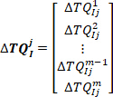
 中的脉冲,估计扩展内部模型 (EIM)29的固有刚度参数
- 将 jbin中的所有内在扭矩响应串联以形成矢量:


其中 ith
ith  ( ) 组 j 中的内在扭矩响应。
注: 同样,串联脚角、速度和加速度,以及步骤2.2.11.2 中使用的 j 组内在扭矩的第一和第二导数。 - 将脚角、速度、加速度和抽搐以及组 j 扭矩的第一和第二导数放在一起,形成回归矩阵:

- 使用反斜杠 (*) 运算符查找j组的内在刚度参数:

- 提取第
 四个元素作为低频内在刚度。
- 将 jbin中的所有内在扭矩响应串联以形成矢量:
- 对所有组(箱)执行第 2.2.2.11 节中的步骤,并估计相应的低频内在刚度。
- 将所有估计的低频刚度值除以受试者的临界刚度:

其中m是受试者的质量,g是重力加速度,是 身体质量中心的高度,在脚踝轴上方的旋转,派生自人体测量数据30。这给出了标准化刚度 (
 )。
)。 - 通过将脚踝背景扭矩与相应的垂直力分开,将
 脚踝背景扭矩转换为脚踝背景 COP 位置 ( )。
 - 绘图
 作为压力中心的函数。
其中

- 区分脚信号一次,以获得脚的速度(,
- 实验
Access restricted. Please log in or start a trial to view this content.
结果
伪随机三元序列 (PRTS) 和 TrapZ 信号
图 2A显示了通过集成伪随机速度配置文件生成的 PRTS 信号。对于每个采样时间, 信号速度可能等于零,或获取预定义的正值或
 负值。通过
负值。通过
Access restricted. Please log in or start a trial to view this content.
讨论
有几个步骤是执行这些实验研究人类姿势控制的关键。这些步骤与信号的正确测量相关,包括:1) 旋转齿形轴与踏板轴的正确对齐,以便正确测量脚踝扭矩。2) 正确设置测距仪,确保它们在其范围内工作,且在实验期间不会饱和。3) 测量EMG具有良好的质量和最小的相声。4) 应用适当的扰动,引起足够的反应,但不中断正常的姿势控制。5) 根据预期分析选择适当的试验长度,同时避免身体换?...
Access restricted. Please log in or start a trial to view this content.
披露声明
作者没有什么可透露的。
致谢
本文由卡塔尔国家研究#6-463-2-189的NPRP赠款和加拿大卫生研究院的MOP赠款#81280。
Access restricted. Please log in or start a trial to view this content.
材料
| Name | Company | Catalog Number | Comments |
| 5K potentiometer | Maurey | 112P19502 | Measures actuator shaft angle |
| 8 channel Bagnoli surface EMG amplifiers and electrodes | Delsys | Measures the EMG of ankle muscles | |
| AlienWare Laptop | Dell Inc. | P69F001-Rev. A02 | VR-ready PC laptop |
| Data acquisition card | National instruments | 4472 | Samples the analogue signals from the sensors |
| Directional valve | REXROTH | 4WMR10C3X | Bypasses the flow if the angle of actuator shaft goes beyond ±20° |
| Full body harness | Jelco | 740 | Protect the subjects from falling |
| Laser range finder | Micro-epsilon 1302-100 | 1507307 | Measures shank linear displacement |
| Laser range finder | Micro-epsilon 1302-200 | 1509074 | Measures body linear displacement |
| Load cell | Omega | LC302-100 | Measures vertical reaction forces |
| Proportional servo-valve | MOOG | D681-4718 | Controls the hydraulic flow to the rotary actuators |
| Rotary actuator | Rotac | 26R21VDEISFTFLGMTG | Applies mechanical perturbations |
| Torque transducer | Lebow | 2110-5k | Measures ankle torque |
| Virtual Environment Motion Trackers | HTC inc. | 1551984681 | Tracks the head motion |
| Virtual Reality Headset | HTC inc. | 1551984681 | Provides visual perturbations |
参考文献
- Horak, F. B. Postural orientation and equilibrium: what do we need to know about neural control of balance to prevent falls? Age and Ageing. 35, 7-11 (2006).
- Morasso, P. G., Schieppati, M. Can muscle stiffness alone stabilize upright standing? Journal of Neurophysiology. 82 (3), 1622-1626 (1999).
- Kearney, R. E., Hunter, I. W. System identification of human joint dynamics. Critical Reviews in Biomedical Engineering. 18 (1), 55-87 (1990).
- Mirbagheri, M. M., Barbeau, H., Kearney, R. E. Intrinsic and reflex contributions to human ankle stiffness: variation with activation level and position. Experimental Brain Research. 135 (4), 423-436 (2000).
- Weiss, P. L., Hunter, I. W., Kearney, R. E. Human ankle joint stiffness over the full range of muscle activation levels. Journal of Biomechanics. 21 (7), 539-544 (1988).
- Golkar, M. A., Sobhani Tehrani, E., Kearney, R. E. Linear Parameter Varying Identification of Dynamic Joint Stiffness during Time-Varying Voluntary Contractions. Frontiers in Computational Neuroscience. 11, 35(2017).
- Weiss, P. L., Kearney, R. E., Hunter, I. W. Position dependence of ankle joint dynamics--I. Passive mechanics. Journal of Biomechanics. 19 (9), 727-735 (1986).
- Weiss, P. L., Kearney, R. E., Hunter, I. W. Position dependence of ankle joint dynamics--II. Active mechanics. Journal of Biomechanics. 19 (9), 737-751 (1986).
- Engelhart, D., Boonstra, T. A., Aarts, R. G. K. M., Schouten, A. C., van der Kooij, H. Comparison of closed-loop system identification techniques to quantify multi-joint human balance control. Annual Reviews in Control. 41, 58-70 (2016).
- Kiemel, T., Elahi, A. J., Jeka, J. J. Identification of the plant for upright stance in humans: multiple movement patterns from a single neural strategy. Journal of Neurophysiology. 100 (6), 3394-3406 (2008).
- Loram, I. D., Lakie, M. Direct measurement of human ankle stiffness during quiet standing: the intrinsic mechanical stiffness is insufficient for stability. Journal of Physiology-London. 545 (3), 1041-1053 (2002).
- Fitzpatrick, R., Burke, D., Gandevia, S. C. Loop gain of reflexes controlling human standing measured with the use of postural and vestibular disturbances. Journal of Neurophysiology. 76 (6), 3994-4008 (1996).
- Dakin, C. J., Son, G. M. L., Inglis, J. T., Blouin, J. S. Frequency response of human vestibular reflexes characterized by stochastic stimuli. The Journal of Physiology. 583 (3), 1117-1127 (2007).
- Vlutters, M., Boonstra, T. A., Schouten, A. C., vander Kooij, H. Direct measurement of the intrinsic ankle stiffness during standing. Journal of Biomechanics. 48 (7), 1258-1263 (2015).
- Casadio, M., Morasso, P. G., Sanguineti, V. Direct measurement of ankle stiffness during quiet standing: implications for control modelling and clinical application. Gait and Posture. 21 (4), 410-424 (2005).
- Sakanaka, T. E. Causes of Variation in Intrinsic Ankle Stiffness and the Consequences for Standing. , University of Birmingham. Doctoral dissertation (2017).
- Sakanaka, T. E., Lakie, M., Reynolds, R. F. Sway-dependent changes in standing ankle stiffness caused by muscle thixotropy. Journal of Physiology. 594 (3), 781-793 (2016).
- Peterka, R. J., Murchison, C. F., Parrington, L., Fino, P. C., King, L. A. Implementation of a Central Sensorimotor Integration Test for Characterization of Human Balance Control During Stance. Frontiers in Neurology. 9, 1045(2018).
- Engelhart, D., Schouten, A. C., Aarts, R. G., van der Kooij, H. Assessment of Multi-Joint Coordination and Adaptation in Standing Balance: A Novel Device and System Identification Technique. IEEE Transactions on Neural Systems and Rehabilitation Engineering. 23 (6), 973-982 (2015).
- Boonstra, T. A., Schouten, A. C., van der Kooij, H. Identification of the contribution of the ankle and hip joints to multi-segmental balance control. Journal of Neuroengineering and Rehabilitation. 10, 23(2013).
- Forster, S. M., Wagner, R., Kearney, R. E. A bilateral electro-hydraulic actuator system to measure dynamic ankle joint stiffness during upright human stance. Proceedings of the 25th Annual International Conference of the IEEE Engineering in Medicine and Biology Society. , Cancun, Mexico. (2003).
- Davis, J., Hsieh, Y. -H., Lee, H. -C. Humans perceive flicker artifacts at 500 Hz. Scientific Reports. 5, 7861(2015).
- Amiri, P., Kearney, R. E. Ankle intrinsic stiffness changes with postural sway. Journal of Biomechanics. 85, 50-58 (2019).
- van der Kooij, H., van Asseldonk, E., van der Helm, F. C. Comparison of different methods to identify and quantify balance control. Journal of Neuroscience Methods. 145 (1-2), 175-203 (2005).
- Amiri, P., MacLean, L. J., Kearney, R. E. Measurement of shank angle during stance using laser range finders. International Conference of the IEEE Engineering in Medicine and Biology. , Orlando, FL. (2016).
- The SENIAM project. , Available from: http://www.seniam.org/ (2019).
- Jalaleddini, K., Tehrani, E. S., Kearney, R. E. A Subspace Approach to the Structural Decomposition and Identification of Ankle Joint Dynamic Stiffness. IEEE Transactions on Biomedical Engineering. 64 (6), 1357-1368 (2017).
- Amiri, P., Kearney, R. E. A Closed-loop Method to Identify EMG-Ankle Torque Dynamic Relation in Human Balance Control. Conference Proceedings of the Annual International Conference of the IEEE Engineering in Medicine and Biology Society. , Berlin, Germany. (2019).
- Sobhani Tehrani, E., Jalaleddini, K., Kearney, R. E. Ankle Joint Intrinsic Dynamics is More Complex than a Mass-Spring-Damper Model. IEEE Transactions on Neural Systems and Rehabilitation Engineering. 25 (9), 1568-1580 (2017).
- NASA. Anthropometry and biomechanics. , Available from: http://msis.jsc.nasa.gov/sections/section03.htm (1995).
- Peterka, R. J. Sensorimotor integration in human postural control. Journal of Neurophysiology. 88 (3), 1097-1118 (2002).
- Amiri, P., Kearney, R. E. Ankle intrinsic stiffness is modulated by postural sway. Conference Proceedings of the Annual International Conference of the IEEE Engineering in Medicine and Biology Society. , Seogwipo, South Korea. (2017).
- Jeka, J. J., Allison, L. K., Kiemel, T. The dynamics of visual reweighting in healthy and fall-prone older adults. Journal of Motor Behavior. 42 (4), 197-208 (2010).
- Jilk, D. J., Safavynia, S. A., Ting, L. H. Contribution of vision to postural behaviors during continuous support-surface translations. Experimental Brain Research. 232 (1), 169-180 (2014).
- Winter, D. A., Patla, A. E., Prince, F., Ishac, M., Gielo-Perczak, K. Stiffness control of balance in quiet standing. Journal of Neurophysiology. 80 (3), 1211-1221 (1998).
- Pasma, J. H., Boonstra, T. A., van Kordelaar, J., Spyropoulou, V. V., Schouten, A. C. A Sensitivity Analysis of an Inverted Pendulum Balance Control Model. Frontiers in Computational Neuroscience. 11, 99(2017).
- Pasma, J. H., et al. Changes in sensory reweighting of proprioceptive information during standing balance with age and disease. Journal of Neurophysiology. 114 (6), 3220-3233 (2015).
- Pasma, J. H., et al. Impaired standing balance: The clinical need for closing the loop. Neuroscience. , 157-165 (2014).
- Engelhart, D., et al. Impaired Standing Balance in Elderly: A New Engineering Method Helps to Unravel Causes and Effects. Journal of the American Medical Directors Association. 15 (3), (2014).
- Pasma, J. H., Boonstra, T. A., Campfens, S. F., Schouten, A. C., Van der Kooij, H. Sensory reweighting of proprioceptive information of the left and right leg during human balance control. Journal of Neurophysiology. 108 (4), 1138-1148 (2012).
- Goodworth, A. D., Peterka, R. J. Sensorimotor integration for multisegmental frontal plane balance control in humans. Journal of Neurophysiology. 107 (1), 12-28 (2012).
- Kiemel, T., Zhang, Y., Jeka, J. J. Identification of neural feedback for upright stance in humans: stabilization rather than sway minimization. Journal of Neuroscience. 31 (42), 15144-15153 (2011).
- van der Kooij, H., van Asseldonk, E. H. F., Geelen, J., van Vugt, J. P. P., Bloem, B. R. Detecting asymmetries in balance control with system identification: first experimental results from Parkinson patients. Journal of Neural Transmission. 114 (10), 1333(2007).
- Fujisawa, N., et al. Human standing posture control system depending on adopted strategies. Medical and Biological Engineering and Computing. 43 (1), 107-114 (2005).
- Johansson, R., Magnusson, M., Fransson, P. A., Karlberg, M. Multi-stimulus multi-response posturography. Mathematical Biosciences. 174 (1), 41-59 (2001).
- Jeka, J., Oie, K., Schöner, G., Dijkstra, T., Henson, E. Position and Velocity Coupling of Postural Sway to Somatosensory Drive. Journal of Neurophysiology. 79 (4), 1661-1674 (1998).
- Peterka, R. J., Benolken, M. S. Role of somatosensory and vestibular cues in attenuating visually induced human postural sway. Experimental Brain Research. 105 (1), 101-110 (1995).
- Maki, B. E., Fernie, G. R. A system identification approach to balance testing. Progress in Brain Research. 76, 297-306 (1988).
- Johansson, R., Magnusson, M., Akesson, M. Identification of human postural dynamics. IEEE Transactions on Biomedical Engineering. 35 (10), 858-869 (1988).
- Maki, B. E., Holliday, P. J., Fernie, G. R. A Posture Control Model and Balance Test for the Prediction of Relative Postural Stability. IEEE Transactions on Biomedical Engineering. BME-34. 10 (10), 797-810 (1987).
- Werness, S. A., Anderson, D. J. Parametric analysis of dynamic postural responses. Biological Cybernetics. 51 (3), 155-168 (1984).
- Hwang, S., Agada, P., Kiemel, T., Jeka, J. J. Identification of the Unstable Human Postural Control System. Frontiers in Systems Neuroscience. 10, 22(2016).
- Ishida, A., Imai, S., Fukuoka, Y. Analysis of the posture control system under fixed and sway-referenced support conditions. IEEE Transactions on Biomedical Engineering. 44 (5), 331-336 (1997).
- Ishida, A., Miyazaki, S. Maximum likelihood identification of a posture control system. IEEE Transactions on Biomedical Engineering. 34 (1), 1-5 (1987).
- Ljung, L. System Identification: Theory for the User. , Prentice-Hall, Inc. Upper Saddle River, NJ. (1986).
- Forssell, U., Ljung, L. Closed-loop identification revisited. Automatica. 35 (7), 1215-1241 (1999).
- Horak, F. B., Nashner, L. M. Central programming of postural movements: adaptation to altered support-surface configurations. Journal of Neurophysiology. 55 (6), 1369-1381 (1986).
Access restricted. Please log in or start a trial to view this content.
转载和许可
请求许可使用此 JoVE 文章的文本或图形
请求许可探索更多文章
This article has been published
Video Coming Soon
版权所属 © 2025 MyJoVE 公司版权所有,本公司不涉及任何医疗业务和医疗服务。