Abdominaluntersuchung I: Inspektion und Auskultation
Überblick
Quelle: Alexander Goldfarb, MD, Assistant Professor für Medizin, Beth Israel Deaconess Medical Center, MA
Magen-Darm-Krankheit entfallen jährlich Millionen von Bürobesuchen und Krankenhauseinweisungen. Körperliche Untersuchung des Bauches ist ein wichtiges Instrument bei der Diagnose von Erkrankungen des Magen-Darm-Trakt; Darüber hinaus kann es helfen, pathologische Prozesse im Herz-Kreislauf-, Harn- und andere Systeme zu identifizieren. Als körperliche Untersuchung ist im Allgemeinen die Prüfung der Bauchregion wichtig für die Arzt-Patient-Kontaktaufnahme für erreichen die vorläufige Diagnose und Auswahl der nachfolgenden Labor und bildgebende Tests und Bestimmung der Dringlichkeit der Versorgung.
Wie bei den anderen Teilen einer körperlichen Untersuchung, Sichtprüfung und Auskultation des Abdomens systematisch erfolgt so, dass keine möglichen Befunde übersehen werden. Besondere Aufmerksamkeit sollte auf mögliche Probleme bereits anhand der Krankengeschichte. Hier wir gehen davon aus, dass der Patient bereits identifiziert worden, und hat Geschichte genommen, Symptome besprochen und mögliche Problembereiche identifiziert hatte. In diesem Video werden wir nicht überprüfen, der Krankengeschichte; Stattdessen fahren wir direkt nach der körperlichen Untersuchung.
Bevor wir zur Untersuchung kommen, betrachten wir kurz Oberfläche Wahrzeichen der Bauchregion, Abdominal-Anatomie und Topographie. Hier ist eine Liste von nützlichen Sehenswürdigkeiten: costal Margen, Xiphoid Prozeß, Rectus Bauchmuskel, Linea Alba, Nabel, Beckenkamm, leisten-Ligament und After Schambein. Die Abdominal-Prüfung deckt den Bereich nach unten von den Rändern xiphoid und kostalen souverän für das After-Schambein inferior.
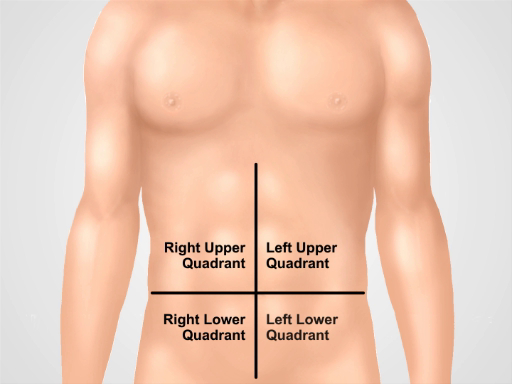
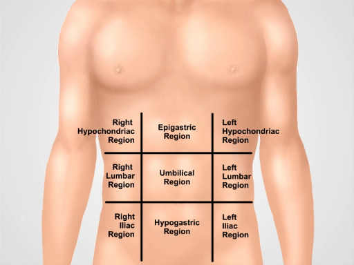
Diagnose- und beschreibenden Zwecken der Bauch gliedert sich in vier Quadranten: rechten oberen Quadranten (oft als RUQ bezeichnet), linken, oberen Quadranten (LUQ), rechten unteren Quadranten (RLQ) und linken unteren Quadranten (LLQ) (Abbildung 1). Die detailliertere Topographie des Bauches teilt es in 9 Regionen: rechten und linken Hypochonder, rechts und links der Lendenwirbelsäule, rechts und links Beckenkamm und auch epigastrische, Nabelschnur und Unterbauch Regionen in der Mitte (Abbildung 2).
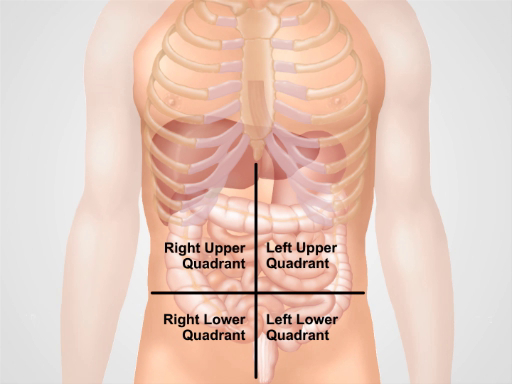
Denken Sie daran, welche Organe in der Regel in jeder Bauchregion ()Abbildung 3projizieren). Es ist wichtig zu wissen, der Region Anatomie und Topographie auch angemessen dokumentieren und interpretieren ein Patient Beschwerden und Symptome sowie körperliche Befunde während der Untersuchung.

Abbildung 1. Vier Abdominal-Quadranten. Der Bauch kann in vier Regionen unterteilt werden, von zwei imaginären Linien schneiden um den Nabel: rechten oberen Quadranten (oft als RUQ bezeichnet), links oberen Quadranten (LUQ), rechten unteren Quadranten (RLQ) und linken unteren Quadranten (LLQ) gezeigt.

Abbildung 2: Neun Regionen Bauch. Midclavicular Linien und subcostal und intertubercular Flächen trennen das Abdomen in neun Regionen: epigastrischen Region, rechts Hypochonder Region linken Hypochonder Region, umbilical Region, rechten Lendengegend, linken Lendenwirbelbereich, Unterbauch Region, rechts inguinalen Region und linken inguinalen Region. Bedingungen für epigastrische, Nabelschnur und Unterbauch und suprapubischen Regionen sind die am häufigsten verwendeten in der klinischen Praxis.

Abbildung 3. Lage der verschiedenen Organe in den vier Abdominal-Regionen. Organe in der Bauchhöhle und ihrer Lage in Bezug auf vier Abdominal-Quadranten.
Verfahren
1. Vorbereitung
- Bevor Sie die körperliche Untersuchung des Bauches beginnen, achten Sie darauf, dass der Patient komfortabel ist und hat seine/ihre Blase entleert.
- Bequem stellen der Patient in Rückenlage, möglicherweise mit dem Patienten unterstützt durch ein Kissen Kopf und Knie leicht gebeugt. Der Patient sollte an der Seite und nicht hinter dem Kopf gefaltet, wie dies die Bauchdecke spannt.
- Bitte um Erlaubnis, aussetzen der Patient Bauchbereich ("ist es ok wenn ich mich das Blatt beweg
Anwendung und Zusammenfassung
In diesem Video wir überprüft die Anatomie des Bauches und gelernt, wie man die ersten beiden Schritte der Abdominal-Prüfung durchführen: Inspektion und Auskultation. Stellen Sie vor Beginn der Prüfung sicher, dass der Patient bequem, gut aufgestellt und ausreichend drapiert. Nie untersuchen Sie einen Patienten durch ein Kleid. Stellen Sie sicher, dass Ihre Hände gewaschen werden und warm. Bitten Sie Patienten für die Erlaubnis, die Prüfung durchzuführen und erklären jeden Schritt des Verfahrens immer. Beginnen...
Tags
pringen zu...
Videos aus dieser Sammlung:

Now Playing
Abdominaluntersuchung I: Inspektion und Auskultation
Physical Examinations II
202.5K Ansichten

Augenuntersuchung
Physical Examinations II
77.0K Ansichten

Ophthalmoskopie
Physical Examinations II
67.7K Ansichten

Untersuchung der Ohren
Physical Examinations II
54.9K Ansichten

Untersuchung der Nase, Nebenhöhlen, Mundhöhle und Rachen
Physical Examinations II
65.6K Ansichten

Untersuchung der Schilddrüse
Physical Examinations II
104.8K Ansichten

Überprüfung der Lymphknoten
Physical Examinations II
386.8K Ansichten

Abdominaluntersuchung III: Palpation
Physical Examinations II
248.0K Ansichten

Abdominal-Prüfung III: Palpation
Physical Examinations II
138.5K Ansichten

Abdominaluntersuchung IV: Beurteilung akuter abdominaler Schmerzen
Physical Examinations II
67.2K Ansichten

Männliche Rektaluntersuchung
Physical Examinations II
114.3K Ansichten

Umfassende Brustuntersuchung
Physical Examinations II
87.4K Ansichten

Gynäkologische Untersuchung I: Beurteilung der äußeren Genitalien
Physical Examinations II
306.4K Ansichten

Gynäkologische Untersuchung II: Spekulumuntersuchung
Physical Examinations II
150.2K Ansichten

Gynäkologische Untersuchung III: Bimanuelle und rektovaginale Untersuchung
Physical Examinations II
147.5K Ansichten
Copyright © 2025 MyJoVE Corporation. Alle Rechte vorbehalten