Method Article
Descompresión quirúrgica mínimamente invasiva de los nervios occipitales
En este artículo
Resumen
El manuscrito presenta un protocolo quirúrgico mínimamente invasivo de preservación de nervios y músculos para descomprimir los nervios occipitales con el objetivo de mejorar la neuralgia occipital.
Resumen
La neuralgia occipital (ON) se destaca como una de las formas más angustiantes de trastornos de dolor de cabeza, que se distingue por dolor persistente en la base del cráneo, dolores de cabeza occipitales recurrentes y disestesia o alodinia del cuero cabelludo. ON es conocida por su implacable agonía, que afecta gravemente a la vida de los afectados. El dolor incesante, que a menudo se irradia hacia arriba desde la base del cráneo hasta el cuero cabelludo, puede ser profundamente debilitante. Los pacientes con frecuencia soportan dolores de cabeza occipitales insoportables, lo que hace que incluso las actividades diarias rutinarias sean un desafío formidable. La carga añadida de la disestesia o alodinia del cuero cabelludo, en la que estímulos aparentemente inocuos provocan un dolor intenso, agrava el sufrimiento. Esta neuralgia surge principalmente de la compresión mecánica ejercida sobre los nervios occipitales a lo largo de la línea nucal. En este trabajo, presentamos una técnica mínimamente invasiva de preservación de nervios y músculos destinada a aliviar esta compresión sobre los nervios occipitales. El diagnóstico preciso y el tratamiento eficaz son primordiales para brindar alivio a las personas que luchan contra esta afección. Los bloqueos nerviosos con anestesia local se han convertido en una piedra angular del diagnóstico, ya que sirven como confirmación de la neuralgia occipital y como una posible intervención terapéutica. Estos procedimientos ofrecen información crucial sobre la fuente del dolor, al tiempo que ofrecen un respiro transitorio. Sin embargo, el verdadero avance radica en la técnica innovadora que proponemos, un procedimiento que aborda la compresión mecánica en la línea nucal, que es un factor importante que contribuye a la neuralgia occipital. Al descomprimir cuidadosamente los nervios occipitales afectados, al tiempo que se preserva su integridad y el tejido muscular circundante, este enfoque mínimamente invasivo ofrece a los pacientes un camino potencial hacia un alivio sostenido. Sorprendentemente, el procedimiento se puede realizar bajo anestesia local, lo que reduce la invasividad de las cirugías tradicionales y minimiza el tiempo de inactividad del paciente.
Introducción
La neuralgia occipital (ON) es una afección crónica de dolor de cabeza que causa un dolor sordo persistente en la parte posterior dela cabeza. El dolor, que difiere de las migrañas típicas, suele ser resistente a los tratamientos estándar para la migraña debido a la compresión mecánica de los nervios occipitales2, especialmente a lo largo de su curso a través de la línea nucal3. Por otro lado, las opciones quirúrgicas pueden ser efectivas, pero implican procedimientos invasivos y tiempos de recuperación prolongados 4,5. Presentamos aquí un enfoque novedoso de los nervios occipitales, que permite una descompresión mínimamente invasiva, un tiempo de inactividad mínimo y la preservación de las ramas musculares y nerviosas sensibles6.
El diagnóstico de la ON se basa en bloqueos nerviosos específicos, que reducen temporalmente el dolor y ayudan a identificar el área exacta de compresión nerviosa7, guiando la descompresión quirúrgica 8,9. A diferencia del tratamiento típico de la migraña, nuestro enfoque se centra en la causa mecánica raíz de la ON, proporcionando una opción terapéutica viable más allá de la medicación.
Numerosos estudios clínicos y anatómicos han conducido a la técnica de descompresión del nervio occipital descrita en 2,3,10,11,12,13. Aunque se ha demostrado que esta técnica es segura y eficaz, las ventajas de la técnica mínimamente invasiva que se presenta aquí incluyen la reducción de la morbilidad del paciente, la reducción de los períodos de recuperación postoperatoria y la reducción de los riesgos de dolor inducido por la iatrogenia debido a la posible formación de neuromas. En particular, la preservación de las estructuras neurales y musculares contribuye a obtener resultados rápidos y favorables. Los nervios occipitales mayor y menor pueden exponerse y descomprimirse a través del abordaje descrito. Para el propósito de este trabajo, solo se describe una mayor descompresión del nervio occipital para mejorar la neuralgia occipital, que se debe a una neuralgia occipital menor. El tercer nervio occipital es responsable de casos raros de neuralgia occipital en nuestra práctica, que se abordan con un enfoque separado debido a su localización medial. La técnica descrita incluye la exploración sistemática del paso del nervio occipital mayor a través del semiespinoso de la cabeza, que puede representar un punto de compresión. Se justifica la investigación adicional y la validación clínica para determinar el alcance total de su eficacia y seguridad.
Protocolo
La recolección de datos se llevó a cabo como un estudio retrospectivo de evaluación de la calidad y el análisis de los resultados fue aprobado por la junta de revisión interna de la Universidad de Padua. Todos los procedimientos se llevaron a cabo de acuerdo con las normas éticas del comité nacional de investigación y la Declaración de Helsinki de 1964 y sus enmiendas posteriores. Todos los pacientes han firmado un consentimiento informado que permite a los autores utilizar los datos retrospectivos de forma anónima. Para este estudio se incluyeron 87 pacientes.
1. Selección de candidatos
- Seleccionar a los pacientes en función de una constelación de síntomas compatibles con la neuralgia occipital, que incluya al menos tres de las siguientes características: dolor, ardor y dolor punzante, que comienza en la fosa entre la inserción del músculo trapecio y el músculo esternocleidomastoideo (aquí definido como triángulo occipital); El dolor viaja en el cuero cabelludo a lo largo de la trayectoria de los nervios occipitales desde la parte posterior de la cabeza hasta las sienes y el frente en uno o ambos lados de la cabeza; el dolor puede parecerse a una descarga eléctrica; el dolor puede ser provocado/empeorado por posiciones particulares de la cabeza (hiperextensión del cuello, rotación de la cabeza, etc.) también mientras se duerme; El dolor a menudo se siente detrás de los ojos; el cuero cabelludo puede exhibir alodinia; Los pacientes pueden tener migrañas y cefaleas en racimo, además de neuralgia occipital.
- Para ser incluidos, asegúrese de que los pacientes respondan con al menos un 50% de disminución del dolor después de bloqueos selectivos de los nervios occipitales.
- Utilice los siguientes criterios de exclusión: dolor iniciado después de un traumatismo como una lesión por latigazo cervical; dolor iniciado después de una cirugía o radioterapia con lesión nerviosa directa o indirecta; pacientes que no responden a los bloqueos nerviosos selectivos.
2. Procedimiento de bloqueo nervioso
- Prepare la mezcla de infiltración como 1,5 mL de lidocaína al 1% con epinefrina (1% Rapidocaína 10 mg/mL) y 1 cc de 40 mg de Triamcinolona (40 mg/mL) en una jeringa de 5 cc preparada con una aguja de 30 G.
- Transfiera la mezcla a una jeringa de 5 ml (Luer Lock) con una aguja de 30 g (0,3 mm x 13 mm).
- Identificar el lugar de la inyección por palpación. Identificar el borde lateral de la inserción del trapecio proximal y el borde medial de la inserción del músculo esternocleidomastoideo proximal (SCM), justo debajo de la línea nucal. El pozo entre estas estructuras corresponde al punto de inyección objetivo.
- Palpar el conducto de la arteria occipital por encima de la línea nucal. Confirmar la posición del haz neurovascular occipital (nervio occipital mayor y menor y arteria occipital) por debajo de la línea nucal mediante Doppler (10 MHz). Mover la sonda Doppler caudalmente hacia el triángulo occipital y confirmar el paso de la arteria en este lugar mediante una señal arterial.
- Realice una aspiración suave con la jeringa antes de la inyección (si se aspira sangre, la aguja se retrae unos mm) para evitar la inyección intravascular. Si el paciente siente descargas eléctricas a lo largo de la trayectoria del nervio occipital, retire la aguja unos mm para evitar la inyección intraneural y el daño al nervio.
- Para asegurarse de que el bloqueo nervioso se ha realizado correctamente, palpe el cuero cabelludo posterior para permitir que el paciente confirme la disminución de la sensibilidad.
3. Preparación de la mesa de instrumentación
- Cubra la mesa de instrumentación con flujo laminar con ropa estéril. Prepare los siguientes instrumentos: Bisturí quirúrgico con hoja No. 15, pinzas quirúrgicas Hudson, portaagujas, tijeras de disección, pinzas bipolares, retractor iluminado, sutura de nylon 5/0, guantes estériles, desinfección con clorhexidina, cinta de papel estéril.
4. Preparación del paciente
- Identifique el punto más sensible con el paciente sentado erguido mediante la palpación dentro del área donde se realizó el bloqueo nervioso.
- Marque el sitio de la incisión con una línea de incisión oblicua de 2 a 3 cm a lo largo del área sensible. Afeitar el área que comprende la línea de incisión y 1 cm alrededor con una afeitadora quirúrgica.
5. Preparación del cirujano
- El cirujano usa asas de aumento de 2.5x a 3.5x, gorro quirúrgico y mascarilla. Lávese las manos con jabón y desinfecte las manos con una solución antiséptica.
6. Técnica quirúrgica
- Posicionamiento del paciente: Coloque a los pacientes lateralmente (en el caso de la ON unilateral) o boca abajo (en el caso de la ON bilateral) en la mesa quirúrgica.
- Anestesia local: Administrar anestesia local, que consiste en 5 mL de lidocaína con epinefrina, inyectada a lo largo de la línea de incisión a través de una aguja de 30G.
- Incisión: Realizar una incisión oblicua, biselada, de 2,5 a 3,5 cm, centrada en una región definida como el triángulo occipital, bordeada cranealmente por la línea nucal, medialmente por el borde lateral del trapecio y lateralmente por el borde medial del músculo esternocleidomastoideo utilizando una cuchilla quirúrgica No. 15.
- Exposición de la línea nucal: Incidir la capa superficial de la línea nucal con el bisturí y diseccionar con tijeras de disección para exponer el nervio occipital mayor (GON), la arteria occipital y los ganglios linfáticos.
- Exploración nerviosa: Siga el GON meticulosamente a lo largo de todos los puntos de posible compresión utilizando tijeras de disección para crear el espacio. Realizar la liberación proximal de la fascia inferior del músculo trapecio con tijeras de disección, fascia del músculo semiespinal y fibras de la línea nucal distal.
- Estructuras vasculares y linfáticas: En los casos en que la arteria occipital y los ganglios linfáticos entran en contacto con los nervios, reposicione o extirpe estas estructuras delicadamente con las pinzas quirúrgicas de Hudson. Desenganchar suavemente los tejidos adventiciales y periarteriales con las pinzas quirúrgicas de Hudson, abundantes en fibras aferentes y eferentes del sistema nervioso autónomo, en todos los casos (arteriolisis).
- Abordar los puntos de contacto neurovascular: Cuando se encuentra un conflicto que no se puede abordar de otra manera (como una rama de la arteria que pasa a través de las fibras nerviosas), divida este segmento arterial (arteriotomía).
- Bloqueos nerviosos: Antes del cierre, realice bloqueos nerviosos con lidocaína al 1% con epinefrina rociada directamente sobre las ramas nerviosas a través de una aguja de 30 G. Pida al paciente que mueva la cabeza y hable para confirmar la descompresión completa.
- Cierre: Repare la piel con puntos individuales de suturas de nailon 5-0. Cubra la abertura con un apósito en aerosol permeable a la humedad y gasas estériles, sujetas con cinta de papel.
7. Protocolo postoperatorio
- Movimientos de la cabeza postoperatorios: Después del procedimiento, pida al paciente que mueva suavemente la cabeza en todas las direcciones al menos 3 veces al día para evitar la formación de adherencias entre las fibras nerviosas y la cicatriz durante un período de 2 a 3 semanas.
- Extracción de suturas: Retire las suturas 10 días después del procedimiento.
- Pida a los pacientes que continúen con sus medicamentos en caso de dolores de cabeza. Dígale al paciente que espere una disminución de la sensibilidad o de la anestesia en el territorio del nervio occipital mayor durante un período variable entre unas pocas horas y unos días después de la operación.
- Indique a los pacientes que tomen ibuprofeno 400-600 mg, 3 veces al día durante 3 días después del procedimiento.
Resultados
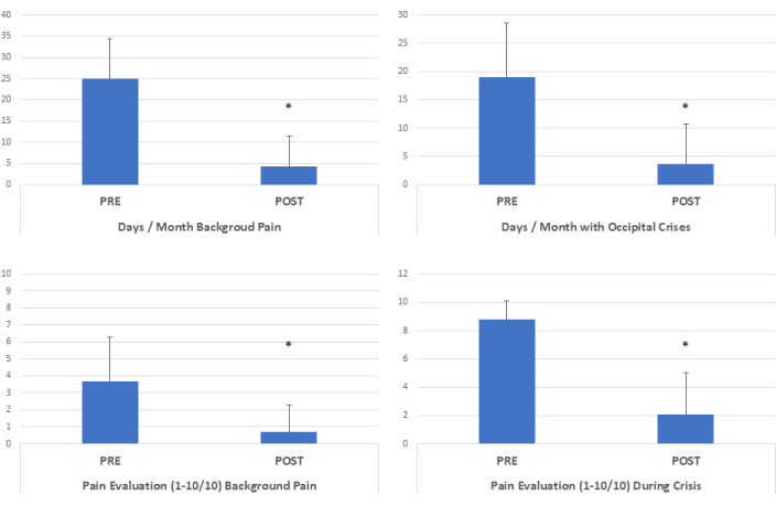
Al año de la descompresión quirúrgica, hubo una notable reducción de los días de dolor crónico, con una disminución sustancial de un promedio inicial de 25 días a 4,3 días (p <0,01), reflejando una reducción del 80,5% (disminución de 5,8 veces) en la frecuencia de dolor crónico (Figura 1)6. Además, el número de días de crisis de dolor al mes mostró una disminución notable, pasando de 19 días a 3,7 días (p <0,01), lo que significa una reducción del 82,8% (disminución de 5,1 veces) en la frecuencia de crisis de dolor (Figura 1 y Figura 2)6.
Los pacientes informaron una intensidad media del dolor de fondo de 3,7 en una escala de 10 antes de la cirugía, que mejoró sustancialmente a 0,7 (p <0,01) después de la cirugía, lo que corresponde a una impresionante reducción del 76,1% (una disminución de 5,2 veces) en la intensidad del dolor de fondo6. Además, la intensidad máxima del dolor experimentada durante las crisis disminuyó significativamente de 8,8/10 a 2,1/10 después de la cirugía (p <0,01), lo que refleja una reducción sustancial del 81,1% (una disminución de 4,2 veces) en la intensidad máxima del dolor (Figura 1 y Figura 2)6.
Sorprendentemente, hubo una reducción significativa en la utilización de todo tipo de fármacos, incluidos AINE, triptanos y fármacos modificadores de la enfermedad, después de la intervención quirúrgica6 (no mostrado).

Figura 1: Evolución clínica tras la descompresión quirúrgica mínimamente invasiva. Después de la descompresión quirúrgica, los días de dolor crónico disminuyeron 5,8 veces. Los días de crisis de dolor/mes disminuyeron 5,1 veces. La intensidad del dolor de fondo disminuyó 5,2 veces después de la cirugía, y los picos de intensidad del dolor durante las crisis disminuyeron 4,2 veces. (*p <0.01, Pruebas t pareadas de dos colas, n=87). Esta cifra ha sido modificada de6. Haga clic aquí para ver una versión más grande de esta figura.

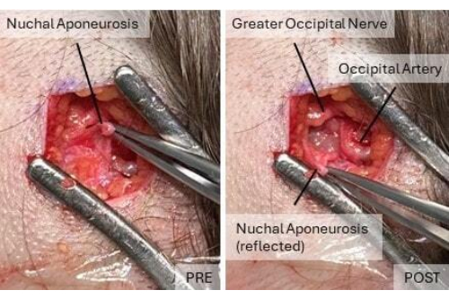
Figura 2: Ilustración de la anatomía del área occipital antes y después de la descompresión. En el cuadro previo a la descompresión, la capa externa de la aponeurosis nucal cubre los nervios occipitales y los vasos sanguíneos. Después de la descompresión, el nervio occipital mayor y la arteria occipital son visibles. Haga clic aquí para ver una versión más grande de esta figura.
Discusión
La neuralgia occipital es una de las formas más debilitantes de dolores de cabeza, principalmente debido al dolor crónico que es incesante. Un estudio sobre la prevalencia del dolor facial en 2009, que a menudo se utiliza como referencia para la neuralgia occipital, encontró una prevalencia de ON de 3,2 por 100.00014. Estas estadísticas subestiman en gran medida el problema, sabiendo que la ON es responsable del dolor facial solo en el 8,3% de los casos y que hasta el 25% de los casos de ingreso en urgencias son por cefaleas debidas a neuralgia occipital15.
Encontramos en nuestra práctica que la ON sola o en combinación con otras migrañas es una de las formas más prevalentes de dolores de cabeza crónicos, posiblemente debido a una postura mórbida con el cuello en flexión frente a computadoras y teléfonos inteligentes varias horas al día, un estilo de vida sedentario y un tiempo limitado al aire libre.
El abordaje quirúrgico descrito aquí ofrece un medio altamente eficiente de acceder a los nervios occipitales bajo anestesia local. La neuralgia occipital mayor puede coexistir con la neuralgia occipital menor, ya que estos nervios tienen ramas comunicantes y sus territorios se superponen. Al utilizar el mismo abordaje quirúrgico, ambos nervios pueden ser explorados y descomprimidos cuando está indicado6. La aceptación del procedimiento por parte de los pacientes ha sido favorable, con una duración promedio de aproximadamente 45 a 60 min requeridos para su finalización por lado.
La meticulosa identificación y preservación de las fibras nerviosas constituye un sello distintivo de este enfoque. Gracias a la naturaleza mínimamente invasiva del procedimiento que se puede realizar con anestesia local, el operador evalúa la eficacia de la descompresión al final del procedimiento instruyendo al paciente para que realice movimientos de cabeza y conversación, asegurando así la ausencia de puntos de compresión residuales.
Una faceta crucial de este procedimiento radica en el énfasis en la movilización temprana y frecuente de la cabeza, realizada varias veces al día. Esta práctica sirve para disuadir la formación de adherencias entre las fibras nerviosas y la cicatriz quirúrgica, que de otro modo podrían impedir la recuperación.
Es fundamental reconocer que no todos los pacientes son candidatos adecuados para esta técnica. En particular, las personas con fragilidad o niveles elevados de ansiedad pueden no tolerar el procedimiento cómodamente bajo anestesia local pura. En ciertos casos, los pacientes pueden experimentar molestias repentinas, ya que incluso la más mínima manipulación de un nervio occipital inflamado puede desencadenar la activación del nervio. En estos casos, la anestesia local se rocía directamente sobre las fibras nerviosas con un alivio inmediato.
Este abordaje quirúrgico representa una alternativa menos invasiva en comparación con las técnicas de descompresión propuestas anteriormente. Su capacidad para preservar tanto las fibras nerviosas como las musculares contribuye a una notable reducción de las tasas de complicaciones. Postulamos que esta metodología mínimamente invasiva, pero altamente efectiva, ampliará la accesibilidad de la descompresión quirúrgica como una opción de tratamiento definitiva para la neuralgia occipital, ofreciendo esperanza a un espectro más amplio de pacientes.
Divulgaciones
Los autores declaran no tener intereses financieros contrapuestos.
Agradecimientos
Los autores agradecen la asistencia técnica de Alexandra Curchod, Yuliethe Martins y el equipo de Filmatik Global. Este trabajo fue financiado en su totalidad por el Global Medical Institute.
Materiales
| Name | Company | Catalog Number | Comments |
| 30G Needle | 0.3x13 mm, BD Microlance 3, Spain | ||
| Bipolar Forceps | McPerson, bipolar forceps, Erbe, Switzerland | 20195 | |
| Chlorhexidine | Hibidil, Chlorhexidini digluconas 0.5 mg/mL, Switzerland | 120099 | |
| dissection scissors | Jarit supercut, Integra Lifescience, USA | 323720 | |
| Doppler | Dopplex DMX Digital Doppler, High Sensitivity 10MHz probe, Huntleigh Healthcare, Wales, United Kingdom | ||
| Ethilon 5/0 Suture | Ethicon, USA | 698 G | |
| Lidocaine ephinephrine 1% | Rapidocain 1% 10 mg/mL, Sintetica, Switzerland | ||
| Lighted retractor | Electro Surgical Instrument Company, Rochester, NY | 08-0195 | |
| Magnifying loops | Design for vision, USA | ||
| Opsity spray | Smith & Nephew, USA | ||
| Sterile gloves | Sempermed sintegra IR, Ireland | ||
| Sterillium | Sterillium disinfectant, Switzerland | ||
| Surgical blade n.15 | Carbon steel surgical blades, Swann-Morton, England) | 205 | |
| Surgical drapes and gauzes | Halyard Universal pack, USA | 88761 | |
| Surgical instruments | Bontempi medical Italy | ||
| Surgical shaver | Carefusion, USA | ||
| Syringe 5cc | BBraun, Omnifix Luer Lock Solo, Switzerland | ||
| Triamcinolone 10mg | Triamcort depot 40 mg/mL, Zentiva Czech Republic |
Referencias
- IHS. The international classification of headache disorders, 3rd edition. Cephalalgia. 38 (1), 1-211 (2018).
- Mosser, S. W., et al. The anatomy of the greater occipital nerve: implications for the etiology of migraine headaches. Plast Reconstr Surg. 113 (2), 693-697 (2004).
- Janis, J. E., et al. Neurovascular compression of the greater occipital nerve: implications for migraine headaches. Plast Reconstr Surg. 126 (6), 1996-2001 (2010).
- Guyuron, B., et al. Five-year outcome of surgical treatment of migraine headaches. Plast Reconstr Surg. 127 (2), 603-608 (2011).
- Blake, P., et al. Tracking patients with chronic occipital headache after occipital nerve decompression surgery: A case series. Cephalalgia. 39 (4), 556-563 (2019).
- Pietramaggiori, G., Scherer, S. Minimally invasive nerve- and muscle-sparing surgical decompression for occipital neuralgia. Plast Reconstr Surg. 151 (1), 169-177 (2023).
- Tobin, J., Flitman, S. Treatment of migraine with occipital nerve blocks using only corticosteroids. Headache. 51 (1), 155-159 (2011).
- Seyed Forootan, N. S., Lee, M., Guyuron, B. Migraine headache trigger site prevalence analysis of 2590 sites in 1010 patients. J Plast Reconstr Aesthet Surg. 70 (2), 152-158 (2017).
- Pietramaggiori, G., Scherer, S. . Minimally invasive surgery for chronic pain management. , (2020).
- Janis, J. E., et al. The anatomy of the greater occipital nerve: Part II. Compression point topography. Plast Reconstr Surg. 126 (5), 1563-1572 (2010).
- Ducic, I., Moriarty, M., Al-Attar, A. Anatomical variations of the occipital nerves: implications for the treatment of chronic headaches. Plast Reconstr Surg. 123 (3), 859-863 (2009).
- Junewicz, A., Katira, K., Guyuron, B. Intraoperative anatomical variations during greater occipital nerve decompression. J Plast Reconstr Aesthet Surg. 66 (10), 1340-1345 (2013).
- Scherer, S. S., et al. The greater occipital nerve and obliquus capitis inferior muscle: Anatomical interactions and implications for occipital pain syndromes. Plast Reconstr Surg. 144 (3), 730-736 (2019).
- Koopman, J. S., et al. Incidence of facial pain in the general population. Pain. 147 (1-3), 122-127 (2009).
- Mathew, P. G., et al. Prevalence of occipital neuralgia at a community hospital-based headache clinic. Neurol Clin Pract. 11 (1), 6-12 (2021).
Reimpresiones y Permisos
Solicitar permiso para reutilizar el texto o las figuras de este JoVE artículos
Solicitar permisoExplorar más artículos
This article has been published
Video Coming Soon
ACERCA DE JoVE
Copyright © 2025 MyJoVE Corporation. Todos los derechos reservados