Detección de bacteriófagos en muestras ambientales
Visión general
Fuente: Laboratorios del Dr. Ian Pepper y el Dr. Charles Gerba - Universidad de Arizona
Demostrando autor: Alex Wassimi
Los virus son un grupo de entidades biológicas que infectan a los organismos eucariotas y procariotas. Son parásitos obligados que no capacidad metabólica y con el fin de replicar, se basan sobre el metabolismo del huésped para producir piezas virales que uno mismo-montan dentro de las células del huésped.
Los virus son ultramicroscópicos, demasiado pequeños para ser vistos con el microscopio de luz, visible sólo con la mayor resolución del microscopio electrónico. Una partícula viral consiste en un genoma de ácido nucleico, DNA o RNA, rodeado por una cubierta proteica, denominada cápside, compuesta por subunidades proteicas o capsómeros. En algunos virus más complejos, la cápside está rodeada por una envoltura de lípidos adicionales, y algunas tienen apéndices superficiales espiga o cola.
Virus que infectan el tracto intestinal de humanos y animales son conocidos como virus entéricos. Son excretados en las heces y puede ser aislados de las aguas residuales domésticas. Virus que infectan bacterias se denominan bacteriófagos, y los que infectan a las bacterias coliformes se llaman colífagos (figura 1). Los fagos de las bacterias coliformes se encuentran en cualquier lugar se encuentran las bacterias coliformes.

Figura 1. Colifago T2.
Procedimiento
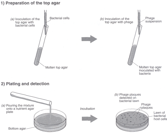
- Obtener una muestra de aguas residuales o aguas que contienen colifago.
- Diluir la muestra 1:10 y 1: 100 utilizando tampón Tris. Para ello, transferir 1,0 mL de cultivo a 9 mL de tampón Tris y luego hacer una dilución de 10 veces en segundo lugar.
- Fundir los tres tubos de agar blando (0,7% de agar nutritivo o agar de soja trypticase por tubo de 3 mL) colocándolos en un baño de vapor o autoclave.
- Ponga el agar en un baño de agua a 45-48 ° C por 15 min para permitir que la temperatura del agar a 45 ° C.
Resultados
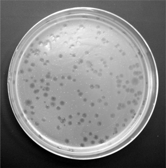
Dilución de muestras de aguas residuales = 10-1
Número de placas obtenidas = 9
Por lo tanto, la concentración de fagos en muestra de aguas residuales
= 10 x 9 ÷ 1 mL
= 90 unidades formadoras de placas / mL
Aguas residuales normalmente contienen 103 – 104 colifago por mL, con una gama de 102 – 108 / mL.
Aplicación y resumen
Hay muchas aplicaciones potenciales de colífagos como indicadores ambientales. Estos incluyen su uso como indicadores de contaminación de las aguas residuales, eficiencia de agua y tratamiento de aguas residuales y la supervivencia de virus entéricos y bacterias en el medio ambiente. El uso de bacteriófagos como indicadores de la presencia y comportamiento de las bacterias entéricas y virus animal siempre ha sido atractivo debido a la facilidad de detección y bajo costo relacionados con phage ensayos. Además, pued...
Tags
Saltar a...
Vídeos de esta colección:

Now Playing
Detección de bacteriófagos en muestras ambientales
Environmental Microbiology
40.9K Vistas

Determinación del contenido de humedad en el suelo
Environmental Microbiology
359.8K Vistas

Técnica aséptica en ciencias ambientales
Environmental Microbiology
126.5K Vistas

Tinción de Gram de la Bacteria de fuentes ambientales
Environmental Microbiology
100.5K Vistas

Visualizarse los microorganismos del suelo mediante el contacto Deslice análisis y microscopia
Environmental Microbiology
42.4K Vistas

Hongos filamentosos
Environmental Microbiology
57.7K Vistas

Extracción de ADN de la comunidad de colonias bacterianas
Environmental Microbiology
28.9K Vistas

Detección de microorganismos ambientales con la reacción en cadena de polimerasa y la electroforesis en Gel de
Environmental Microbiology
44.6K Vistas

Análisis de muestras ambientales mediante RT-PCR del ARN
Environmental Microbiology
40.6K Vistas

Cuantificación de microorganismos ambientales y virus utilizando qPCR
Environmental Microbiology
47.9K Vistas

Análisis de la calidad del agua a través de organismos indicadores
Environmental Microbiology
29.6K Vistas

Aislamiento de bacterias fecales de muestras de agua por filtración
Environmental Microbiology
39.4K Vistas

Cultivo y enumeración de bacterias a partir de muestras de suelo
Environmental Microbiology
184.9K Vistas

Análisis de la curva de crecimiento bacteriano y sus aplicaciones ambientales
Environmental Microbiology
296.3K Vistas

Enumeración de las algas mediante metodología cultivable
Environmental Microbiology
13.8K Vistas
ACERCA DE JoVE
Copyright © 2025 MyJoVE Corporation. Todos los derechos reservados

