Ensayo de placa: Un método para determinar los títulos virales como unidades formadoras de placas (UFP)
Visión general
Fuente: Tilde Andersson1, Rolf Lood1
1 Departamento de Ciencias Clínicas Lund, División de Medicina de Infecciones, Centro Biomédico, Universidad de Lund, 221 00 Lund, Suecia
Los virus que infectan los organismos procaloicos, llamados bacteriófagos o simplemente fagos, fueron identificados a principiosdel siglo XX por Twort (1) y d'Hérelle (2) de forma independiente. Desde entonces, los fagos han sido ampliamente reconocidos por su valor terapéutico (3) y su influencia en los ecosistemas humanos (4), así como en los ecosistemas globales (5). Las preocupaciones actuales han impulsado un renovado interés en el uso de fagos como alternativa a los antibióticos modernos en el tratamiento de enfermedades infecciosas (6). Esencialmente toda la investigación de fagose se basa en la capacidad de purificar y cuantificar virus, también conocido como un titer viral. Descrito inicialmente en 1952, este fue el propósito del ensayo de placa (7). Décadas y múltiples avances tecnológicos más tarde, el ensayo de placa sigue siendo uno de los métodos más fiables para la determinación del titer viral (8).
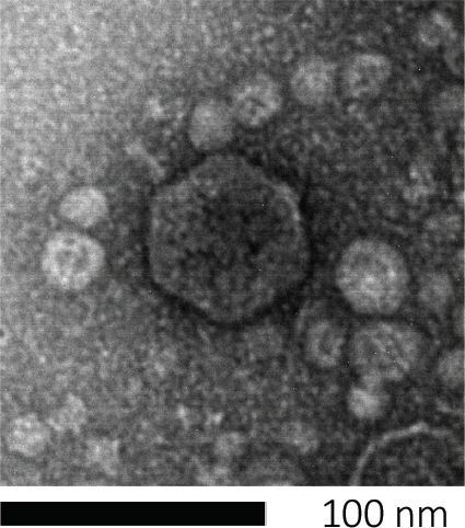
Los bacteriófagos subsisten inyectando su material genético en las células huésped, secuestrando las máquinas para la producción de nuevas partículas de fago, y eventualmente causando que el huésped libere numerosos viriones de progenie a través de la lisis celular. Debido a su tamaño minúsmico, los bacteriófagos no se pueden observar utilizando únicamente microscopía de luz; por lo tanto, se requiere microscopía electrónica de barrido (Figura 1). Además, los fagos no se pueden cultivar en placas de agar nutricionales como bacterias, ya que necesitan células huésped para abeste.

Figura 1: La morfología de un bacteriófago, aquí ejemplificada por un fago E. coli, se puede estudiar mediante microscopía electrónica de barrido. La mayoría de los bacteriófagos pertenecen a Caudovirales (bacteriófagos de cola). Este fago en particular tiene una estructura de cola muy corta y una cabeza icosahedral, colocándola en la familia de Podovirus.
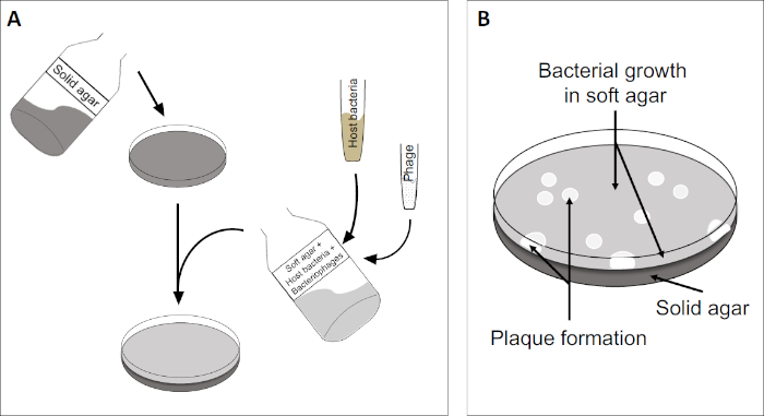
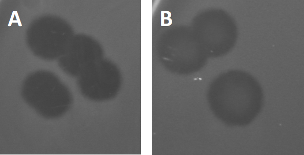
El ensayo de placa (Figura 2) se basa en la incorporación de células huésped, preferentemente en el crecimiento de la fase log, en el medio. Esto crea una capa densa y turbia de bacterias capaces de mantener el crecimiento viral. Un fago aislado puede infectar, replicar y oxidar una célula. Con cada célula lysed, varias de las adyacentes se infectan inmediatamente. Varios ciclos en, una zona clara (una placa) se pueden observar en la placa de otro modo turbia (Figura 2B/Figura 3A),lo que indica la presencia de lo que inicialmente era una sola partícula de bacteriófago. Por lo tanto, el número de unidades formadoras de placas por volumen(es decir, PFU/ml) de una muestra puede determinarse a partir del número de placas generadas.

Figura 2: Las pruebas para las unidades formadoras de placas (PFU) son un método común para determinar el número de bacteriófagos en una muestra. (A) La base de una placa estéril de Petri está cubierta con un medio de nutrientes sólido adecuado, seguido de una mezcla de medios blandos, células huésped susceptibles y una dilución de la muestra original de bacteriófagos. Tenga en cuenta que la suspensión del fago podría, en algunos casos, también extenderse uniformemente a través de la superficie del agar blando ya solidificado. (B) El crecimiento de las bacterias anfitrionas forma un césped de células en la capa superior de agar. La replicación de bacteriófagos genera zonas claras, o placas, causadas por la lisis de la célula huésped.

Figura 3: Los resultados de las pruebas de PFU muestran múltiples placas generadas por bacteriófagos. Debido a la lisis de las células huésped susceptibles, las placas pueden ser vistas como zonas de despeje en el césped bacteriano, ya sea con (A) aclaramiento completo, o (B) re-crecimiento parcial causado por la generación de bacterias resistentes (o posiblemente por fagos templados en el lisogénico).
Ciertos fagos templados pueden adoptar lo que se conoce como un ciclo de vida lisogénico, además del crecimiento lítico descrito anteriormente. En la lisógena, el virus asume un estado latente mediante la incorporación de su material genético en el genoma de la célula huésped (9), a menudo confiriendo resistencia a más infecciones por fagos. Esto a veces se revela a través de una ligera opacidad de la placa (Figura 3B). Vale la pena señalar sin embargo, que las placas también pueden aparecer borrosas debido al re-crecimiento de bacterias que han evolucionado resistencia al fago independiente de las infecciones anteriores de fago.
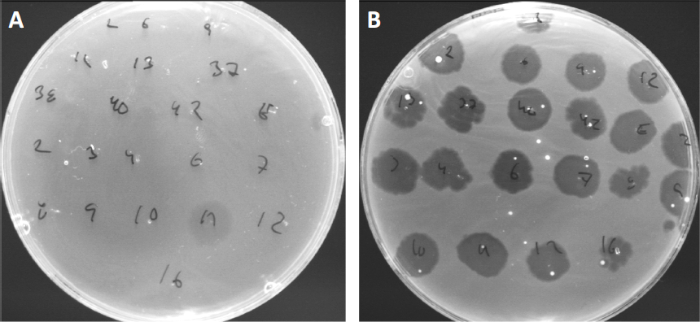
Los virus pueden adjuntarse, o adsorgar, a sólo un rango limitado de bacterias anfitrionas (10). Los rangos de acogida están aún más limitados por estrategias antivirales intracelulares como el sistema CRISPR-Cas (11). La resistencia/sensibilidad hacia fagos específicos mostrados por subgrupos bacterianos se ha utilizado históricamente para clasificar las cepas bacterianas en diferentes tipos de fagos (Figura 4). Aunque la eficacia de este método ha sido ahora superada por nuevas técnicas de secuenciación, la tipificación de fagos todavía puede proporcionar información valiosa sobre las interacciones entre bacterias y fagos, por ejemplo, facilitando el diseño de un cóctel de fago para uso clínico .

Figura 4: Sensibilidad del fago de diferentes cepas bacterianas. Las placas de agar blandas con cepa Cutibacterium acnes (A) AD27 y (B) AD35, fueron manchadas con 21 bacteriófagos Diferentes de C. acnes. Sólo el fago 11 fue capaz de infectar y matar AD27 mientras que la cepa AD35 mostró sensibilidad hacia todos los fagos. Esta técnica, que se denomina tipificación de fagos, se puede utilizar para dividir especies bacterianas y cepas en diferentes subgrupos basados en la susceptibilidad del fago.
Procedimiento
1. Configuración
- Antes de empezar cualquier trabajo que involucre microbios, asegúrese de que el espacio de trabajo esté esterilizado(por ejemplo, limpiado con 70% de etanol). Use siempre una capa de laboratorio y guantes, mantenga el cabello largo atado hacia atrás y asegúrese de que las heridas estén particularmente bien protegidas.
- Cuando haya terminado, esterilizar todas las superficies y lavar/esterilizar a fondo las manos y las muñecas.
<
Aplicación y resumen
A pesar de los múltiples avances tecnológicos, los ensayos de placas siguen siendo el estándar de oro para la determinación del titer viral (como PFU) y esenciales para el aislamiento de las poblaciones de bacteriófagos puros. Las células huésped susceptibles se cultivan en la capa superior de una placa de agar de dos capas, formando un lecho homogéneo que permite la replicación viral. El evento inicial donde un bacteriófago aislado en el ciclo de vida lítico infecta una célula, se replica dentro de ella, y f...
Referencias
- Twort, F. An investigation on the nature of ultra-microscopic viruses. Lancet. 186 (4814): 1241-1243. (1915)
- d'Hérelle, F. An invisible antagonist microbe of dysentery bacillus. Comptes Rendus Hebdomadaires Des Seances De L Academie Des Sciences. 165: 373-375. (1917)
- Cisek AA, Dąbrowska I, Gregorczyk KP, Wyżewski Z. Phage Therapy in Bacterial Infections Treatment: One Hundred Years After the Discovery of Bacteriophages. Current Microbiology. 74 (2):277-283. (2017)
- Mirzaei MK, Maurice CF. Ménage à trois in the human gut: interactions between host, bacteria and phages. Nature Reviews Microbiology. 15 (7):397. (2017)
- Breitbart M, Bonnain C, Malki K, Sawaya NA. Phage puppet masters of the marine microbial realm. Nature Microbiology. 3 (7):754-766. (2018)
- Leung CY, Weitz JS. Modeling the synergistic elimination of bacteria by phage and the innate immune system. Journal of Theoretical Biology. 429:241-252. (2017)
- Dulbecco R. Production of Plaques in Monolayer Tissue Cultures by Single Particles of an Animal Virus. Proceedings of the National Academy of Sciences of the United States of America. 38 (8):747-752. (1952)
- Juarez D, Long KC, Aguilar P, Kochel TJ, Halsey ES. Assessment of plaque assay methods for alphaviruses. J Virol Methods. 187 (1):185-9. (2013)
- Clokie MRJ, Millard AD, Letarov AV, Heaphy S. 2011. Phages in nature. Bacteriophage. 1 (1):31-45. (2011)
- Moldovan R, Chapman-McQuiston E, Wu XL. On kinetics of phage adsorption. Biophys J. 93 (1):303-15. (2007)
- Garneau JE, Dupuis M-È, Villion M, Romero DA, Barrangou R, Boyaval P, Fremaux C, Horvath P, Magadán AH, Moineau S.. The CRISPR/Cas bacterial immune system cleaves bacteriophage and plasmid DNA. Nature. 468 (7320):67. (2010)
Saltar a...
Vídeos de esta colección:

Now Playing
Ensayo de placa: Un método para determinar los títulos virales como unidades formadoras de placas (UFP)
Microbiology
186.0K Vistas

Creación de la columna de Winogradsky: Un método que sirve para enriquecer las especies microbianas en una muestra de sedimento
Microbiology
128.9K Vistas

Diluciones en serie y enchapado: enumeración microbiana
Microbiology
315.4K Vistas

Cultivos de enriquecimiento: Cultivo de bacterias aerobias y anaerobias en medios selectivos y diferenciales
Microbiology
132.0K Vistas

Cultivos puros y siembra por estrías: aislamiento de colonias bacterianas únicas de una muestra mixta
Microbiology
166.0K Vistas

Secuenciación del ARNr 16s: Una técnica basada en PCR para identificar especies bacterianas
Microbiology
188.6K Vistas

Curvas de crecimiento: Generación de curvas de crecimiento utilizando unidades formadoras de colonias y mediciones de densidad óptica
Microbiology
294.9K Vistas

Pruebas de susceptibilidad a los antibióticos: Pruebas con epsilometro para determinar los valores de la CMI de dos antibióticos y evaluar la sinergismos
Microbiology
93.6K Vistas

Microscopía y tinción: Tinción de Gram, cápsula y endosporas
Microbiology
363.0K Vistas

Transformación de células E. coli mediante un procedimiento con cloruro de calcio
Microbiology
86.6K Vistas

Conjugación: Un método para transferir resistencia a ampicilina de un donante a una E. Coli receptora
Microbiology
38.2K Vistas

Transducción de fagos: Un método para transferir resistencia a ampicilina de un donante una E. coli receptora
Microbiology
29.0K Vistas
ACERCA DE JoVE
Copyright © 2025 MyJoVE Corporation. Todos los derechos reservados
 decir)