
Characterization of Amyloid Structures in Aging C. Elegans Using Fluorescence Lifetime Imaging
March 27th, 2020
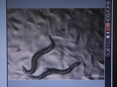
•Fluorescence lifetime imaging monitors, quantifies and distinguishes the aggregation tendencies of proteins in living, aging, and stressed C. elegans disease models.
Tags
Videos relacionados

Imaging Amyloid Tissues Stained with Luminescent Conjugated Oligothiophenes by Hyperspectral Confocal Microscopy and Fluorescence Lifetime Imaging

Structural Characterization of Mannan Cell Wall Polysaccharides in Plants Using PACE
A Simple Method for High Throughput Chemical Screening in Caenorhabditis Elegans

Growing Protein Crystals with Distinct Dimensions Using Automated Crystallization Coupled with In Situ Dynamic Light Scattering

Use of Two Dimensional Semi-denaturing Detergent Agarose Gel Electrophoresis to Confirm Size Heterogeneity of Amyloid or Amyloid-like Fibers

Calibration-free In Vitro Quantification of Protein Homo-oligomerization Using Commercial Instrumentation and Free, Open Source Brightness Analysis Software

Rapid Fluorescence-based Characterization of Single Extracellular Vesicles in Human Blood with Nanoparticle-tracking Analysis

In Vivo Calcium Imaging in C. elegans Body Wall Muscles

Interactions with and Membrane Permeabilization of Brain Mitochondria by Amyloid Fibrils

SUMO-Binding Entities (SUBEs) as Tools for the Enrichment, Isolation, Identification, and Characterization of the SUMO Proteome in Liver Cancer
ACERCA DE JoVE
Copyright © 2024 MyJoVE Corporation. Todos los derechos reservados