
A subscription to JoVE is required to view this content. Sign in or start your free trial.
Method Article
מדידה של פעילות פרונטו-הלימבית שימוש מוזר משימה רגשית בילדים בסיכון גבוה לסכיזופרניה משפחתי
In This Article
Summary
This paper describes how to use the emotional oddball task and fMRI to measure brain activation in children and adolescents at familial high risk for schizophrenia (FHR). FMRI was used to measure differences in fronto-striato-limbic regions during an emotional oddball task. Children with FHR exhibited abnormal functional activation during adolescence.
Abstract
Adolescence is a critical developmental period where the early symptoms of schizophrenia frequently emerge. First-degree relatives of people with schizophrenia who are at familial high risk (FHR) may show similar cognitive and emotional changes. However, the neurological changes underlying the emergence of these symptoms remain unclear. This study sought to identify differences in frontal, striatal, and limbic regions in children and adolescents with FHR using functional magnetic resonance imaging. Groups of 21 children and adolescents at FHR and 21 healthy controls completed an emotional oddball task that relied on selective attention and the suppression of task-irrelevant emotional information. The standard oddball task was modified to include aversive and neutral distractors in order to examine potential group differences in both emotional and executive processing. This task was designed specifically to allow for children and adolescents to complete by keeping the difficulty and emotional image content age-appropriate. Furthermore, we demonstrate a technique for suitable fMRI registration for children and adolescent participants. This paradigm may also be applied in future studies to measure changes in neural activity in other populations with hypothesized developmental changes in executive and emotional processing.
Introduction
Schizophrenia is a neurodevelopmental disorder with a known genetic component1,2 and with symptoms including deficits in both executive and emotional processing3,4. First-degree relatives are thought to be at an increased risk of developing schizophrenia, and have been shown to share some of these same neurocognitive deficits in both cognitive and social-emotional domains5. We therefore expect that brain activity in regions associated with executive and emotional processing may be altered in at-risk family members preceding the onset of clinical symptoms.
Previous studies have indicated that both adults with schizophrenia and adults at familial high risk show aberrant activity within executive and emotional processing networks; however it remains unclear how these changes come about during development. Demonstrating that these changes occur early in life will be a critical first step in understanding the pathophysiology of the disorder. Therefore, this study utilizes an emotional oddball paradigm during functional MRI (fMRI) scanning in order to measure brain activity during the completion of a task that requires both executive and emotional processing in adolescents who are at risk for developing schizophrenia. Oddball paradigms are frequently used to examine the function of fronto-striate circuitry in schizophrenia6 and in individuals with familial high risk7 by measuring selective attention processes allocated to task-relevant target stimuli. Here, a standard oddball task has been modified to include task-irrelevant aversive and neutral stimuli that have been shown to elicit changes in brain activity in patients with schizophrenia8.
This paper measures functional differences between healthy adolescents and adolescents at high familial risk for schizophrenia using an emotional oddball task. The task design is similar to that used by Fichtenholtz and colleagues9, but the selection of aversive emotional images has been modified to be appropriate for children between the ages of 9-18. The use of this task during functional MRI allowed for the identification of specific brain regions that showed patterns of hyperactivation and hypoactivation in children and adolescents with FHR for schizophrenia, in addition to age-related changes in neural activity during adolescent development.
Access restricted. Please log in or start a trial to view this content.
Protocol
טכניקות המחקר השתמשו במחקר זה אושרו על ידי לוחות הסקירה המוסדיים (IRB) של אוניברסיטת דיוק ואוניברסיטת צפון קרוליינה - צ'אפל היל.
1. הדמיה משימה עיצוב
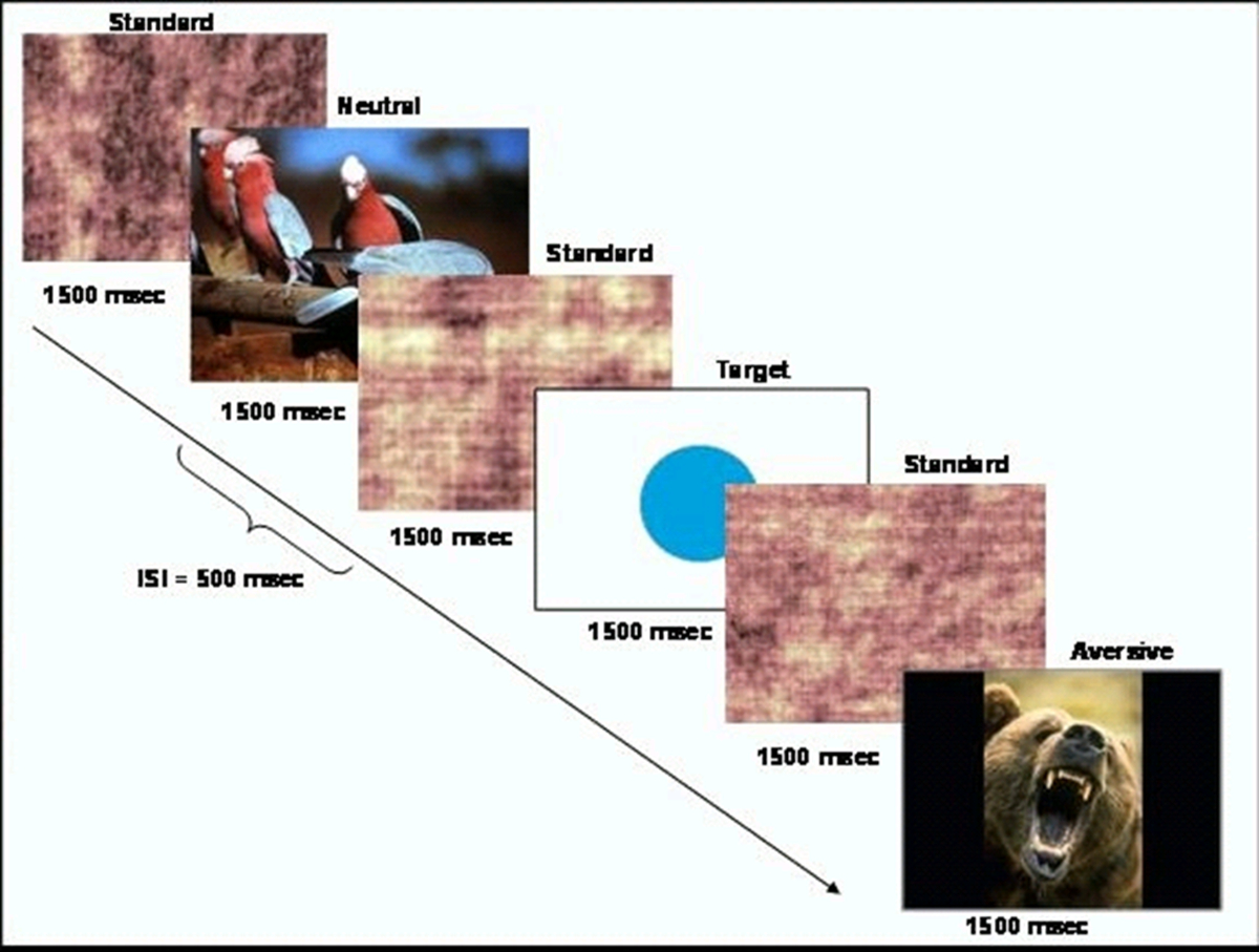
- צור משימה התנהגותית המבוסס על אירוע שמציגה גירויים נדירים יעד (עיגול) בתוך רצף של גירויים סטנדרטיים יותר תכופים (תמונות מקושקשות). סכמטי של המשימה מוצג באיור 1. הצג את המשימה באמצעות תוכנת CIGAL 10.

איור 1. סכמטי של משימות עיצוב. נתון זה שונה מהארט et al. 20, באישור. אנא לחץ כאן כדי לצפות בגרסה גדולה יותר של דמות זו.
- בחר כאח של גירויים מרתיעים וסט של גירויים ניטראליים מבסיס נתוני מערכת תמונה הרגשית הבינלאומית (IAPS). תמונות IAPS מדורגות בסולם של 1-9, על מנת לשקף את הרמות של עוררות וערכיות 11. מספרים גבוהים מצביעים ערכיות חיוביות גבוה יותר והתעוררות. בחר סט של תמונות שמתאימות לגיל לקבוצת המחקר כגון תמונות של נחשים, עכבישים, או בעלי חיים אחרים.
הערה: תמונות גירויים מרתיעים המשימה-רלוונטית המשמשות למחקר זה היה דירוג ממוצע של 3.38 ערכיות (SD = 1.78) ודירוג ממוצע של 6.14 עוררות (SD = 2.08). היו לי תמונות גירויים ניטרליות ערכיות ממוצעת של 6.21 (SD = 0.26) ודירוג ממוצע של 3.72 עוררות (SD = 2.15). - תכנית תסריט המשימה כזו שתמונות מוצגות בצו-אקראי מדומה ל1,500 מילים-שני עם 500 אלפיות שניים אומרת מרווח בין גירוי. גירויי הווה יעד ותמונות ניטרליות משימה-לא רלוונטית לא בתדירות גבוהה יותר מאשר בכל 15 שניות ולעשות כל כ -4% מהגירויים. ג'יפעמים תחילת אירוע tter על מנת לספק רזולוציה טובה יותר של פונקצית התגובה המודינמית.
- צור 8 סטים של תמונות, אחת לכל אחת מריצות 8 פונקציונליות כגון שהמשתתפים מוצגים עם סך של 40 מטרות ו -40 תמונות ניטרליות משימה-לא רלוונטית במהלך כל 8 הריצות.
2. הגדרת משתתף וסריקה
- לגייס ילדים ובני נוער בין גילאי 9 ו -18 שהם או אנשים בקרה בריאים או שנמצאים בסיכון גבוה למשפחתי פסיכוזה.
- ודא שיש לי אנשים בריאים אין מחלות פסיכיאטריות או כל בני משפחה מדרגה ראשונה עם מחלה פסיכיאטרית. ודא שיש לי משתתפי סיכון משפחתיים יחסי לפחות אחד מהדרגה הראשונה (הורה או אח) עם סכיזופרניה. לא לכלול אותם לנוכחות של מחלות פסיכיאטריות אחרות בקרובים מהדרגה הראשונה.
- משתתפים בריאים גיל ומין משחק עם משתתף קבוצה משפחתי סיכוןים.
- לרכוש הסכמה מדעת של משתתפים מעל גיל 18. לקטינים, לרכוש הסכמה מדעת מהורים / אפוטרופוסים חוקיים. בנוסף, תרכוש הסכמה בכתב מקטינים שהם לוקחים חלק במחקר.
- מניחים את המשתתפים בסורק MRI מדומה כדי קראתי אותם עם הסביבה. שחק הקלטת שמע רעש של הסורק ובהם להשלים ריצה בפועל של המשימה התנהגותיות כדי להבטיח שהם מבינים את הוראות המשימה.
- מניחים את המשתתף בסורק MRI ולרכוש כל סריקות המוח הכרחיות לוקליזציה ו / או תמונות אנטומיים.
- שימוש בתיבת קלט MRI-בטוח, לספר משתתפים ללחוץ על כפתור אחד עם האצבע שלהם בתגובה לכל גירויי היעד ועוד כפתור עם האצבע האמצעית שלהם לכל גירויים האחרים.
- בעקבות סריקת fMRI, לאסוף דירוגים סובייקטיבית של עוררות וערכיות לתמונות השתמשו במחקר מקבוצת משנה של משתתפים. Currenמחקר לא השיג דירוגים מ -15 בקרות ועם 13 משפחתי סיכון גבוה.
3. תמונת רכישה
- משתתפי מקום לסורק MRI טסלה 3.0. ראשית, לרכוש סט של תמונות מבניות כולל 3D coplanar תמונת T1-ניגוד אנטומיים באמצעות רצף דופק מפונק-נזכר שיפוע רכישה (TR: 5.16 אלפיות השני; TE: 2.04 אלפיות השני; FOV: 24 סנטימטר; מטריצת תמונה: 256 × 256; Flip זווית: 20; גודל voxel: 0.94 מ"מ × 0.94 מ"מ × 1.9 מ"מ; 68 פרוסות ציריות).
- רוכשים את נתוני הדמיה תפקודיות באמצעות רצף הדמיה שיפוע הד הד מישורי עם כיסוי מלא של מוח (TR: 2,000 אלפיות השני; TE: 27 אלפיות שני; FOV: 24 סנטימטר; מטריצת תמונה: 64 × 64; Flip זווית: 60; גודל voxel: 3.75 מ"מ × 3.75 מ"מ × 3.8 מ"מ; 34 פרוסות ציריות) כך שפעילות המוח ניתן למדוד בזמן ההופעה של המשימה התנהגותיות. הפעל רצף הדמיה זו לכל ריצה של המשימה התנהגותיות. כל ריצה צריכה להיות מורכבת של 120 נקודות זמן הדמיה.
- להציג את ת"אSK ב 8 ריצות פונקציונליות, כל שנמשך כ -4 דקות.
4. ניתוח
- עיבוד מקדים תמונה: להרחיב fMRI מומחה כלי ניתוח (feat) ב -12 FSL. בחר ניתוח ברמה הראשונה וטרום-סטטיסטי.
- בכרטיסייה "נתונים", בחר את מספר תמונות קלט והזן את הנתיב לכל אחת מתמונות MR אתה הולך לעבד. הגדר את ספריית הפלט. הזן את סה"כ הכרכים, מספר הרכישות שהושלכו, וTR.
- בכרטיסייה "פרה-סטטיסטי", קבע תיקון תנועה לMCFLIRT, מרחבי החלקת FWHM 5 MMS, ו" תיקון עיתוי Slice ". בחר באפשרות" חילוץ BET מוח "וסינון זמני" Highpass "אך לא תבחר unwarping B0 (uness יש לך מפת שדה שיפוע) או" אני ntensitiy normaization ". 12,14.
- בכרטיסייה "רישום", בחר "stru ראשיתמונת ctural ". הזן את הנתיב לתמונה משוקלל T1-הפשיט את גולגולתו של הנושא. השתמש בחיפוש רגיל ליניארי עם לפחות 6 DOF. בחר בתיבת סימון השטח רגילה. הזן את הנתיב לתמונת אטלס משרד התשתיות הלאומיות. השתמש נורמלי, ליניארי חיפוש עם 12 DOF. לחץ על בצע.
- תכלול משתתפים עם תנועה גדולה יותר מ -3 מ"מ הראש בX, Y, Z או כיוונים.
- רמת 1: השוואת נתונים בין תנאי משימה בתוך טווח אחת. FEAT הפתוח. בחר "ניתוח ברמה הראשונה" ו- "סטטיסטיקה + פוסט-סטטיסטי".
- בכרטיסייה נתונים, להגדיר את מספר הכניסות והזן את הנתיב לכל אחת מתמונות MR. הזן את נתיב עבור "ספריית הפלט". הזן "סה"כ הכרכים", מספר הרכישות שהושלכו, וTR.
- בכרטיסייה "סטטיסטיקה", בחר את "להשתמש בסרט prewhitening" תיבת סימון 16. לחץ על "המודל מלא seכפתור איל ". הגדר את "מספר של כלי רכב חשמליים המקוריים" למספר תנאי משימה. עבור כל מצב, בחר "(פורמט 3 טור) מותאמים אישית" מתפריט הצורה הבסיסית הנפתח ו" פעמיים גאמה HRF מ" "התפריט הנפתח פיתול 17,18 ובחר קובץ טקסט המכיל את עיתוי המשימה.
- פורמט קובץ טקסט זה ב 3 עמודים בכניסה אחת לכל "אירוע" מהסוג המסוים. העמודה הראשונה צריכה להכיל את זמן תחילת (בשניות), השני צריך להכיל את המשך (בשניות), ושלישית צריך להכיל את משקל האירוע. בכרטיסייה הניגודים & F-בדיקות, ליצור ניגוד אחד לכל מצב משימה ואחד לכל השוואה.
- על "פוסט-הסטטיסטיים" כרטיסייה, בחר "האשכול" בתפריט "ערכי סף" הנפתח ולהגדיר את "סף Z" ולא P אשכולhreshold 2.3 ו 0.05 בהתאמה 8,19.
- בכרטיסייה "רישום", בחר "תמונה מבנית עיקרית". הזן את הנתיב לתמונה משוקלל T1-הפשיט את גולגולתו של הנושא. השתמש בחיפוש רגיל ליניארי עם לפחות 6 DOF. בחר בתיבת הסימון "המרחב רגילה". הזן את הנתיב לתמונת אטלס משרד התשתיות הלאומיות. השתמש בחיפוש רגיל, ליניארי עם עומק שדה 12. לחץ על "Go".
- רמה 2: השוואת נתונים בין ריצות לכל מצב משימה. FEAT הפתוח. בחר "ניתוח ברמה גבוהה" ו- "סטטיסטיקה + פוסט-סטטיסטי" מתפריט הנפתח.
- בכרטיסייה נתונים, בחר "תשומות מדריכים FEAT ברמה נמוכה יותר". הגדר את מספר הכניסות והזן את הנתיב לכל אחת מתמונות MR. הזן את נתיב עבור "ספריית הפלט".
- בכרטיסייה "סטטיסטיקה", לשנות את "השפעות מעורבות: FLAME1" תיבת בחירה ל" קבוע Effects ". לחץ על" דגם אשף הגדרה "הכפתור. בחר" ממוצע קבוצה אחת "ולחץ על הכפתור" תהליך ".
- בכרטיסייה "פוסט-סטטיסטי", בחר "אשכול" בתפריט "ערכי סף" הנפתח ולהגדיר את "סף Z" וסף "אשכול P" 2.3 ו 0.05 בהתאמה 8,19. לחץ על "Go".
- רמה 3: השוואת נתונים בין נושאים לכל מצב משימה בכל הריצות. FEAT הפתוח. בחר "ניתוח ברמה גבוהה" ו- "סטטיסטיקה + פוסט-סטטיסטי" מתפריט הנפתח.
- בכרטיסייה נתונים, בחר "תשומות הן 3D להתמודד תמונות מספריות Feat." הגדר את מספר הכניסות והזן את הנתיב לכל אחת מתמונות MR. הזן את נתיב עבור "ספריית הפלט".
- על T "סטטיסטיקה"ab, לחץ על "התקנת מודל מלאה". הגדר את מספר כלי הרכב החשמליים שווה למספר משתני קבוצה ומשתנים כגון קבוצת אבחון, גיל, מין, וכו 'הזינו את הערכים לכל נושא (קלט 1 - n קלט) עבור כל EV. אתה יכול להשתמש בחלון "הדבק" כדי להעתיק גיליון אלקטרוני של ערכים אלה.
- בכרטיסייה "הניגודים & F-בדיקות", מוסיף ניגוד לכל משתנה מבחן ולכל ניגוד (למשל, קבוצת אבחון). לכל אחד ממשתני בדיקה, להגדיר את הניגוד ידי בחירת הערך 1 בעמודה תחת EV המתאים. לכל לעומת זאת, לקבוע את הערך הראשון 1 והשני ל-1. בחר "בוצע".
- בכרטיסייה "פוסט-סטטיסטי", בחר "האשכול" בתפריט "ערכי סף" הנפתח ולהגדיר את "סף Z" וסף "אשכול P" 2.3 ו 0.05 בהתאמה 8,19 . לחץ על "Go".
Access restricted. Please log in or start a trial to view this content.
תוצאות
לא היו הבדלים בין קבוצות על בסיס מאפיינים דמוגרפיים 20. הנתונים מצביעים על כך שהתנהגות משימת איתור היעד היא ברמה מתאימה של קושי לילדים ובני נוער בגילאי 9-18. במחקר הנוכחי, בקרות זיהו 82.36% מיעדים (SD = 0.14), וקבוצת הסיכון המשפחתית זיהתה 76.8% מיעדים (SD = 0.17). שני הק?...
Access restricted. Please log in or start a trial to view this content.
Discussion
The modified emotional oddball paradigm in the current study has been shown to elicit differences in neural recruitment during selective attention and emotional processing in children and adolescents at risk for schizophrenia. While existing paradigms using the emotional oddball task have been used to investigate neural changes in adult populations with psychiatric illness8, the current paradigm may be particularly useful for measurement of vulnerability markers in younger age groups.
Access restricted. Please log in or start a trial to view this content.
Disclosures
Dr. Perkins is currently receiving grant support from Janssen, and is a consultant to Dainippon. In the past, Dr. Perkins is or has been on speaker's bureau for Eli Lilly and AstraZeneca. Dr. Perkins has previously received grants from AstraZeneca, Bristol-Myers Squibb, Otsuka, Eli Lilly, Janssen and Pfizer; and consulting and educational fees from AstraZeneca, Bristol-Myers Squibb, Eli Lilly, Janssen, Glaxo Smith Kline, Forest Labs, Pfizer and Shire. All other authors declare no biomedical financial interests or potential conflict of interest.
Acknowledgements
We thank Erin Douglas, Anna Evans, and Carolyn Bellion for their contributions to participant recruitment and clinical assessment. We also thank Michael Casp, Zoe Englander, Justin Woodlief, and James Carter for their contributions to data collection and analysis, and Robert M. Hamer for consultation on statistical analysis and editing of the manuscript. Finally, we thank the individuals and their families who participated in this study.
This study was supported by Conte center grant P50 MH064065 from the National Institute of Mental Health. Dr. Hart was supported by T32 HD040127 from the National Institute of Child Health and Human Development.
Access restricted. Please log in or start a trial to view this content.
Materials
| Name | Company | Catalog Number | Comments |
| 3T MRI scanner | GE | BIAC 3T scanner (replaced) |
References
- Kety, S. S., Rosenthal, D., Wender, P. H., Schulsinger, F. Mental illness in the biological and adoptive families of adpoted schizophrenics. Am J Psyc. 128, 302-306 (1971).
- Weinberger, D. R. Implications of normal brain development for the pathogenesis of schizophrenia. Arch Gen Psychia. 44, 660-669 (1987).
- Nuechterlein, K. H., Dawson, M. E. Information processing and attentional functioning in the developmental course of schizophrenic disorders. Schizophr Bul. 10, 160-203 (1984).
- Nuechterlein, K. H. The vulnerability/stress model of schizophrenic relapse: a longitudinal study. Acta Psychiatr Scand, Supp. 382, 58-64 Forthcoming.
- Keshavan, M. S. Premorbid cognitive deficits in young relatives of schizophrenia patients. Front Hum Neurosc. 3 (62), (2010).
- Kiehl, K. A., Liddle, P. F. An event-related functional magnetic resonance imaging study of an auditory oddball task in schizophrenia. Schizophr Re. 48, 159-171 (2001).
- Bramon, E. Is the P300 wave an endophenotype for schizophrenia? A meta-analysis and a family study. Neuroimag. 27, 960-968 (2005).
- Dichter, G. S., Bellion, C., Casp, M., Belger, A. Impaired modulation of attention and emotion in schizophrenia. Schizophr Bul. 36, 595-606 (2010).
- Fichtenholtz, H. M. Emotion-attention network interactions during a visual oddball task. Brain Res Cogn Brain Re. 20, 67-80 (2004).
- Voyvodic, J. T. Real-time fMRI paradigm control, physiology, and behavior combined with near real-time statistical analysis. Neuroimag. 10, 91-106 (1999).
- International affective picture system (IAPS): Digitized photographs, instruction manual and affective ratings. Technical Report A-6. , The Center for Research in Psychophysiology, University of Florida. (2005).
- Smith, S. M. Advances in functional and structural MR image analysis and implementation as FSL. Neuroimag. 23, 208-219 (2004).
- Smith, S. M. Fast robust automated brain extraction. Hum Brain Map. 17, 143-155 (2002).
- Jenkinson, M., Bannister, P., Brady, M., Smith, S. Improved optimization for the robust and accurate linear registration and motion correction of brain images. Neuroimag. 17, 825-841 (2002).
- Jenkinson, M., Smith, S. A global optimisation method for robust affine registration of brain images. Med Image Ana. 5, 143-156 (2001).
- Woolrich, M. W., Ripley, B. D., Brady, M., Smith, S. M. Temporal autocorrelation in univariate linear modeling of FMRI data. Neuroimag. 14, 1370-1386 (2001).
- Beckmann, C. F., Jenkinson, M., Smith, S. M. General multilevel linear modeling for group analysis in FMRI. Neuroimag. 20, 1052-1063 (2003).
- Woolrich, M. W., Behrens, T. E., Beckmann, C. F., Jenkinson, M., Smith, S. M. Multilevel linear modelling for FMRI group analysis using Bayesian inference. Neuroimag. 21, 1732-1747 (2004).
- Genovese, C. R., Lazar, N. A., Nichols, T. Thresholding of statistical maps in functional neuroimaging using the false discovery rate. Neuroimag. 15, 870-878 (2002).
- Hart, S. J. Altered fronto-limbic activity in children and adolescents with familial high risk for schizophrenia. Psychiatry Re. 212, 19-27 (2013).
- Hariri, A. R., Bookheimer, S. Y., Mazziotta, J. C. Modulating emotional responses: effects of a neocortical network on the limbic system. Neurorepor. 11, 43-48 (2000).
- Gottesman, I. I., Gould, T. D. The endophenotype concept in psychiatry: etymology and strategic intentions. Am J Psyc. 160, 636-645 (2003).
- Glahn, D. C., Thompson, P. M., Blangero, J. Neuroimaging endophenotypes: strategies for finding genes influencing brain structure and function. Hum Brain Map. 28, 488-501 (2007).
Access restricted. Please log in or start a trial to view this content.
Reprints and Permissions
Request permission to reuse the text or figures of this JoVE article
Request PermissionThis article has been published
Video Coming Soon
Copyright © 2025 MyJoVE Corporation. All rights reserved