
このコンテンツを視聴するには、JoVE 購読が必要です。 サインイン又は無料トライアルを申し込む。
Method Article
統合失調症のための家族ハイリスク児の感情オドボール課題を用いて前頭辺縁系の活性の測定
要約
This paper describes how to use the emotional oddball task and fMRI to measure brain activation in children and adolescents at familial high risk for schizophrenia (FHR). FMRI was used to measure differences in fronto-striato-limbic regions during an emotional oddball task. Children with FHR exhibited abnormal functional activation during adolescence.
要約
Adolescence is a critical developmental period where the early symptoms of schizophrenia frequently emerge. First-degree relatives of people with schizophrenia who are at familial high risk (FHR) may show similar cognitive and emotional changes. However, the neurological changes underlying the emergence of these symptoms remain unclear. This study sought to identify differences in frontal, striatal, and limbic regions in children and adolescents with FHR using functional magnetic resonance imaging. Groups of 21 children and adolescents at FHR and 21 healthy controls completed an emotional oddball task that relied on selective attention and the suppression of task-irrelevant emotional information. The standard oddball task was modified to include aversive and neutral distractors in order to examine potential group differences in both emotional and executive processing. This task was designed specifically to allow for children and adolescents to complete by keeping the difficulty and emotional image content age-appropriate. Furthermore, we demonstrate a technique for suitable fMRI registration for children and adolescent participants. This paradigm may also be applied in future studies to measure changes in neural activity in other populations with hypothesized developmental changes in executive and emotional processing.
概要
Schizophrenia is a neurodevelopmental disorder with a known genetic component1,2 and with symptoms including deficits in both executive and emotional processing3,4. First-degree relatives are thought to be at an increased risk of developing schizophrenia, and have been shown to share some of these same neurocognitive deficits in both cognitive and social-emotional domains5. We therefore expect that brain activity in regions associated with executive and emotional processing may be altered in at-risk family members preceding the onset of clinical symptoms.
Previous studies have indicated that both adults with schizophrenia and adults at familial high risk show aberrant activity within executive and emotional processing networks; however it remains unclear how these changes come about during development. Demonstrating that these changes occur early in life will be a critical first step in understanding the pathophysiology of the disorder. Therefore, this study utilizes an emotional oddball paradigm during functional MRI (fMRI) scanning in order to measure brain activity during the completion of a task that requires both executive and emotional processing in adolescents who are at risk for developing schizophrenia. Oddball paradigms are frequently used to examine the function of fronto-striate circuitry in schizophrenia6 and in individuals with familial high risk7 by measuring selective attention processes allocated to task-relevant target stimuli. Here, a standard oddball task has been modified to include task-irrelevant aversive and neutral stimuli that have been shown to elicit changes in brain activity in patients with schizophrenia8.
This paper measures functional differences between healthy adolescents and adolescents at high familial risk for schizophrenia using an emotional oddball task. The task design is similar to that used by Fichtenholtz and colleagues9, but the selection of aversive emotional images has been modified to be appropriate for children between the ages of 9-18. The use of this task during functional MRI allowed for the identification of specific brain regions that showed patterns of hyperactivation and hypoactivation in children and adolescents with FHR for schizophrenia, in addition to age-related changes in neural activity during adolescent development.
Access restricted. Please log in or start a trial to view this content.
プロトコル
チャペルヒル - この試験中に使用される研究技術は、デューク大学とノースカロライナ大学の機関審査委員会(IRB)により承認されました。
1.イメージングタスクデザイン
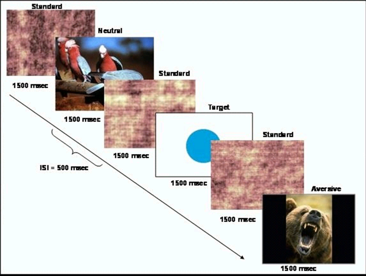
- より頻度の標準刺激(スクランブル画像)のシーケンス内のまれなターゲット刺激を提示し、イベントベースの行動のタスク(円)を生成します。タスクの概略を図1に示されている。CIGALソフトウェア10を使用してタスクを提示します。

タスク設計の図1の回路図は、この図は、許可を得て、ハートら 20から変更されている。 この図の拡大版をご覧になるにはこちらをクリックしてください。
- 選択嫌悪刺激のらと国際情動写真システムデータベース(IAPS)から中性刺激のセット。 IAPS画像は覚醒と価11のレベルを反映させるために1-9のスケールで評価されています。高い数値は、より高い正の価数と覚醒を示します。そのようなヘビ、クモ、または他の動物の絵のように年齢に応じた研究グループにある画像のセットを選択します。
注意:この研究のために使用されるタスク無関係な嫌悪刺激画像は3.38(SD = 1.78)および6.14(SD = 2.08)の平均覚醒評価の平均価数の評価を持っていました。中性刺激画像は6.21(SD = 0.26)および3.72(SD = 2.15)の平均覚醒評価の平均価数を持っていました。 - 画像は500ミリ秒と1500ミリ秒のための擬似ランダム化された順に表示されるように、プログラムのタスクスクリプトは、刺激間の間隔を意味します。現在の目標刺激とタスク無関係な中立画像なしより頻繁に15秒毎とは、刺激の各約4%を占めています。チ血行動態応答関数の良好な分解能を提供するためにtterイベント開始時間。
- 画像の8セット、参加者は全8回の過程で40の目標と40のタスク無関係な中立画像の合計が提示されるように、8の機能の実行ごとに1つを作成します。
2.参加者の設定とスキャン
- 精神病のための家族リスクが高い健康な対照個体またはいずれかである9歳と18との間に子供や青年を募集。
- 健康な個体は全く精神疾患や精神疾患を持つ任意の第一度家族を持っていないことを確認してください。家族のリスク参加者は統合失調症との少なくとも1つの第1度近親者(親や兄弟を)持っていることを確認してください。第一度近親における他の精神疾患の存在のためにそれらを除外しないでください。
- 家族リスクグループの参加者との年齢と性別が一致する健康参加秒。
- 未成年者のために18歳以上の参加者からインフォームドコンセントを取得し、親/法定保護者からインフォームドコンセントを取得してください。さらに、この研究に参加している未成年者からの書面による同意を取得します。
- 環境でそれらを理解するために、モックMRIスキャナの参加者を配置します。スキャナノイズの録音を再生し、彼らがタスクの手順を理解していることを確実にするために行動タスクの練習走行を完了しています。
- MRIスキャナの参加者を配置し、必要な脳のローカリゼーションスキャンおよび/または解剖学的画像を取得します。
- MRIセーフ入力ボックスを使用して、すべてのターゲット刺激とすべての他の刺激のための彼らの中指と別のボタンに応じて、その人差し指で1ボタンを押して、参加者に伝えます。
- fMRIのスキャンに続いて、参加者のサブセットからの研究に使用されている画像のための覚醒と価電子の主観的な評価を集めます。カレントンの研究では、15コントロールと家族リスクの高い13から評価を得ました。
3.画像取得
- 3.0テスラのMRIスキャナに参加者を配置します。 TE; 5.16ミリ秒:2.04ミリ秒; FOV:まず、(TRスポイル勾配リコール取得パルスシーケンスを使用して、解剖学的T1コントラスト画像に同一平面上に3Dを含む構造画像のセット取得24センチメートルを、画像マトリクス:256×256を、フリップ角度:20;ボクセルサイズ:1.9ミリメートル×0.94ミリメートル×0.94ミリメートル; 68の軸方向のスライス)。
- フル脳のカバレッジと傾斜エコーエコープラナーイメージングシーケンスを使用して、機能画像データを取得し(TR:2000ミリ秒、TE:27ミリ秒; FOV:24センチメートル;画像マトリクス:64×64;フリップ角:60;ボクセルサイズ:3.75脳活動は、行動のタスクの実行中に測定することができるように34の軸方向スライス); MMは3.8ミリメートル×3.75ミリメートル×。行動のタスクを実行するたびにこの撮影シーケンスを実行します。各ランは120撮影時間点で構成する必要があります。
- TAを提示SKは8機能の実行で、それぞれが約4分を持続します。
4.分析
- 画像前処理:FSL 12で開くfMRIのエキスパート解析ツール(FEAT)。第1レベルの分析と事前統計を選択します。
- 「データ」タブで、入力画像の数を選択し、処理しようとしているMR画像のそれぞれへのパスを入力します。出力ディレクトリを設定します。 トータルボリューム、廃棄された買収の数、およびTRを入力してください。
- 「前の統計情報 」タブで、5 MMSにFWHMを平滑MCFLIRT、空間に動き補正を設定し、 そして、「スライスタイミング補正」。「ベット脳抽出」と「 ハイパス 」時間フィルタリングを選択しますが、B0のunwarpingを選択しない(あなたは傾斜磁場マップを持っているuness)または「私はnormaizationをntensitiy」。12,14。
- 「登録」タブで、「メインSTRUを選択ctural画像」。。被験者の頭蓋骨ストリッピングT1強調画像へのパスを入力し、少なくとも6自由度で線形通常の検索を使用します。 標準領域のチェックボックスをオンにします。MNIアトラスイメージへのパスを入力します。通常の線形を使用12 DOF。押し移動して検索します。
- X、Y、またはZ方向に3mmより大きいヘッドの動きに参加者を除外する。
- レベル1:単一のランに作業条件との間でデータを比較してください。オープンFEAT。 「第1レベルの分析」と「統計+ポスト統計」を選択します。
- [データ]タブで 、入力の数を設定し、MR画像のそれぞれへのパスを入力します。 。「出力ディレクトリ」のパスを入力して「トータルボリューム」、廃棄された買収の数、およびTRを入力してください。
- 「 統計 」タブで、「使用フィルムはプリホワイトニング 」のチェックボックス16を選択します。 「フルモデルSEを押しますTUP」ボタンを押します。タスク条件の数に「 元の電気自動車の数」に設定します。各条件について、 コンボリューション 」ドロップダウン・メニュー17,18」からダブルガマHRF「基本形状]ドロップダウンメニューから「 カスタム(3列形式)」を選択し、タスクのタイミングを含むテキストファイルを選択します。
- 指定されたタイプの各「イベント」ごとに1つのエントリを持つ3列に、このテキストファイルをフォーマットします。最初の列は、第二の時間(秒単位)が含まれている必要があり(秒)開始時間が含まれている必要があり、3番目は、イベントの重みを含める必要があります。 コントラスト&F検定]タブで 、各比較のために、各タスクの状態と1のための1つのコントラストを作成します。
- 「ポスト・統計」タブで、「しきい」ドロップダウン・メニューの 「クラスタ」を選択し、「Zしきい値」とクラスタP Tを設定それぞれ2.3および0.05 8,19にhreshold。
- 「登録」タブで、「メイン構造画像」を選択します。被験者の頭蓋骨ストリッピングT1強調画像へのパスを入力します。少なくとも6自由度で線形通常の検索を使用します。 「標準容量」チェックボックスを選択します。 MNIアトラスイメージへのパスを入力します。 12自由度ノーマル、線形検索を使用します。押し、「移動」。
- レベル2:各タスク条件の実行間のデータを比較してください。オープンFEAT。ドロップダウンメニューから「より高いレベルの分析」と「統計+ポスト統計」を選択します。
- [データ]タブで、「入力が低レベルのFEATのディレクトリです 」を選択します。入力数を設定し、MR画像のそれぞれへのパスを入力します。 「出力ディレクトリ」のパスを入力します。
- 選択ボックス"Eを修正するには:「統計」タブで、「FLAME1は混合効果」に変更ffects「押し」モデルセットアップウィザード単一のグループの平均を"ボタンを押します。選択」」をクリックして「プロセス」ボタンをクリックします。
- 「ポスト・統計 」タブで、「しきい 」ドロップダウン・メニューの 「クラスタ」を選択し、8,19はそれぞれ2.3と0.05に「Zしきい値 」と「クラスタP」のしきい値を設定します。押し、「移動」。
- レベル3:すべての実行間で、各タスクの状態のための科目間のデータを比較してください。オープンFEAT。ドロップダウンメニューから「より高いレベルの分析 」と「 統計+ポスト統計」を選択します。
- [データ]タブで、[入力の数を設定し、MR画像のそれぞれへのパスを入力し、「 入力がFEATディレクトリから画像を対応させていただきます。3Dです 」。 「出力ディレクトリ 」のパスを入力します。
- 「統計」T ONAB、押して「フルモデルのセットアップ」。各EVのために - このような診断等のグループ、年齢、性別、各被験者の値(入力■入力1)を入力し、グループ変数と共変量の数に等しい電気自動車の数を設定します。あなたはこれらの値のスプレッドシートをコピーするために「貼り付け」ウィンドウを使用することができます。
- 「コントラスト&F検定 」タブで、各テスト変数と各コントラスト( 例えば、診断グループ)のコントラストを追加します。各試験変数については、該当するEVの下欄に値1を選択することにより、コントラストを設定します。各コントラストのために、1〜-1の第1の値と第2のセット。 「完了」を選択します。
- 「ポスト・統計 」タブで、「しきい」ドロップダウン・メニューの 「クラスタ」を選択し、2.3と0.05に「Zしきい値 」と「クラスタP」のしきい値を設定し、それぞれ8,19 。押し、「移動」。
Access restricted. Please log in or start a trial to view this content.
結果
人口統計学的特性20に基づいてグループ間の差は認められませんでした。行動データは、ターゲット検出タスクは9-18歳の小児および青年のための難しさの適切なレベルであることを示しました。現在の研究では、正しくターゲット(SD = 0.14)の82.36パーセントを同定し、および家族のリスクグループは、正しくターゲット(SD = 0.17)の76.8パーセントを同定し制御します?...
Access restricted. Please log in or start a trial to view this content.
ディスカッション
The modified emotional oddball paradigm in the current study has been shown to elicit differences in neural recruitment during selective attention and emotional processing in children and adolescents at risk for schizophrenia. While existing paradigms using the emotional oddball task have been used to investigate neural changes in adult populations with psychiatric illness8, the current paradigm may be particularly useful for measurement of vulnerability markers in younger age groups.
Access restricted. Please log in or start a trial to view this content.
開示事項
Dr. Perkins is currently receiving grant support from Janssen, and is a consultant to Dainippon. In the past, Dr. Perkins is or has been on speaker's bureau for Eli Lilly and AstraZeneca. Dr. Perkins has previously received grants from AstraZeneca, Bristol-Myers Squibb, Otsuka, Eli Lilly, Janssen and Pfizer; and consulting and educational fees from AstraZeneca, Bristol-Myers Squibb, Eli Lilly, Janssen, Glaxo Smith Kline, Forest Labs, Pfizer and Shire. All other authors declare no biomedical financial interests or potential conflict of interest.
謝辞
We thank Erin Douglas, Anna Evans, and Carolyn Bellion for their contributions to participant recruitment and clinical assessment. We also thank Michael Casp, Zoe Englander, Justin Woodlief, and James Carter for their contributions to data collection and analysis, and Robert M. Hamer for consultation on statistical analysis and editing of the manuscript. Finally, we thank the individuals and their families who participated in this study.
This study was supported by Conte center grant P50 MH064065 from the National Institute of Mental Health. Dr. Hart was supported by T32 HD040127 from the National Institute of Child Health and Human Development.
Access restricted. Please log in or start a trial to view this content.
資料
| Name | Company | Catalog Number | Comments |
| 3T MRI scanner | GE | BIAC 3T scanner (replaced) |
参考文献
- Kety, S. S., Rosenthal, D., Wender, P. H., Schulsinger, F. Mental illness in the biological and adoptive families of adpoted schizophrenics. Am J Psyc. 128, 302-306 (1971).
- Weinberger, D. R. Implications of normal brain development for the pathogenesis of schizophrenia. Arch Gen Psychia. 44, 660-669 (1987).
- Nuechterlein, K. H., Dawson, M. E. Information processing and attentional functioning in the developmental course of schizophrenic disorders. Schizophr Bul. 10, 160-203 (1984).
- Nuechterlein, K. H. The vulnerability/stress model of schizophrenic relapse: a longitudinal study. Acta Psychiatr Scand, Supp. 382, 58-64 Forthcoming.
- Keshavan, M. S. Premorbid cognitive deficits in young relatives of schizophrenia patients. Front Hum Neurosc. 3 (62), (2010).
- Kiehl, K. A., Liddle, P. F. An event-related functional magnetic resonance imaging study of an auditory oddball task in schizophrenia. Schizophr Re. 48, 159-171 (2001).
- Bramon, E. Is the P300 wave an endophenotype for schizophrenia? A meta-analysis and a family study. Neuroimag. 27, 960-968 (2005).
- Dichter, G. S., Bellion, C., Casp, M., Belger, A. Impaired modulation of attention and emotion in schizophrenia. Schizophr Bul. 36, 595-606 (2010).
- Fichtenholtz, H. M. Emotion-attention network interactions during a visual oddball task. Brain Res Cogn Brain Re. 20, 67-80 (2004).
- Voyvodic, J. T. Real-time fMRI paradigm control, physiology, and behavior combined with near real-time statistical analysis. Neuroimag. 10, 91-106 (1999).
- International affective picture system (IAPS): Digitized photographs, instruction manual and affective ratings. Technical Report A-6. , The Center for Research in Psychophysiology, University of Florida. (2005).
- Smith, S. M. Advances in functional and structural MR image analysis and implementation as FSL. Neuroimag. 23, 208-219 (2004).
- Smith, S. M. Fast robust automated brain extraction. Hum Brain Map. 17, 143-155 (2002).
- Jenkinson, M., Bannister, P., Brady, M., Smith, S. Improved optimization for the robust and accurate linear registration and motion correction of brain images. Neuroimag. 17, 825-841 (2002).
- Jenkinson, M., Smith, S. A global optimisation method for robust affine registration of brain images. Med Image Ana. 5, 143-156 (2001).
- Woolrich, M. W., Ripley, B. D., Brady, M., Smith, S. M. Temporal autocorrelation in univariate linear modeling of FMRI data. Neuroimag. 14, 1370-1386 (2001).
- Beckmann, C. F., Jenkinson, M., Smith, S. M. General multilevel linear modeling for group analysis in FMRI. Neuroimag. 20, 1052-1063 (2003).
- Woolrich, M. W., Behrens, T. E., Beckmann, C. F., Jenkinson, M., Smith, S. M. Multilevel linear modelling for FMRI group analysis using Bayesian inference. Neuroimag. 21, 1732-1747 (2004).
- Genovese, C. R., Lazar, N. A., Nichols, T. Thresholding of statistical maps in functional neuroimaging using the false discovery rate. Neuroimag. 15, 870-878 (2002).
- Hart, S. J. Altered fronto-limbic activity in children and adolescents with familial high risk for schizophrenia. Psychiatry Re. 212, 19-27 (2013).
- Hariri, A. R., Bookheimer, S. Y., Mazziotta, J. C. Modulating emotional responses: effects of a neocortical network on the limbic system. Neurorepor. 11, 43-48 (2000).
- Gottesman, I. I., Gould, T. D. The endophenotype concept in psychiatry: etymology and strategic intentions. Am J Psyc. 160, 636-645 (2003).
- Glahn, D. C., Thompson, P. M., Blangero, J. Neuroimaging endophenotypes: strategies for finding genes influencing brain structure and function. Hum Brain Map. 28, 488-501 (2007).
Access restricted. Please log in or start a trial to view this content.
転載および許可
このJoVE論文のテキスト又は図を再利用するための許可を申請します
許可を申請さらに記事を探す
This article has been published
Video Coming Soon
Copyright © 2023 MyJoVE Corporation. All rights reserved