
JoVE 비디오를 활용하시려면 도서관을 통한 기관 구독이 필요합니다. 전체 비디오를 보시려면 로그인하거나 무료 트라이얼을 시작하세요.
Method Article
정신 분열증에 대한 가족 성 위험이 높은 아동의 정서적 괴짜 작업을 사용하여 이마 관자 - 변연계의 활동의 측정
요약
This paper describes how to use the emotional oddball task and fMRI to measure brain activation in children and adolescents at familial high risk for schizophrenia (FHR). FMRI was used to measure differences in fronto-striato-limbic regions during an emotional oddball task. Children with FHR exhibited abnormal functional activation during adolescence.
초록
Adolescence is a critical developmental period where the early symptoms of schizophrenia frequently emerge. First-degree relatives of people with schizophrenia who are at familial high risk (FHR) may show similar cognitive and emotional changes. However, the neurological changes underlying the emergence of these symptoms remain unclear. This study sought to identify differences in frontal, striatal, and limbic regions in children and adolescents with FHR using functional magnetic resonance imaging. Groups of 21 children and adolescents at FHR and 21 healthy controls completed an emotional oddball task that relied on selective attention and the suppression of task-irrelevant emotional information. The standard oddball task was modified to include aversive and neutral distractors in order to examine potential group differences in both emotional and executive processing. This task was designed specifically to allow for children and adolescents to complete by keeping the difficulty and emotional image content age-appropriate. Furthermore, we demonstrate a technique for suitable fMRI registration for children and adolescent participants. This paradigm may also be applied in future studies to measure changes in neural activity in other populations with hypothesized developmental changes in executive and emotional processing.
서문
Schizophrenia is a neurodevelopmental disorder with a known genetic component1,2 and with symptoms including deficits in both executive and emotional processing3,4. First-degree relatives are thought to be at an increased risk of developing schizophrenia, and have been shown to share some of these same neurocognitive deficits in both cognitive and social-emotional domains5. We therefore expect that brain activity in regions associated with executive and emotional processing may be altered in at-risk family members preceding the onset of clinical symptoms.
Previous studies have indicated that both adults with schizophrenia and adults at familial high risk show aberrant activity within executive and emotional processing networks; however it remains unclear how these changes come about during development. Demonstrating that these changes occur early in life will be a critical first step in understanding the pathophysiology of the disorder. Therefore, this study utilizes an emotional oddball paradigm during functional MRI (fMRI) scanning in order to measure brain activity during the completion of a task that requires both executive and emotional processing in adolescents who are at risk for developing schizophrenia. Oddball paradigms are frequently used to examine the function of fronto-striate circuitry in schizophrenia6 and in individuals with familial high risk7 by measuring selective attention processes allocated to task-relevant target stimuli. Here, a standard oddball task has been modified to include task-irrelevant aversive and neutral stimuli that have been shown to elicit changes in brain activity in patients with schizophrenia8.
This paper measures functional differences between healthy adolescents and adolescents at high familial risk for schizophrenia using an emotional oddball task. The task design is similar to that used by Fichtenholtz and colleagues9, but the selection of aversive emotional images has been modified to be appropriate for children between the ages of 9-18. The use of this task during functional MRI allowed for the identification of specific brain regions that showed patterns of hyperactivation and hypoactivation in children and adolescents with FHR for schizophrenia, in addition to age-related changes in neural activity during adolescent development.
Access restricted. Please log in or start a trial to view this content.
프로토콜
채플 힐 -이 연구 기간 동안 사용 된 연구 기술은 제도적 검토 보드 듀크 대학 (IRB)와 노스 캐롤라이나 대학에 의해 승인되었다.
1. 이미징 작업 디자인
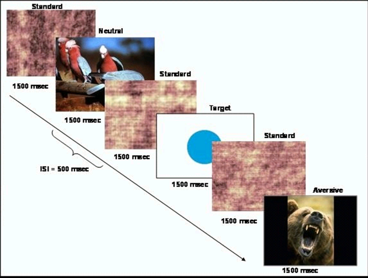
- 더 - 자주 표준 자극 (스크램블 이미지)의 순서 내에서 드문 대상 자극 (원)을 제시 이벤트 기반 행동 작업을 생성합니다. 태스크의 개략도는도 1에 도시된다. CIGAL 소프트웨어 (10)를 이용하여 작업을 제시한다.

작업 디자인의 그림 1. 도식은.이 그림은 허가, 하트 등. (20)에서 수정되었습니다. 이 그림의 더 큰 버전을 보려면 여기를 클릭하십시오.
- 등의 선택혐오 자극 등 국제 정서 그림 시스템 데이터베이스 (IAPS)에서 중립 자극의 집합. IAPS 이미지는 각성 및 가전 (11)의 수준을 반영하기 위해 1-9의 규모로 평가된다. 높은 숫자는 더 높은 긍정적 인 원자가과 각성을 나타냅니다. 뱀, 거미, 또는 다른 동물의 사진으로 연령에 적합한 연구 그룹에있는 이미지의 집합을 선택합니다.
참고 : 본 연구에 사용 된 태스크 관련이없는 혐오 자극 이미지는 3.38 (SD = 1.78)와 6.14 (SD = 2.08)의 평균 각성 등급의 평균 원자가 평가했다. 중립 자극 이미지는 6.21 (SD = 0.26)와 3.72 (SD = 2.15)의 평균 각성 등급의 평균 원자가 있었다. - 이미지는 500 밀리 1500 밀리 초 동안 의사 무작위 순서로 표시되도록 프로그램 태스크 스크립트 자극 간 간격을 의미한다. 현재 목표 자극과 태스크 관련이없는 중립적 인 이미지를 더 자주보다 매 15 초와는 자극의 각각의 약 4 %를합니다. 지혈역학 응답 함수의 더 나은 해상도를 제공하기 위하여 라 자세 히 이벤트 개시 시간.
- 이미지의 8 세트, 참가자 모두 8 실행의 과정을 통해 40 목표와 40 태스크 관련이없는 중립적 인 이미지의 총되게됩니다 있도록 8 기능 실행의 각 하나를 만듭니다.
2. 참가자 설정 및 스캔
- 정신병에 대한 가족의 위험이 높은 건강한 제어 개인 또는 중 하나 인 9 세에서 18 사이의 어린이와 청소년을 모집.
- 건강한 사람이 정신 질환을 앓고있는 어떤 정신 질환 또는 일급 가족이 없는지 확인하십시오. 가족 위험 참가자가 정신 분열증에 적어도 하나의 제 1도 상대 (부모 또는 형제 자매)가 있는지 확인합니다. 먼저도 친척의 다른 정신 질환의 존재를 배제하지 마십시오.
- 가족 위험 그룹 참가자와 나이와 성별이 일치 건강한 참가자에스.
- 미성년자의 경우 18 세 이상 참가자의 동의를 획득 부모 / 법적 보호자로부터 동의를 획득. 또한, 연구에 참여하고있다 미성년자의 서면 동의를 획득.
- 환경을 숙지하기 위해 모의 MRI 스캐너에서 참가자를 놓습니다. 스캐너 노이즈의 오디오 녹음을 재생하고 그들이 작업 지침을 이해했는지 확인하기 위해 행동 작업의 연습 실행을 완료해야합니다.
- MRI 스캐너의 참가자를 놓고 필요한 뇌 현지화 검사 및 / 또는 해부학 적 영상을 획득.
- MRI 안전 입력 상자를 사용하여 모든 대상 자극과 다른 자극에 대한 자신의 가운데 손가락에 다른 버튼에 대한 응답으로 자신의 검지 손가락으로 하나의 버튼을 눌러 참가자를 말한다.
- fMRI를 스캔 후, 참가자의 하위 집합에서 연구에 사용 된 이미지에 대한 각성과 원자가의 주관적 평가를 수집합니다. CURRENT 연구는 15 컨트롤과 가족 위험이 높은 13 등급을 획득했습니다.
3. 이미지 인식
- 3.0 테슬라 MRI 스캐너에 배치 할 참가자. TE, 5.16 밀리 초 : 2.04 밀리 초, FOV : 첫째, (TR 버릇 그라데이션 리콜 획득 펄스 시퀀스를 사용하여 해부학 T1 대비 이미지 동일 평면 3D를 포함한 구조 이미지 세트 취득 24cm를, 이미지 매트릭스 : 256 × 256, 플립 각도 : 20; 복셀의 크기 : 0.94 mm × 0.94 mm mm 1.9 × 68 축 조각).
- 전체 뇌 따르면 그라데이션 에코 에코 평면 촬상 시퀀스를 이용하여 기능성 촬상 데이터를 취득 (TR : 2,000 밀리; TE : 27 밀리; FOV : 24cm, 화상 매트릭스 : 64 × 64; 각도 플립 : 60; 복셀의 크기 : 3.75 mm 3.8 mm × 3.75 mm × 34 축선 슬라이스)되도록 뇌 활성 행동 태스크의 수행 동안 측정 될 수있다. 행동 작업의 각 실행이 영상 시퀀스를 실행합니다. 각각의 실행은 120 이미징 시점으로 구성해야합니다.
- 타 제시SK는 8 기능 실행에서, 각각 약 4 분 동안 지속.
4. 분석
- 이미지 전처리 : FSL 12에서 열기의 fMRI 전문가 분석 도구 (FEAT). 첫 번째 수준의 분석 및 사전 통계를 선택합니다.
- "데이터"탭에서, 입력 이미지의 수를 선택하고 처리하려고하는 자기 공명 영상의 각각의 경로를 입력합니다. 출력 디렉토리를 설정합니다. 전체 볼륨, 폐기 인수의 수와 TR을 입력합니다.
- "사전 통계"탭에서, 5 MMS에 MCFLIRT, FWHM을 부드럽게 공간에 모션 보정을 설정 와 "슬라이스 타이밍 보정". "BET 뇌 추출"와 "하이 패스"시간 필터링을 선택하지만 B0의 뒤 틀어짐 방지를 선택하지 (당신이 그라데이션 필드 맵이 uness) 또는 "나는 normaization을 ntensitiy."(12, 14).
- "등록"탭에서 "메인 STRU을 선택ctural 이미지 ".. 피사체의 두개골 제거 T1 강조 영상의 경로를 입력 최소 6 DOF와 선형 일반 검색을 사용하십시오. 표준 공간 확인란을 선택합니다. MNI 아틀라스 이미지의 경로를 입력합니다. 보통의 선형을 사용하여 12 DOF.를 눌러 이동하여 검색 할 수 있습니다.
- X, Y보다 큰 3mm 머리의 움직임, 또는 Z 방향으로 참가자를 제외합니다.
- 레벨 1 : 단일의 런 작업 조건 사이의 데이터를 비교. 열기 FEAT. "첫 번째 수준의 분석"과 "통계 + 후 통계"를 선택합니다.
- 데이터 탭에서의 입력 번호를 설정하고 MR 이미지의 각각의 경로를 입력한다. . "출력 디렉토리"에 대한 경로를 입력 "전체 볼륨", 폐기 인수의 수와 TR을 입력합니다.
- "통계"탭에서 "사용 필름은 사전 백색 화"체크 상자 (16)을 선택합니다. "전체 모델을 누르면 그 자체TUP "버튼을 누릅니다. 작업 조건의 수에 "원래의 전기차 번호"를 설정한다. 각 조건의 경우, 회선 "드롭 다운 메뉴 17, 18"에서 두 번 가마 HRF "기본 모양의 드롭 다운 메뉴에서"사용자 지정 (3 열 형식) "을 선택하고 작업시기를 포함하는 텍스트 파일을 선택합니다.
- 주어진 유형의 각 "이벤트"에 대한 하나의 항목으로 3 열에서이 텍스트 파일을 포맷합니다. 첫 번째 열은 두 번째 시간 (초) 지속 기간을 포함한다 (초) 시작 시간을 포함해야하고, 세 번째 이벤트 중량을 함유한다. 대조 및 F-테스트 탭에서 한 각 작업 조건에 대한 대비와 각각 비교를 만듭니다.
- "후 통계"탭을 선택하고 '형 임계 값 "드롭 다운 메뉴에서"클러스터 "및"Z 임계 값 "및 클러스터 P (T)를 설정각각 2.3과 0.05 8,19에 hreshold.
- "등록"탭에서. "기본 구조 이미지"를 선택 피사체의 두개골 제거 T1 강조 영상의 경로를 입력합니다. 최소 6 DOF와 선형 일반 검색을 사용합니다. "표준 공간"확인란을 선택합니다. MNI 아틀라스 이미지의 경로를 입력합니다. 12 DOF와 정상, 선형 검색을 사용합니다. 를 눌러 "이동".
- 2 단계 : 각 작업 조건에 대해 실행 사이의 데이터를 비교. 열기 FEAT. 드롭 다운 메뉴에서 "높은 수준의 분석"과 "통계 + 후 통계"를 선택합니다.
- 데이터 탭에서 "입력은 낮은 수준의 묘기 디렉토리입니다"를 선택합니다. 입력의 수를 설정하고 자기 공명 영상의 각각의 경로를 입력합니다. "출력 디렉토리"에 대한 경로를 입력합니다.
- "통계"탭에서 "혼합 효과 : FLAME1"변경 선택 상자하는 "E 고정하기효과 적용 ".을 눌러"모델 설정 마법사 하나의 그룹 평균 "버튼을 누릅니다. 선택" "를 클릭"프로세스 "버튼을 클릭합니다.
- "후 통계"탭에서 "형 임계 값"드롭 다운 메뉴에서 "클러스터"를 선택하고 각각 2.3과 0.05 8,19에 "Z 임계 값"과 "클러스터 P"임계 값을 설정합니다. 를 눌러 "이동".
- 레벨 3 : 모든 실행에서 각 작업 조건에 대한 주제 사이의 데이터를 비교. 열기 FEAT. 드롭 다운 메뉴에서 "높은 수준의 분석"과 "통계 + 후 통계"를 선택합니다.
- 데이터 탭에서 입력의 수를 설정합니다. "입력은 FEAT 디렉토리에서 이미지를 극복 차원이다"를 선택하고 자기 공명 영상의 각각의 경로를 입력합니다. "출력 디렉토리"에 대한 경로를 입력합니다.
- "통계"T에AB, 눌러 "전체 모델 설정". 각 EV 용 - 같은 진단 등 그룹, 나이, 성별, 각 주제에 대한 값 (입력 N 입력 1)을 입력으로 그룹 변수와 공변량의 수와 같은 전기 자동차의 수를 설정합니다. 이러한 값의 스프레드 시트를 복사하려면 "붙여 넣기"창을 사용할 수 있습니다.
- "대조 및 F-테스트"탭에서, 각 시험 변수에 대해 각각의 대비 (예를 들어, 진단 그룹)에 대한 대비를 추가 할 수 있습니다. 각각의 실험 변수에 대한 적절한 EV 아래 열의 값 하나를 선택하여 명암을 설정한다. 각각의 대비, 1 첫 번째 값과 -1로 두 번째를 설정합니다. "완료"를 선택합니다.
- "후 통계"탭을 선택하고 '형 임계 값 "드롭 다운 메뉴에서"클러스터 "를 각각 2.3과 0.05 8,19에"Z 임계 값 "과"클러스터 P "임계 값을 설정 . 를 눌러 "이동".
Access restricted. Please log in or start a trial to view this content.
결과
인구 통계 학적 특성 (20)에 따라 그룹 사이에 차이가 없었다. 행동 데이터는 표적 탐지 작업이 9-18 세 사이의 어린이와 청소년을위한 어려움이 적절한 수준에 있음을 지적했다. 현재의 연구에서 제대로 대상 (SD = 0.14)의 82.36 %를 식별하고 가족 위험군은 제대로 목표 (SD = 0.17)의 76.8 %를 식별 제어합니다. 두 그룹은 중립 사진에 비해 감정적 인 사진을 식별 할 때 정확성이 감소 ?...
Access restricted. Please log in or start a trial to view this content.
토론
The modified emotional oddball paradigm in the current study has been shown to elicit differences in neural recruitment during selective attention and emotional processing in children and adolescents at risk for schizophrenia. While existing paradigms using the emotional oddball task have been used to investigate neural changes in adult populations with psychiatric illness8, the current paradigm may be particularly useful for measurement of vulnerability markers in younger age groups.
Access restricted. Please log in or start a trial to view this content.
공개
Dr. Perkins is currently receiving grant support from Janssen, and is a consultant to Dainippon. In the past, Dr. Perkins is or has been on speaker's bureau for Eli Lilly and AstraZeneca. Dr. Perkins has previously received grants from AstraZeneca, Bristol-Myers Squibb, Otsuka, Eli Lilly, Janssen and Pfizer; and consulting and educational fees from AstraZeneca, Bristol-Myers Squibb, Eli Lilly, Janssen, Glaxo Smith Kline, Forest Labs, Pfizer and Shire. All other authors declare no biomedical financial interests or potential conflict of interest.
감사의 말
We thank Erin Douglas, Anna Evans, and Carolyn Bellion for their contributions to participant recruitment and clinical assessment. We also thank Michael Casp, Zoe Englander, Justin Woodlief, and James Carter for their contributions to data collection and analysis, and Robert M. Hamer for consultation on statistical analysis and editing of the manuscript. Finally, we thank the individuals and their families who participated in this study.
This study was supported by Conte center grant P50 MH064065 from the National Institute of Mental Health. Dr. Hart was supported by T32 HD040127 from the National Institute of Child Health and Human Development.
Access restricted. Please log in or start a trial to view this content.
자료
| Name | Company | Catalog Number | Comments |
| 3T MRI scanner | GE | BIAC 3T scanner (replaced) |
참고문헌
- Kety, S. S., Rosenthal, D., Wender, P. H., Schulsinger, F. Mental illness in the biological and adoptive families of adpoted schizophrenics. Am J Psyc. 128, 302-306 (1971).
- Weinberger, D. R. Implications of normal brain development for the pathogenesis of schizophrenia. Arch Gen Psychia. 44, 660-669 (1987).
- Nuechterlein, K. H., Dawson, M. E. Information processing and attentional functioning in the developmental course of schizophrenic disorders. Schizophr Bul. 10, 160-203 (1984).
- Nuechterlein, K. H. The vulnerability/stress model of schizophrenic relapse: a longitudinal study. Acta Psychiatr Scand, Supp. 382, 58-64 Forthcoming.
- Keshavan, M. S. Premorbid cognitive deficits in young relatives of schizophrenia patients. Front Hum Neurosc. 3 (62), (2010).
- Kiehl, K. A., Liddle, P. F. An event-related functional magnetic resonance imaging study of an auditory oddball task in schizophrenia. Schizophr Re. 48, 159-171 (2001).
- Bramon, E. Is the P300 wave an endophenotype for schizophrenia? A meta-analysis and a family study. Neuroimag. 27, 960-968 (2005).
- Dichter, G. S., Bellion, C., Casp, M., Belger, A. Impaired modulation of attention and emotion in schizophrenia. Schizophr Bul. 36, 595-606 (2010).
- Fichtenholtz, H. M. Emotion-attention network interactions during a visual oddball task. Brain Res Cogn Brain Re. 20, 67-80 (2004).
- Voyvodic, J. T. Real-time fMRI paradigm control, physiology, and behavior combined with near real-time statistical analysis. Neuroimag. 10, 91-106 (1999).
- International affective picture system (IAPS): Digitized photographs, instruction manual and affective ratings. Technical Report A-6. , The Center for Research in Psychophysiology, University of Florida. (2005).
- Smith, S. M. Advances in functional and structural MR image analysis and implementation as FSL. Neuroimag. 23, 208-219 (2004).
- Smith, S. M. Fast robust automated brain extraction. Hum Brain Map. 17, 143-155 (2002).
- Jenkinson, M., Bannister, P., Brady, M., Smith, S. Improved optimization for the robust and accurate linear registration and motion correction of brain images. Neuroimag. 17, 825-841 (2002).
- Jenkinson, M., Smith, S. A global optimisation method for robust affine registration of brain images. Med Image Ana. 5, 143-156 (2001).
- Woolrich, M. W., Ripley, B. D., Brady, M., Smith, S. M. Temporal autocorrelation in univariate linear modeling of FMRI data. Neuroimag. 14, 1370-1386 (2001).
- Beckmann, C. F., Jenkinson, M., Smith, S. M. General multilevel linear modeling for group analysis in FMRI. Neuroimag. 20, 1052-1063 (2003).
- Woolrich, M. W., Behrens, T. E., Beckmann, C. F., Jenkinson, M., Smith, S. M. Multilevel linear modelling for FMRI group analysis using Bayesian inference. Neuroimag. 21, 1732-1747 (2004).
- Genovese, C. R., Lazar, N. A., Nichols, T. Thresholding of statistical maps in functional neuroimaging using the false discovery rate. Neuroimag. 15, 870-878 (2002).
- Hart, S. J. Altered fronto-limbic activity in children and adolescents with familial high risk for schizophrenia. Psychiatry Re. 212, 19-27 (2013).
- Hariri, A. R., Bookheimer, S. Y., Mazziotta, J. C. Modulating emotional responses: effects of a neocortical network on the limbic system. Neurorepor. 11, 43-48 (2000).
- Gottesman, I. I., Gould, T. D. The endophenotype concept in psychiatry: etymology and strategic intentions. Am J Psyc. 160, 636-645 (2003).
- Glahn, D. C., Thompson, P. M., Blangero, J. Neuroimaging endophenotypes: strategies for finding genes influencing brain structure and function. Hum Brain Map. 28, 488-501 (2007).
Access restricted. Please log in or start a trial to view this content.
재인쇄 및 허가
JoVE'article의 텍스트 или 그림을 다시 사용하시려면 허가 살펴보기
허가 살펴보기더 많은 기사 탐색
This article has been published
Video Coming Soon
Copyright © 2025 MyJoVE Corporation. 판권 소유