このコンテンツを視聴するには、JoVE 購読が必要です。 サインイン又は無料トライアルを申し込む。
Method Article
ハイスループットのアプリケーションのための大人のゼブラフィッシュハーツの回復
要約
Use of zebrafish for cardiovascular research is expanding towards research on adult hearts. For these applications, quick and simple isolation of cardiac tissues is key to avoid post-mortem changes and to obtain an adequate number of samples. Here, we describe a fast and reproducible method for dissecting adult zebrafish hearts.
要約
発達、再生、および疾患を研究するためのゼブラフィッシュモデル系の使用はRNA、DNA、およびタンパク質の細胞分離および精製のための大人の心の使用に向けて拡大している。これらのアプリケーションはすべて、調節遺伝子、代謝、および死後に始まる他の変更を避けるために、ゼブラフィッシュの心のかなりの数の急速な回復を要求する。大人のゼブラフィッシュの心も、突然変異体のさまざまな心臓の構造を研究するため、心臓の再生を研究するために必要とされる。しかし、従来のゼブラフィッシュの心臓解剖が遅いとは困難であり、退屈な大人のゼブラフィッシュの心の大規模な解剖をする、特殊なツールが必要です。伝統的な方法はまた、切開中に心臓を損傷する危険性を保有する。ここでは、高速で再現性があり、心臓のアーキテクチャを保持成体ゼブラフィッシュの心臓の解剖するための方法を記載する。さらに、この方法は、特殊なツールを必要としない、ゼブラフィッシュのために無痛である、新鮮なまたは固定された試料で行うことができ、生後1ヶ月の幼いゼブラフィッシュで行うことができる。記載されたアプローチは、心血管研究のための大人のゼブラフィッシュの使用を拡大する。
概要
Zebrafish are an excellent model for studying heart development and human disease1,2. Specific advantages include the translucent nature of zebrafish embryos, the availability of many genetic mutants and transgenic reporter lines, and the availability of genome editing technologies. In addition to their advantages for studying early heart development, zebrafish are an ideal system for studying vertebrate heart regeneration3.
More recently, adult zebrafish are playing an important part in bioinformatics approaches to studying cardiovascular development and disease, due to their relatively large clutch size and relatively quick and inexpensive breeding compared to other vertebrate models. Promising techniques include ribosome profiling, RNA-Seq, and cell dissociation and FACS sorting4-7. However, for these techniques the quality of the data can depend on obtaining a large number of samples in a rapid, efficient, and reproducible manner, before gene regulatory, metabolic, transcriptional, and other changes occur.
Dissection of adult zebrafish organs has been described in the past8,9. However, previous approaches to dissection of the heart were slow, ran the risk of damaging the heart during dissection, required special tools, and/or required fixation of the zebrafish prior to dissection; for these reasons, past approaches to zebrafish adult heart dissection were not optimized for high-throughput applications and/or applications requiring fresh tissue.
Here, we describe a method for adult zebrafish heart dissection that is simple, fast, efficient, and reproducible, while preserving cardiac morphology. This method does not include cutting into the pericardial space and therefore does not risk damaging the heart during dissection. Instead, this method relies on anatomical landmarks of the zebrafish, and therefore, it is highly reproducible. This dissection method is also versatile in that it can be used on fresh or fixed fish, and on zebrafish as young as one month old. Finally, this method results in minimal suffering to the zebrafish because after anesthesia and/or rapid cooling, the fish is additionally decapitated and pithed in the course of the dissection procedure.
プロトコル
注:常に動物実験委員会または倫理委員会の承認は、ゼブラフィッシュを使用して、任意の実験手順を開始する前に、所定の位置にあることを確認してください。
1.試薬およびセットアップを準備
- 以下のソリューションを準備します。標準のゼブラフィッシュマニュアル10におけるこれらのソリューションのすべてのレシピを検索します。
- 卵水500mlの
- 卵、水で0.03%トリカインの200ミリリットル
- 1×リン酸緩衝生理食塩水(PBS)100ml中
- RBC溶解バッファー(オプション)
- 氷上の1.5mlマイクロチューブ中の選択肢の宛先バッファ(固定液、トリゾール、PBS など )
注:彼らは皮膚に接触した場合に固定液とトリゾールは有毒である。これらの物質を取り扱う際は手袋を着用してください。
- 解剖のセットアップを準備します。
- 解剖顕微鏡上グースネック光源を配置します。解剖顕微鏡のステージ上で9センチメートルシャーレの蓋を置き、中に1×PBSの薄い層を注ぐそれ(ディッシュ自体は簡単に解剖のために深すぎるので、蓋が使用されている)。
- 解剖スコープの近くにカウンター上のペトリ皿自体(いない蓋)を配置し、その中に1×PBSの約15ミリリットルを注ぐ。解剖後に心臓を洗浄するために使用します。また、RBC溶解バッファーを使用しています。
- 置きかみそりの刃、顕微鏡の横に鉗子、microscissors(オプション)、および転送ピペットの2組。急速冷却による安楽死を選択した場合の氷のコンテナを取得します。
- トリカインによって安楽死を選択した場合250mlのガラスビーカーに卵水中の0.03%トリカインを200ml注ぐ。ゼブラフィッシュ死骸の処分のための小さなバイオハザードバッグを準備します。
2.ゼブラフィッシュを準備
- 固定魚の場合は、顕微鏡のセットアップに、PBSですすぎ、固定された魚を、持って来る。
- > 10分間氷で急冷することによって全体の大人のゼブラフィッシュを安楽死させる。
- 腹膜上の皮膚に穴を作るために鉗子を使用して、(心臓から)固定液の浸透を補助するため、その後、4℃でRTまたはO / Nで2時間修正バッファ10に4%パラホルムアルデヒドでそれらを置くために。
- また、新鮮な魚のために、小さな保持タンクに安楽死させ、顕微鏡のセットアップにもたらすことが魚を置く。個々の施設の魚のプロトコルと解剖魚の心のために下流の用途に応じて、トリカインで魚を麻酔または断頭前に急速に冷却することにより安楽死させる。
- 魚は、約5分の移動を停止するまで、氷を使用するには、氷水で魚網と場所で1魚を拾う。
- 鰓の動きが停止するまでまたは、トリカインを使用するために、溶液のビーカー内の魚網と場所で1魚を拾う。
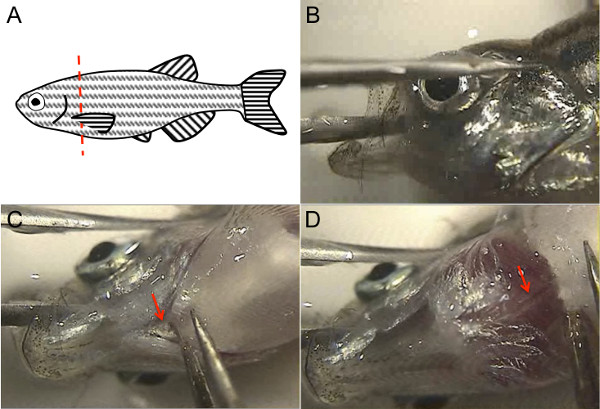
- すぐにそのテールフィンにより安楽死させた魚をピックアップし、1×PBSで満たしたペトリ皿のカバーでその側にそれを置く。 RAZOを使用しながら、片手で胸びれを持ち上げるために鉗子を使用して、Rもう一方の手で魚を斬首するブレード、ちょうど胸びれ( 図1A)の結合を後方。顕微鏡を通して見ることによって、または単に直接顕微鏡ステージ上の魚を視覚化することによってどちらか、この手順を実行します。
注:新鮮な安楽死魚はまだ断頭後に出血します。

図1.ゼブラフィッシュの大人の心臓解剖は、ゼブラフィッシュ、解剖学的ランドマークを利用しています。 (A)は 、魚を斬首ピンセットで胸びれを持ち上げ、図のように赤の点線に沿ってシャープなきれいなカミソリの刃を使用します。(B)、魚の頭を安定魚の口のしばらく鉗子の1歯を配置するには他の歯は、目全体に位置し、腹面がアップしていると鉗子の両方の歯は、ペトリ皿の底に対して安定しているように、その後魚の頭を回す。(C)無料FORCを使用してくださいEPSは蓋(矢印)。(D)は 、このリフティングの添付ファイルをカットする、背側大動脈を管腔血液(矢印)を示すピンクのストライプと白の構造として表示されている。 この図の拡大版をご覧になるにはこちらをクリックしてください。
3.ハートを解剖
- 顕微鏡下で魚の頭を可視化。他の歯は、目( 図1B)全体で、頭部の外にある間に1鉗子で、脳を通して、魚の口の中で鉗子の1歯を配置することにより、魚の頭を安定。鉗子の両方の歯は、ペトリ皿の底に対して安定していることを確認してください。魚の頭をこのように保持し、その腹面を上向きにする必要があります。
- もう一方の手で、体(矢印、 図1C)に蓋の腹側添付ファイルをカットする第二鉗子を使用しています。鉗子で少しこのリフティング、Dorsalの大動脈、大動脈内腔(矢印、 図1D)の血液の赤のストライプと白の構造体としての下に表示されます。大動脈をつまんで上向きに引いて、ピンセットで背側大動脈をカット。代わりに、背側大動脈をカットするmicroscissorsを使用しています。
- さて、フィンのベースのボディ軟骨を含む、そのベースから胸びれを把握することが第二の鉗子を使用し、これをオフに持ち上げます。他の胸鰭を繰り返します。時折、心臓は胸びれで出てくるので、心がそれらを廃棄する前に接続されていないことを確認するこれらの作品を調べます。
注:この時点で、心臓は(時にはそれが胸びれで外れが)まだそのまま、残りの魚の頭に接続され、表示されるはずです。心臓は、その形状、そのピンク色、および色素性心膜に囲まれた人間によって同定することができる。魚の安楽死を迅速に実行されているので、新鮮に安楽死魚の心はまだSLOを破っできるWLY。 - 両手が鉗子を保持し、自由であるように、最初の鉗子で魚の頭を手放す。離れて心膜から心をいじめるために両方の鉗子を使用してください。残りの心膜が引き離されている間背部大動脈の残りの部分によって心をつかみます。
注:固定または新鮮かどうか、心が周囲の結合組織はよりもろい状態で引っ張って穏やかに堅牢である。そのため、周囲の心膜組織は、心臓が無傷で残っていると離れて解剖することができます。 - バイオハザードバッグ内の残りの魚の死体を捨てる。
4.ダウンストリームアプリケーションのハートを準備
- 鉗子を使用して、背側大動脈/動脈球の縁によって心をつかみ、1×PBSの新鮮な皿に心を置く。また、転送ピペットを使用しています。約10倍はできるだけ多くの血を洗い流すために、PBS中で前後に心を移動します。
- それはすべてpossibを除去することが重要である用途のためにルの血液細胞は、同一の往復運動を使用して、代わりにPBSを15mlのRBC溶解バッファーと9センチメートルシャーレに洗う。心臓の構造を維持すること( 例えば、RNAする)、心臓腔を開いて離れ、血液細胞のすすぎ容易にするために、鉗子またはmicroscissorsを使用する下流側のアプリケーションのために重要でない場合。
- 心腔を分離する用途では、所望の動脈球を分析するために鉗子またはmicroscissorsを使用し、心房、および互いに離間心室される。
- 氷の上の宛先バッファに、心臓、または別々のチャンバを転送します。
注:一般的な目的地は、細胞解離、4%パラホルムアルデヒド、またはトリゾールための緩衝液を含む。
結果
この方法を用いて、成体ゼブラフィッシュの心臓は、伝統的な方法8を用いて5分間かけて比較して、1分未満で解剖することができる。ハーツは、この方法を使用して解剖し、従来の方法8は心膜に盲目的に切断が必要なので、一般的にアトリウムまたは球部動脈( 図2B)の損傷または損失の原因となりながら、確実に( 図2A)は無傷である。切開した?...
ディスカッション
成体ゼブラフィッシュの心臓を切開するための方法が記載されているが、これらの方法は時間がかかりました、一般に切開の間に心臓への損傷を引き起こした。成人の心臓の多数必要になることが実験を実行するために、および/または心臓組織の劣化を回避することが下流の適用のために重要である場合、従来の切開技術を使用してかなりの時間が必要である。同様に、再現可能に損傷を受...
開示事項
The authors have no disclosures.
謝辞
The authors would like to thank Dr. Shaun Coughlin for hosting the filming of this procedure in his laboratory, and for general support. R.A. was supported by the NIH (F32HL110489) and the Sarnoff Cardiovascular Research Foundation. S.R. was supported by a Research Fellowship of the Deutsche Forschungsgemeinschaft (DFG) and the American Heart Association (AHA). D.Y.R.S was supported by the NIH (RO1HL54737), the Packard Foundation, and the Max Planck Society.
資料
| Name | Company | Catalog Number | Comments |
| Small tank for transporting fish | Aquaneering | ZHCT100 | |
| Fish net | Petsmart | 36-16731 | |
| 250 ml glass beaker | Kimble | 14005-250 | |
| 9 cm polystyrene Petri dish | Nunc | 172958 | |
| Razor blade | Personna American Safety Razor Company | 94-120-71 | |
| 2 Dumont #5SF forceps | Fine Science Tools | 11252-00 | |
| Dissecting microscope | Olympus | SZX16 | |
| Tricaine | Sigma | A-5040 | |
| Plastic transfer pipette | Thermo Scientific | 202-20S | |
| Gooseneck light source | Dolan-Jenner Industries, Inc | Fiber-Lite 180 Illuminator, 181 Dual Gooseneck System | |
| Fluorescent light source | Lumen Dynamics | X-Cite 120Q | optional |
| Micro-scissors | Biomedical Research Instruments, Inc | 11-1000 | optional |
| RBC lysis buffer | eBioscience | 00-4333-57 | optional |
参考文献
- Arnaout, R., et al. Zebrafish model for human long QT syndrome. Proceedings of the National Academy of Sciences of the United States of America. 104 (27), 11316-11321 (2007).
- Chi, N. C., et al. Genetic and physiologic dissection of the vertebrate cardiac conduction system. PLoS Biology. 6 (5), 109 (2008).
- Poss, K. D. Getting to the heart of regeneration in zebrafish. Seminars in Cell & Developmental Biology. 18 (1), 36-45 (2007).
- Fang, Y., et al. Translational profiling of cardiomyocytes identifies an early Jak1/Stat3 injury response required for zebrafish heart regeneration. Proceedings of the National Academy of Sciences of the United States of America. 110 (33), 13416-13421 (2013).
- Manoli, M., Driever, W. Fluorescence-activated cell sorting (FACS) of fluorescently tagged cells from zebrafish larvae for RNA isolation. Cold Spring Harbor Protocols. 2012 (8), (2012).
- Cannon, J. E., et al. Global analysis of the haematopoietic and endothelial transcriptome during zebrafish development. Mechanisms of Development. 130 (2-3), 122-131 (2013).
- Wang, Z., Gerstein, M., Snyder, M. RNA-Seq: a revolutionary tool for transcriptomics. Nature Reviews Genetics. 10 (1), 57-63 (2009).
- Gupta, T., Mullins, M. C. Dissection of organs from the adult zebrafish. Journal of Visualized Experiments. 37, (2010).
- Singleman, C., Holtzman, N. G. Heart dissection in larval, juvenile and adult zebrafish, Danio rerio. Journal of Visualized Experiments. 55, (2011).
- Westerfield, M. . The zebrafish book: a guide for the laboratory use of zebrafish (Brachydanio rerio. , (1993).
転載および許可
このJoVE論文のテキスト又は図を再利用するための許可を申請します
許可を申請さらに記事を探す
This article has been published
Video Coming Soon
Copyright © 2023 MyJoVE Corporation. All rights reserved