このコンテンツを視聴するには、JoVE 購読が必要です。 サインイン又は無料トライアルを申し込む。
Method Article
ラットにおける上腕と浅大腿動脈のフロー依存性血管拡張の超音波評価
要約
ヒトにおける内皮機能の非侵襲的評価は、フロー媒介拡張技法によって決定することができます。研究の数千人がこの技術を使用しているが、何の研究では、ラットで非侵襲的にこのテクニックを行いませんでした。次の資料は、上腕のフロー依存性血管拡張およびラットの浅大腿動脈の非侵襲的測定を説明します。
要約
Arterial vasodilation to increases in wall shear rate is indicative of vascular endothelial function. In humans, the non-invasive measurement of endothelial function can be achieved by employing the flow-mediated dilation technique, typically performed in the brachial or superficial femoral artery. Briefly, a blood pressure cuff placed distal to an ultrasound probe is inflated to a suprasystolic pressure, which results in limb ischemia. After 5 min of occlusion the cuff is deflated, resulting in reactive hyperemia and increases in wall shear rate that signal vasodilatory molecules to be released from the endothelium eliciting vasodilation. Despite the thousands of studies performing flow-mediated dilation in humans, surprisingly, no studies have performed this technique non-invasively in living rats. Considering the recent shift in focus to translational research, the establishment of guidelines for non-invasive measurement of flow-mediated dilation in rats and other rodents would be extremely valuable. In the following article, a protocol is presented for the non-invasive measurement of flow-mediated dilation in brachial and superficial femoral arteries of rats, as those sites are most commonly measured in humans.
概要
血管内皮は、ライン動脈の内腔の細胞単層であり、血管機能の重要な調節因子です。血管径の調節をもたらす内皮細胞から放出され、多数の分子が存在します。これらの分子、一酸化窒素(NO)の中で、刺激に応答(せん断応力で例えば、インスリン、アセチルコリン、または変更)1における血管内皮から放出された主要な血管拡張性分子であると思われます。血管内皮では、NOは、NOシンターゼ(eNOSの)を内皮ない酵素によって生成され、続いて、内皮細胞2から放出されます。 NOは、リラクゼーションと増加し、血管径3を引き起こす血管平滑筋に拡散します。
内皮機能不全は、フロー媒介性拡張(FMD)技術4,5を用いて、ヒトにおいて非侵襲的に評価することができます。 FMDは、内皮由来ための機能バイオアッセイを表現することが提案されていますヒトでのNO生物学的利用能は、典型的には約5分肢閉塞6次反応性充血に応じて、上腕や浅大腿動脈で評価されていません。反応性充血はNO 8のリリースをシグナリング、内皮細胞7に形質導入された層状剪断力を増大させます。近年では、NO放出によって開始血管拡張の割合は9,10議論してきたが、FMDは、内皮依存性拡張を示すものであると一貫して心血管イベント11-13を予測することが示されています。
現在までに、研究の数千人は、ヒトにおける内皮機能の非侵襲的測定のためのFMD技術を採用しています。トランスレーショナルリサーチへのフォーカスの最近のシフトを考慮すると、げっ歯類における口蹄疫の非侵襲的測定のためのガイドラインは非常に有益であろう。翻訳アプローチを踏まえ、このプロトコルは、上腕とsupeにFMDの測定のために設立されましたこれらのサイトのようなラットのrficial大腿動脈は、最も一般的にヒトで測定されます。ラットにおける堅牢かつ反復FMD応答におけるこのプロトコルの結果は、しかし、ラットでの口蹄疫の測定は技術的に厳しいですし、他の研究者は、デモビデオずに複製するのは難しいかもしれません。したがって、以下の記事では、上腕とラットの浅大腿動脈における口蹄疫の非侵襲的測定のための方法を紹介します。
プロトコル
すべての動物の手順は、実験動物の管理と使用14用のガイドに準拠し、ユタ大学、ソルトレイクシティ退役軍人医療センター動物実験によって承認されました。
1.動物の準備
- 100%の酸素中3%のイソフルランを含む麻酔導入室で動物を置きます。それは、外部刺激に応答しなくなるまで誘導室で動物を残します。
- 誘導チャンバーから動物を削除し、心電図(ECG)電極を備えた加熱された診察台の上に置きます。酸素100%の3%イソフルランで麻酔を維持します。上腕と浅大腿動脈FMDを同時に行うことはできません。したがって、各測定のための準備手順は以下のとおりです。
2.上腕動脈の準備
- 仰臥位動物を置き、左上肢とANの各下肢を拘束外科手術用テープで診察台にimal。
- 上肢の下部が若干プラットフォーム上に(〜0.2センチメートル)に上昇するように、動物の右上肢を抑制する。
- 髪を除去するために、動物の右上肢に脱毛剤(例えば、Nairさん)を適用します。
- 肘に右上肢遠位に位置閉塞カフ(直径標準血管閉塞器内腔10ミリメートル)。膨張/収縮が手足を動かし、超音波画像を乱すように、プラットフォームに閉塞器を休まないでください。
- 超音波のキーボードを使用して、Bモードに超音波マシンを設定します。
- 閉塞カフの近位に、動物の上肢に超音波ゲルの少量を適用します。
- 手動で上肢と定位ホルダーに取り付けられた超高周波リニアアレイトランスデューサを合わせます。上腕動脈は2〜3ミリメートルの深見えるはずです。
- 上腕動脈ではなく、上腕静脈は、されていることを確認するために、画像化され、超音波のキーボードを使用してPW-モードに切り替えます。連続血流を持つことになり、隣接する静脈とは対照的に、動脈は拍動の血流を持っています。
3.浅大腿動脈の準備
- 位置動物仰臥位とサージカルテープで検査テーブルに上肢と左下肢を拘束。
- パッド(例えば、折り畳まれた紙タオル)を使用してプラットフォーム上に高い位置(〜0.5〜1センチメートル)に動物の右下肢を拘束。
- 髪を除去するために、動物の右下肢に脱毛剤(例えば、Nairさん)を適用します。脱毛後に大腿静脈は、上部太ももの内側にはっきりと見えるはずです。
- 右足首に閉塞カフ(直径標準血管閉塞器内腔10ミリメートル)近位の位置を決めます。膨張/収縮が下肢を動かし、超音波画像を乱すように、プラットフォームに閉塞器を休まないでください。
- に超音波マシンを設定しますBモード。
- 閉塞カフの近位に、動物の下肢に超音波ゲルの少量を適用します。
- 手動で皮膚を通して見える大腿静脈、と定位ホルダーに取り付けられた超高周波リニアアレイトランスデューサを合わせます。浅大腿動脈は、1ミリメートルの深<表示されるはずです。
- 浅大腿動脈ではなく、大腿静脈は、画像化されていることを確認するために、PW-モードに切り替えます。連続血流を持つことになり、隣接する静脈とは対照的に、動脈は拍動の血流を持っています。
4.ベースラインフェーズ
- それは人間の15で行われることになるのと同じようにBモード画像を、最適化します。両壁に可視化し、内膜 - メディアと容器の横、縦方向の画像が観察されていることを確認。少しできるだけ動脈の限りがキャプチャウィンドウに表示可能であることを保証するために、超音波プローブの配置を調整することにより、画像を最適化します。
- あるいは、明るさ/コントラスト、焦点域、周波数、ダイナミック・レンジ、及び線密度を変化させることで良好な画像を得るための超音波設定を調整します。そこに超音波画像を最適化するための他の方法があるが、それらの詳細な説明は、このプロトコルの範囲を超えています。
- 動脈画像の最適化の後、唯一の直径フレームが収集されることを保証するために、R波の間に撮影した画像のみを表示するには、ECGゲーティングをオンにする心周期の各拡張期部分の間にあります。
注:ECGゲーティングは、生理的な設定オプションの下にECGゲーティングを選択することで、このプロトコルで使用される超音波マシン上で利用可能である、しかし、この機能は、すべての超音波マシンで利用できない場合があります。画像が最適化された後、ECGゲーティングは、より低いフレームレートで画像を得ることが困難であるように、ターンオンされるべきである(すなわち、一度R波あたり)。 ECGゲーティング、ラットおよび高フレームの要求の高い心拍数の組み合わせなし心周期の拡張期部分だけ〜10-20秒のクリップを可能にしますキャプチャするレート。各クリップのデータの面倒な大きさと量は、実質的に分析負担を増大させます。 - 録音Bモードを使用したベースライン・データの60秒。
注:超音波クリップに記録することができるフレーム数に制限があるような超音波装置は常に記録され、ただし、すべての画像は、超音波装置に格納されています。クリップの長さ(すなわち、フレーム数)の設定を調整することができます。クリップあたりのフレーム数の最大値に設定されたことが示唆されています。記録は、クリップ(すなわち、到達したフレームの最大数)の末尾にある場合、記録は継続しますが、クリップは、最新のフレームをキャプチャするロールフォワード。この場合、最大フレームリミットの外で捕獲された以前のフレームはその後削除されます。記録中のこれらの複雑さは、マシン間で異なるが、記録長の調整が必要であり得ます。 - SWIPW-モードにTCH。内腔の中央にカーソルを置きます。サンプルゲートは、カーソルを参照して自動的に配置されるが、超音波のキーボードを使用して、幅のために調整することができます。 ≤60°の超音波照射角を維持します。
- ドップラービーム角度を変えることによって、超音波照射角度を調整します。超音波のキーボードを使用して角度を微調整してください。これらのいずれも測定に適した角度を提供する場合は、手動でより最適な角度に動脈を傾けることによって超音波プローブを調整します。超音波の角度の任意の調整が行われた場合、Bモード画像を取り戻します。
- 速度データの10秒を記録します。
5.オクルージョンフェーズ
- 空気で満たされた10ミリリットル注射器を用いて血管の閉塞器を膨らませます。血管の閉塞器内の空気の圧力を一定に保つために、自分自身にチューブを折ると、折り畳まれたチューブ上にバインダークリップを配置します。
- によって証明されるように、カフ閉塞を確認するために、PW-モードに切り替え血流速度の大幅な削減。
- 閉塞の午前4時45分分まで、60秒のクリップにBモードと記録データに切り替えます。
- PW-モードに切り替えます。心拍数および分析のための各超音波クリップの時間の記録を保管してください。
6.充血相
- PW-モードで録画中に折り畳まれた管からのバインダークリップを削除することによってカフを解放します。録音の前に5秒とカフリリース後5秒。
- 閉塞後3分まで、60秒のクリップにBモードと記録データに切り替えます。心拍数および分析のための各超音波クリップの時間の記録を保管してください。
- FMDの完了後診察台から動物を削除し、それが胸骨横臥位を維持するのに十分な意識を取り戻したまで監視します。
7.解析
- 分析のために、公平を可能にするエッジ検出ソフトウェアを搭載したオフラインコンピュータへのDICOMファイルとしてエクスポートする超音波を抑止します各フレームでの動脈直径のmination。それは非常に時間がかかり、研究者バイアスの対象となるように分析は、超音波マシン上で可能である、しかし、それは、お勧めできません。
- ベースラインと、閉塞段階の間に60秒のセグメントにおける動脈径データを分析し、充血相中に10秒のセグメントインチ
- 自動エッジ検出ソフトウェアのフロー解析機能を用いて血流速度データを分析します。ベースラインと、閉塞段階の間に均一な外観の5以上の連続した波形を測定することにより、平均血流速度を決定します。すぐにカフリリース後に血液速度のための反応性充血の間の平均血流速度を決定します。最高血流速度の波形は、ピーク血流速度と考えられています。
結果
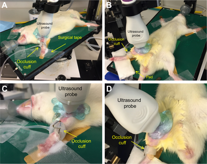
フロー媒介拡張8 Wistarラットの上腕および浅大腿動脈で行いました。ラットの位置決めは、図1に示されています。
浅大腿動脈の代表的な超音波画像は、 図2に示されています。

図1.
ディスカッション
本研究では、FMDの非侵襲的測定は、上腕およびラットの浅大腿動脈に実証されました。ヒト6と同様に、5分間の閉塞期間の後、それによって動脈のその後の血管拡張をもたらし、動脈壁のせん断速度を増加させる血流速度(すなわち、反応性充血)の急激な増加がありました。 FMDは、上腕と浅大腿動脈の両方で観察されました。また、動脈の間FMDに強い関係がありました。ピーク剪?...
開示事項
None.
謝辞
All animal imaging was performed at the Small Animal Imaging Core Facility, University of Utah.
This study was funded in part by grants from the National Institutes of Health (R21 AG043952, R01 AG040297, K01 AG046326, K02 AG045339, and R01 DK100505).
資料
| Name | Company | Catalog Number | Comments |
| Vevo 2100 High Resolution Micro-Ultrasound Imaging System | VisualSonics, Toronto, ON, CAN | ||
| MicroScan Ultra-High Frequency Linear Array Transducer - MS-700 30-70 MHz | VisualSonics, Toronto, ON, CAN | ||
| Vevo Imaging Station | VisualSonics, Toronto, ON, CAN | ||
| Thermasonic gel warmer | Parker Laboratories, Fairfield, NJ, USA | 82-03 | Optional |
| Signacreme electrode cream | Parker Laboratories, Fairfield, NJ, USA | 17-05 | |
| Transpore surgical tape | 3M, Maplewood, MN, USA | 1527-1 | |
| Depilatory cream (e.g., Nair) | General supply | ||
| Cotton swabs | General supply | ||
| Ultrasound gel | General supply | ||
| Standard vascular occluder, 10 mm lumen diameter | Harvard Apparatus, Holliston, MA, USA | 62-0115 | |
| 10 ml syringe with Luer-Lok tip | General Supply | Used for occlusion cuff apparatus | |
| Paperclip | General Supply | Used for occlusion cuff apparatus | |
| Hypodermic needle – 18 gauge | General Supply | Used for occlusion cuff apparatus | |
| Medium binder clip | General Supply | Used for occlusion cuff apparatus |
参考文献
- Smits, P., et al. Endothelial release of nitric oxide contributes to the vasodilator effect of adenosine in humans. Circulation. 92, 2135-2141 (1995).
- Forstermann, U., et al. Nitric oxide synthase isozymes. Characterization, purification, molecular cloning, and functions. Hypertension. 23, 1121-1131 (1994).
- Gardiner, S. M., Compton, A. M., Bennett, T., Palmer, R. M., Moncada, S. Control of regional blood flow by endothelium-derived nitric oxide. Hypertension. 15, 486-492 (1990).
- Harris, R. A., Nishiyama, S. K., Wray, D. W., Richardson, R. S. Ultrasound assessment of flow-mediated dilation. Hypertension. 55, 1075-1085 (2010).
- Corretti, M. C., et al. Guidelines for the ultrasound assessment of endothelial-dependent flow-mediated vasodilation of the brachial artery: a report of the International Brachial Artery Reactivity Task Force. J Am Coll Cardiol. 39, 257-265 (2002).
- Celermajer, D. S., et al. Non-invasive detection of endothelial dysfunction in children and adults at risk of atherosclerosis. Lancet. 340, 1111-1115 (1992).
- Niebauer, J., Cooke, J. P. Cardiovascular effects of exercise: role of endothelial shear stress. J Am Coll Cardiol. 28, 1652-1660 (1996).
- Sessa, W. C. eNOS at a glance. J Cell Sci. 117, 2427-2429 (2004).
- Wray, D. W., et al. Does brachial artery flow-mediated vasodilation provide a bioassay for NO?. Hypertension. 62, 345-351 (2013).
- Green, D. J., Dawson, E. A., Groenewoud, H. M., Jones, H., Thijssen, D. H. Is flow-mediated dilation nitric oxide mediated? A meta-analysis. Hypertension. 63, 376-382 (2014).
- Green, D. J., Jones, H., Thijssen, D., Cable, N. T., Atkinson, G. Flow-mediated dilation and cardiovascular event prediction: does nitric oxide matter?. Hypertension. 57, 363-369 (2011).
- Brevetti, G., Silvestro, A., Schiano, V., Chiariello, M. Endothelial dysfunction and cardiovascular risk prediction in peripheral arterial disease: additive value of flow-mediated dilation to ankle-brachial pressure index. Circulation. , 2093-2098 (2003).
- Gokce, N., et al. Predictive value of noninvasively determined endothelial dysfunction for long-term cardiovascular events in patients with peripheral vascular disease. J Am Coll Cardiol. 41, 1769-1775 (2003).
- National Research Council (U.S.). . Guide for the care and use of laboratory animals. , (2011).
- Alley, H., Owens, C. D., Gasper, W. J., Grenon, S. M. Ultrasound assessment of endothelial-dependent flow-mediated vasodilation of the brachial artery in clinical research. J Vis Exp. , e52070 (2014).
- Ghiadoni, L., et al. Assessment of flow-mediated dilation reproducibility: a nationwide multicenter study. J Hypertension. 30, 1399-1405 (2012).
- Thijssen, D. H., et al. Heterogeneity in conduit artery function in humans: impact of arterial size. Am J Physiol Heart Circ. 295, H1927-H1934 (2008).
- Green, D. J., et al. Why isn't flow-mediated dilation enhanced in athletes?. Med Sci Sports. 45, 75-82 (2013).
- Heiss, C., et al. In vivo measurement of flow-mediated vasodilation in living rats using high-resolution ultrasound. Am J Physiol Heart Circ. 294, H1086-H1093 (2008).
- Chen, Q., et al. Pharmacological inhibition of S-nitrosoglutathione reductase improves endothelial vasodilatory function in rats in vivo. J Appl Physiol. 114, 752-760 (2013).
- Pinnamaneni, K., et al. Brief exposure to secondhand smoke reversibly impairs endothelial vasodilatory function. Nicotine Tob Res. 16, 584-590 (2014).
- Liu, J., et al. Impairment of Endothelial Function by Little Cigar Secondhand Smoke. Tob Regul Sci. 2, 56-63 (2016).
- Schuler, D., et al. Measurement of endothelium-dependent vasodilation in mice--brief report. Arterioscler Thromb Vasc Biol. 34, 2651-2657 (2014).
- Erkens, R., et al. Left ventricular diastolic dysfunction in Nrf2 knock out mice is associated with cardiac hypertrophy, decreased expression of SERCA2a, and preserved endothelial function. Free Radic Biol Med. 89, 906-917 (2015).
- Harris, S. A., Billmeyer, E. R., Robinson, M. A. Evaluation of repeated measurements of radon-222 concentrations in well water sampled from bedrock aquifers of the Piedmont near Richmond, Virginia, USA: : effects of lithology and well characteristics. Environmental research. 101, 323-333 (2006).
転載および許可
このJoVE論文のテキスト又は図を再利用するための許可を申請します
許可を申請さらに記事を探す
This article has been published
Video Coming Soon
Copyright © 2023 MyJoVE Corporation. All rights reserved