
4D Microscopy: Unraveling Caenorhabditis elegans Embryonic Development Using Nomarski Microscopy
October 8th, 2020
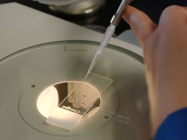
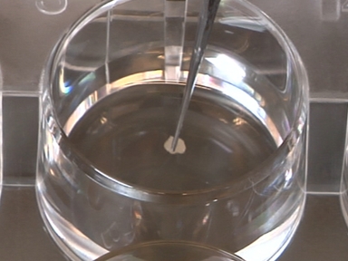
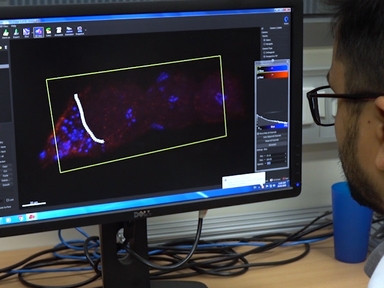
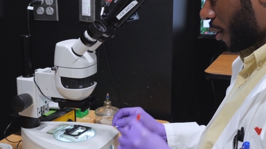
•Here, we present a protocol for preparing and mounting Caenorhabditis elegans embryos, recording development under a 4D microscope and tracing cell lineage.
Related Videos
Comprehensive Assessment of Germline Chemical Toxicity Using the Nematode Caenorhabditis elegans
Analysis of Cardiomyocyte Development using Immunofluorescence in Embryonic Mouse Heart

Transcriptome Profiling of In-Vivo Produced Bovine Pre-implantation Embryos Using Two-color Microarray Platform

Analysis of Zebrafish Kidney Development with Time-lapse Imaging Using a Dissecting Microscope Equipped for Optical Sectioning

Using Light Sheet Fluorescence Microscopy to Image Zebrafish Eye Development

Multimodal Hierarchical Imaging of Serial Sections for Finding Specific Cellular Targets within Large Volumes

Assessing Lysosomal Alkalinization in the Intestine of Live Caenorhabditis elegans
Computational Analysis of the Caenorhabditis elegans Germline to Study the Distribution of Nuclei, Proteins, and the Cytoskeleton
Isotropic Light-Sheet Microscopy and Automated Cell Lineage Analyses to Catalogue Caenorhabditis elegans Embryogenesis with Subcellular Resolution

Isolation of Specific Neuron Populations from Roundworm Caenorhabditis elegans
Copyright © 2024 MyJoVE Corporation. 판권 소유