Aby wyświetlić tę treść, wymagana jest subskrypcja JoVE. Zaloguj się lub rozpocznij bezpłatny okres próbny.
Method Article
Isolation and Culture of Adult Epithelial Stem Cells from Human Skin
W tym Artykule
Podsumowanie
A rapid, robust way of isolating viable adult epithelial stem cells from human skin is described. The method utilizes enzymatic digestion of skin collagen matrix , followed by plucking of hair follicles and isolation of single cell suspensions or tissue fragments for cell culture.
Streszczenie
The homeostasis of all self-renewing tissues is dependent on adult stem cells. As undifferentiated stem cells undergo asymmetric divisions, they generate daughter cells that retain the stem cell phenotype and transit-amplifying cells (TA cells) that migrate from the stem cell niche, undergo rapid proliferation and terminally differentiate to repopulate the tissue.
Epithelial stem cells have been identified in the epidermis, hair follicle, and intestine as cells with a high in vitro proliferative potential and as slow-cycling label-retaining cells in vivo 1-3. Adult, tissue-specific stem cells are responsible for the regeneration of the tissues in which they reside during normal physiologic turnover as well as during times of stress 4-5. Moreover, stem cells are generally considered to be multi-potent, possessing the capacity to give rise to multiple cell types within the tissue 6. For example, rodent hair follicle stem cells can generate epidermis, sebaceous glands, and hair follicles 7-9. We have shown that stem cells from the human hair follicle bulge region exhibit multi-potentiality 10.
Stem cells have become a valuable tool in biomedical research, due to their utility as an in vitro system for studying developmental biology, differentiation, tumorigenesis and for their possible therapeutic utility. It is likely that adult epithelial stem cells will be useful in the treatment of diseases such as ectodermal dysplasias, monilethrix, Netherton syndrome, Menkes disease, hereditary epidermolysis bullosa and alopecias 11-13. Additionally, other skin problems such as burn wounds, chronic wounds and ulcers will benefit from stem cell related therapies 14,15. Given the potential for reprogramming of adult cells into a pluripotent state (iPS cells)16,17, the readily accessible and expandable adult stem cells in human skin may provide a valuable source of cells for induction and downstream therapy for a wide range of disease including diabetes and Parkinson's disease.
Protokół
1. Extract Epithelial Stem Cells from Human Skin
- Before starting the procedure of isolating epithelial stem cells one needs to prepare the respective medias and reagents (see table1).
- Fresh adult human scalp skin from facelift procedures or punch biopsy is collected, then incubate in DMEM / 10% FBS / Dispase (4 mg/ mL) overnight at 4°C. Incubation for 2-4 hr at 37°C is also effective. Skin pieces should be a maximum width of 1 cm to allow for enzyme to penetrate.
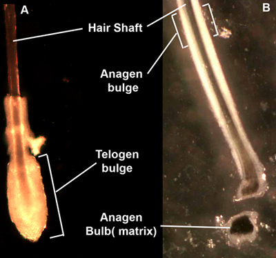
- Transfer the skin into a sterilized Petri dish, pull off each hair from the skin by grasping the hair shaft near the skin surface and pulling firmly and smoothly. Select follicles at telogen stage based on their morphology under a dissecting microscope, cut out the bulge region (Figure 1A) transfer follicles into a 15 mL sterilized tube. Anagen follicles can also be used if cut at upper 1/3 of follicle to obtain hair follicle "bulge" region (Figure 1B).
- Incubate the isolated follicle fragments in a mixture of 0.05% trypsin-EDTA (GIBCO) and Versene (0.53 mM EDTA 4Na, GIBCO) (1:1) for 15-20 min at room temperature with shaking periodically, add 4 mL DMEM + 10% FBS to stop reaction, spin down for 5 min at 800 rpm. Alternatively, follicle tissue fragment explants can be placed in culture without trypsin digest to culture cells from single follicles.
- Discard supernatant carefully (save ~0.2-0.5 mL to avoid losing cells) and re-suspend in 1 mL KCM (keratinocyte medium), without EGF.
2. Culture Primary Epithelial Stem Cells
- Before culturing the epithelial stem cells, mitomycin C-treated 3T3-J2 cells (feeder cells) should be prepared as following. 3T3-J2 cells are cultured in DMEM with 10% FBS before treatment. Remove culturing media and wash cell twice with PBS, treat cell with 15μg / mL mitomycin C in DMEM without serum for 2 hr at 37°C, 5% CO2. Remove the mitomycin C-containing DMEM and wash twice with PBS, the cells are ready to use. (Mitomycin C-treated 3T3-J2 cells can also be prepared previously and stored at -80°C. Thaw and seed the cells one day before culturing primary epithelial stem cells, incubate at 37°C, 5% CO2. 150,000-200,000 3T3-J2 cells per well of 6-well plate is recommended.
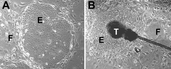
- Seed isolated hair follicle stem cell from step 1.5 on mitomycin C-treated 3T3-J2 cells in KCM without EGF overnight at 37°C, 5% CO2, changed to EGF-containing KCM the next day. Cells are grown at 37°C in a humid atmosphere containing 5% CO2. All keratinocytes are fed with KCM containing EGF every 2 days and grown for 14-20 days. Check cell every day under microscope. Hair follicle stem cells will form colonies (Figure 2A). Hair follicle explants will form outgrowths after 10-14 days (Figure 2B).
3. Passage Epithelial Stem Cells
- Wash cells with PBS once. Remove 3T3-J2 feeder cells by Versene (pre-warmed to RT) treatment. Incubate culture dished in Versene for 5 minutes, then gently shake and aspirate off feeder cells.
- Wash hair follicle stem cells with either Versene or PBS one time.
- Add pre-warmed (to 37°C, critical) trypsin-EDTA (2X) and incubate at 37°C for 7 minutes or longer if necessary for the hair follicle stem cell to detach. No longer than 15 minutes is best.
- Add KCM w/o EGF to stop the trypsin, gently pipette up and down to disperse cells, spin down at 200g, 5 minutes.
- Re-suspend cells with KCM without EGF, Count and re-plate on mitomycin C-treated 3T3-J2 cells layer.
4. Immortalize Epithelial Stem Cells
Primary cells, even adult stem cells, reach senescence after serial passage and months in culture. Immortalization of primary stem cells is an effective way to relieve this problem.
- Plate primary epithelial stem cells (~350,000 cells/well of 6 well plate) at passage 1 on feeder layer (3T3-J2) and culture for 2 days.
- Culture PA317 LXSN 16E6E7 cell line one day after seeding the primary epithelial stem cells with DMEM containing 10% FBS. (This line produces the amphotropic retrovirus LXSN16E6E7 which encodes the HPV16 E6 and E7 open reading frames, and which can be used to stably infect and immortalize many cell types).
- Treat PA317 LXSN 16E6E7 cell line with mitomycin C for 2 hours (Same protocol with 3T3-J2 cells). Add mitomycin C treated PA317 LXSN 16E6E7 cell line to the primary epithelial stem cells, co-culture for 6 days in KCM with EGF. Renew fresh KCM with EGF media every 2 days.
- Remove 3T3-J2 and PA317 LXSN 16E6E7 cell with Versene. Add mitomycin-C treated 3T3-J2 NHP cells (neomycin, hygromycin, puromycin resistant, ~200,000 cells per well) to the epithelial stem cells. Cells are selected under 0.2 mg/ mL of G418 (Gibco) for additional 6 days. Renew fresh G418 containing KCM with EGF media every 2 days.
Surviving epithelial stem cells will be immortalized stem cells. A well of primary epithelial stem cells which are not co-cultured with PA317 LXSN 16E6E7 cell should be set as a control for G418 selection, all the cells in this well should be killed by G418 after 6 days of selection.
5. Representative Results
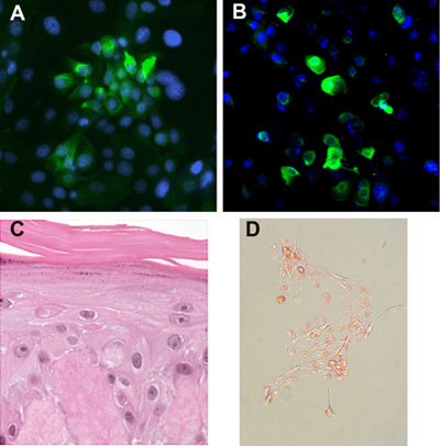
Early passage of skin epithelial stem cells and immortalized epithelial stem cells form tight colonies consisting of small keratinocytes (Figure 2A) surrounded by feeder cells. If cultured in serum free media with defined supplements, without a feeder layer, the stem cells will disperse and not form tight colonies (they grow as single cells and small clusters). Immortalized epithelial stem cells maintain a stable phenotype for >12 months of continuous passage, but tended to form tighter colonies than the primary cells. They do not form anchorage independent colonies in soft agar assays, indicating that these immortalized cells do not possess the characteristics of cancerous cells. Both primary and immortalized epithelial stem cells express hair follicle stem cell marker cytokeratin 15 (Figure 3A), and are able to differentiate into epidermal, hair follicle and sebaceous lineages (Figure 3B-D).

Figure 1. Plucked hairs. After plucking from dispase-treated skin, hair follicles appear as either telogen club hairs (A) with a ball of cells surrounding the bottom of the hair, or anagen hairs (B) with a sleeve of epithelium surrounding the length of the hair. The telogen bulge region forms a distinct morphologic region to micro-dissect, while the anagen bulge is less distinct. The anagen bulb contains matrix keratinocytes, representing transit-amplifying cells that can also be isolated and cultured.

Figure 2. Stem cells cultures. (A) Epithelial stem cells from skin form tight epithelial colonies (designated by E) when cultured in KCM with a feeder layer of cells (designated by F). (B) Hair follicle explants give rise to epithelial outgrowths. The telogen bulge region (designated by T) is attached to the dish and surrounded epithelial cell colony along with feeder cells.

Figure 3. Stem cell differentiation. (A) Stem cells colonies maintain expression of epithelial markers such as cytokeratin 15. (B) Skin epithelial stem cells can be differentiated along the hair follicle lineage as determined by K6Hf expression. (C) The cells can be cultured on de-epidermalized dermis or other matrices at the air-liquid interface and will form a stratified epidermis with cornified layer, granular layer and spinous layer. (D) Stem cells can be induced along the sebaceous lineage with Oil Red positive globules.
Dyskusje
The cell extraction and culture methods described are surprisingly facile and reproducible. We have generated epithelial stem cell cultures from dozens of individuals across a broad age range, including patients with inherited skin defects18. It is best to begin the process on the day of tissue harvest, however cells will remain viable in media on ice for several days, facilitating overnight shipping if needed. Discarded facelift skin yields hundreds of viable follicles for cell extraction as single cell suspe...
Ujawnienia
No conflicts of interest declared.
Podziękowania
This work is funded by NIH/ NCI grant R01CA-118916
Materiały
| Name | Company | Catalog Number | Comments |
| DMEM | GIBCO, by Life Technologies | 11995 | |
| Hams F12 | GIBCO, by Life Technologies | 11765 | |
| Fetal Bovine Serum (FBS) | GIBCO, by Life Technologies | 16000 | |
| Insulin | GIBCO, by Life Technologies | 12585 | |
| T3 | Sigma-Aldrich | T-2752 | |
| Transferrin | Roche Group | 10652202001 | |
| Hydrocortisone | Sigma-Aldrich | H-4001 | |
| Cholera Toxin | Sigma-Aldrich | C8052 | |
| Epidermal growth factor (EGF) | Sigma-Aldrich | E-9644 | |
| Adenine | Sigma-Aldrich | A9795 | |
| Trypsin(10X) | GIBCO, by Life Technologies | 15090 | |
| VERSENE | GIBCO, by Life Technologies | 15040 | |
| G418 Sulfate | Cellgro | 30-234-CR | |
| Hanks’ Balanced Salt solution | Sigma-Aldrich | H6648 | |
| 1X PBS | Cellgro | 21-040-CV | |
| Mitomycin C | Roche Group | 10107409001 | |
| Penicillin/streptomycin | Invitrogen | 15140122 | |
| Dispase | Invitrogen | 17105 | |
| Crystal Violet | Fisher Scientific | C581-25 | |
Keratinocyte media (KCM) [DMEM and Ham s F12 (GIBCO, 3:1), adenine (Sigma, 180 mM), 10% fetal bovine serum (GIBCO), cholera toxin (ICN, 0.1 nM), penicillin/streptomycin (GIBCO, 100 U/ml and 100 mg/ml, respectively), hydrocortisone (Sigma, 0.4 mg/ml, 1.1 mM), T/T3 (transferrin, GIBCO, 5 μg/ml, 649 nM; and triiodo-l-thyronine, Sigma, 2 nM), insulin (Sigma, 5 mg/ml, 862 nM), and EGF (Sigma, 10 ng/ml, 1.6 nM), pH 7.2] | |||
[DMEM and Ham s F12 (GIBCO, 3:1), adenine (Sigma, 180 mM), 10% fetal bovine serum (GIBCO), cholera toxin (ICN, 0.1 nM), penicillin/streptomycin (GIBCO, 100 U/ml and 100 mg/ml, respectively), hydrocortisone (Sigma, 0.4 mg/ml, 1.1 mM), T/T3 (transferrin, GIBCO, 5 μg/ml, 649 nM; and triiodo-l-thyronine, Sigma, 2 nM), insulin (Sigma, 5 mg/ml, 862 nM), and EGF (Sigma, 10 ng/ml, 1.6 nM), pH 7.2] | |||
Odniesienia
- Jones, P. H., Watt, F. M. Separation of human epidermal stem cells from transit amplifying cells on the basis of differences in integrin function and expression. Cell. 73, 713-724 (1993).
- Lyle, S., Christofidou-Solomidou, M., Liu, Y., Elder, D. E., Albelda, S., Cotsarelis, G. The C8/144B monoclonal antibody recognizes cytokeratin 15 and defines the location of human hair follicle stem cells. J. Cell. Sci. 111, 3179-3188 (1998).
- Bac, S. P., Reneha, A. G., Potte, C. S. Stem cells: the intestinal stem cell as a paradigm. Carcinogenesis. 21, 469-476 (2000).
- Slac, J. M. Stem cells in epithelial tissues. Science. 287, 1431-1433 (2000).
- It, M., Li, Y., Yan, Z., Nguye, J., Lian, F., Morri, R. J., Cotsarelis, G. Stem cells in the hair follicle bulge contribute to wound repair but not to homeostasis of the epidermis. Nat Med. 11, 1351-134 (2005).
- Spradlin, A., Drummond-Barbos, D., Kai, T. Stem cells find their niche. Nature. 414, 98-104 (2001).
- Taylo, G., Lehre, M. S., Jense, P. J., Su, T. T., Lavke, R. M. Involvement of follicular stem cells in forming not only the follicle but also the epidermis. Cell. 102, 451-461 (2000).
- Oshim, H., Rocha, A., Kedzi, C., Kobayash, K., Barrandon, Y. Morphogenesis and renewal of hair follicles from adult multipotent stem cells. Cell. 104, 233-245 (2001).
- Morri, R. J., Li, Y., Marle, L., Yan, Z., Trempu, C., L, S., Li, J. S., Sawick, J. A. Cotsarelis G Capturing and profiling adult hair follicle stem cells. Nat. Biotechnol. 22, 411-417 (2004).
- Ro, C., Roch, M., Gu, Z., Photopoulo, C., Ta, Q., Lyle, S. Multi-potentiality of a new immortalized epithelial stem cell line derived from human hair follicles. In vitro Cell. & Dev. Biol. 44, 236-244 (2008).
- Ohyama, M., Vogel, J. C. G. e. n. e. delivery to the hair follicle. J Investig Dermatol Symp Proc. 8, 204-206 (2003).
- Sugiyama-Nakagiri, Y., Akiyama, M., Shimizu, H. Hair follicle stem cell-targeted gene transfer and reconstitution system. Gene Ther. 13, 732-737 (2006).
- Stenn, K. S., Cotsarelis, G. Bioengineering the hair follicle: fringe benefits of stem cell technology. Curr Opin Biotechnol. 16, 493-497 (2005).
- Hoeller, D. An improved and rapid method to construct skin equivalents from human hair follicles and fibroblasts. Exp Dermatol 10. , 264-271 (2001).
- Navsaria, H. A., Ojeh, N. O., Moiemen, N., Griffiths, M. A., Frame, J. D. Reepithelialization of a full-thickness burn from stem cells of hair follicles micrografted into a tissue-engineered dermal template (Integra). Plast Reconstr Surg. 113, 978-981 (2004).
- Werni, M., Meissne, A., Forema, R., Brambrin, T., K, M., Hochedlinge, K., Bernstei, B. E., Jaenisch, R. In vitro reprogramming of fibroblasts into a pluripotent ES-cell-like state. Nature. 448, 318-324 (2007).
- Par, I. H., Zha, R., Wes, J. A., Yabuuch, A., Hu, H., Inc, T. A., Lero, P. H., Lensc, M. W., Dale, G. Q. Reprogramming of human somatic cells to pluripotency with defined factors. Nature. 451, 141-146 (2008).
- Kazantsev, A., Goltso, A., Zinchenk, R., Grigorenk, A. P., Abrukov, A. V., Moliak, Y. K., Kirillo, A. G., Gu, Z., Lyl, S., Ginte, E. K., Rogae, E. I. Human hair growth deficiency is linked to a genetic defect in the phospholipase gene LIPH. Science. 314, 982-985 (2006).
- Tola, J., Ishida-Yamamot, A., Riddl, M., McElmurr, R. T., Osbor, M., Xi, L., Lun, T., Slatter, C., Uitt, J., Christian, A. M., Wagne, J. E., Blaza, B. R. Amelioration of epidermolysis bullosa by transfer of wild-type bone marrow cells. Blood. 113, 1167-1174 (2009).
- Wagne, J. E., Ishida-Yamamot, A., McGrat, J. A., Hordinsk, M., Keen, D. R., Riddl, M. J., Osbor, M. J., Lun, T., Dola, M., Blaza, B. R., Tolar, J. Bone marrow transplantation for recessive dystrophic epidermolysis bullosa. N Engl J Med. 363, 629-639 (2010).
- Muraue, E. M., Gach, Y., Grat, I. K., Klausegge, A., Mus, W., Grube, C., Meneguzz, G., Hintne, H., Baue, J. W. Functional Correction of Type VII Collagen Expression in Dystrophic Epidermolysis Bullosa. J Invest Dermatol. , (2010).
- Y, H., Kuma, S. M., Kossenko, A. V., Show, L., X, X. Stem cells with neural crest characteristics derived from the bulge region of cultured human hair follicles. J Invest Dermatol. 130, 1227-1236 (2010).
- Nishimur, E. K., Grante, S. R., Fishe, D. E. Mechanisms of hair graying: incomplete melanocyte stem cell maintenance in the niche. Science. 307, 720-724 (2005).
Przedruki i uprawnienia
Zapytaj o uprawnienia na użycie tekstu lub obrazów z tego artykułu JoVE
Zapytaj o uprawnieniaPrzeglądaj więcej artyków
This article has been published
Video Coming Soon
Copyright © 2025 MyJoVE Corporation. Wszelkie prawa zastrzeżone