Ressonância Magnética Cardíaca
Visão Geral
Fonte: Frederick W. Damen e Craig J. Goergen, Weldon School of Biomedical Engineering, Purdue University, West Lafayette, Indiana
Neste vídeo, a ressonância magnética de campo alto e pequeno (RM) com monitoramento fisiológico é demonstrada para adquirir laços de cine fechados do sistema cardiovascular murino. Este procedimento fornece uma base para avaliar a função ventricular esquerda, visualizar redes vasculares e quantificar o movimento dos órgãos devido à respiração. As modalidades de imagem cardiovascular animal de pequeno porte comparáveis incluem ultrassom de alta frequência e tomografia microcomputada (TC); no entanto, cada modalidade está associada a compensações que devem ser consideradas. Embora o ultrassom forneça alta resolução espacial e temporal, artefatos de imagem são comuns. Por exemplo, tecido denso (ou seja, esterno e costelas) pode limitar a profundidade de penetração de imagem, e o sinal hiperecóico na interface entre gás e líquido (ou seja, pleura ao redor dos pulmões) pode desfocar o contraste no tecido próximo. A micro-TC em contraste não sofre de tantos artefatos no avião, mas tem menor resolução temporal e contraste limitado de tecido mole. Além disso, a micro-TC usa radiação de raios-X e muitas vezes requer o uso de agentes de contraste para visualizar a vasculatura, ambos conhecidos por causar efeitos colaterais em altas doses, incluindo danos à radiação e lesão renal. A ressonância magnética cardiovascular proporciona um bom compromisso entre essas técnicas, negando a necessidade de radiação ionizante e proporcionando ao usuário a capacidade de imagem sem agentes de contraste (embora agentes de contraste sejam frequentemente usados para ressonância magnética).
Esses dados foram adquiridos com uma sequência de ressonância magnética rápida de ângulo baixo (FLASH) que foi fechada dos picos R no ciclo cardíaco e planaltos expiratórios na respiração. Estes eventos fisiológicos foram monitorados através de eletrodos subcutâneos e um travesseiro sensível à pressão que foi fixado contra o abdômen. Para garantir que o mouse fosse devidamente aquecido, uma sonda de temperatura retal foi inserida e usada para controlar a saída de um ventilador de aquecimento seguro para ressonância magnética. Uma vez que o animal foi inserido no furo do scanner de ressonância magnética e as sequências de navegação foram executadas para confirmar o posicionamento, os planos de imagem FLASH fechados foram prescritos e os dados adquiridos. No geral, a ressonância magnética de alto campo é uma poderosa ferramenta de pesquisa que pode fornecer contraste de tecido mole para o estudo de modelos de pequenas doenças animais.
Procedimento
1. Preparação animal
- Identifique o rato para ser imageado em sua gaiola e transfira-o para a câmara de indução da anestesia.
- Anestesiar o mouse usando isoflurane e confirmar o knockdown usando uma técnica de beliscar o dedo do dedo do dedo do dedo. Pegue a pata entre o polegar e o dedo indicador e belisque firmemente para verificar se há uma resposta. Se o animal retirar o pé, você deve esperar ou se avermelhar com anestesia de acordo com o protocolo aprovado.
- Verifique se todos os funcionár
Resultados
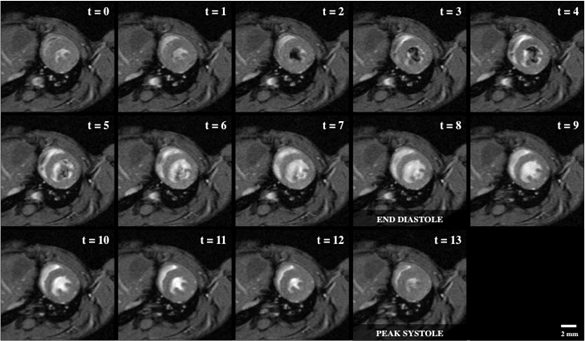
A Figura 1 mostra um laço cine de uma visão de eixo curto do ventrículo esquerdo, que é diretamente perpendicular ao eixo base-ápice do coração e em uma posição que inclui os músculos papilares.

Figura 1: Imagens de cine sanguíneo brilhante de um coração de rato com ...
Aplicação e Resumo
Aqui, a ressonância magnética cardíaca é usada em conjunto com o coração e respiração-gating para adquirir dados de loop cine do coração murino. Enquanto o coração foi o foco da demonstração, regiões adicionais do sistema cardiovascular podem ser imagens seguindo a mesma metodologia. Embora a ressonância magnética não sofra dos mesmos artefatos comumente vistos com outras modalidades de imagem, há uma notável compensação com a resolução espacial alcançada por duração de aquisição. Essa troca ...
Pular para...
Vídeos desta coleção:

Now Playing
Ressonância Magnética Cardíaca
Biomedical Engineering
15.0K Visualizações

Imageamento de amostras biológicas com microscopia ótica e confocal
Biomedical Engineering
36.2K Visualizações

Imageamento por MEV de Amostras Biológicas
Biomedical Engineering
24.0K Visualizações

Biodistribuição de Nano-fármacos Transportadores: Aplicações de MEV
Biomedical Engineering
9.5K Visualizações

Ultrassonografia de alta frequência da aorta abdominal
Biomedical Engineering
14.8K Visualizações

Mapeamento Quantitativo de Tensão de um Aneurisma da Aorta Abdominal
Biomedical Engineering
4.6K Visualizações

Tomografia fotoacústica para imageamento de sangue e lipídios na aorta infrarrenal
Biomedical Engineering
5.9K Visualizações

Simulações computacionais de fluidodinâmica do fluxo sanguíneo em um aneurisma cerebral
Biomedical Engineering
11.9K Visualizações

Imageamento de fluorescência de infravermelho próximo de aneurismas da aorta abdominal
Biomedical Engineering
8.4K Visualizações

Técnicas não invasivas de medição da pressão arterial
Biomedical Engineering
12.1K Visualizações

Aquisição e análise de um sinal de ECG (eletrocardiografia)
Biomedical Engineering
106.7K Visualizações

Resistência à Tração de Biomateriais Reabsorvíveis
Biomedical Engineering
7.5K Visualizações

Imagem de micro-TC de uma medula espinal de camundongo
Biomedical Engineering
8.3K Visualizações

Visualização da degeneração da articulação do joelho após lesão não invasiva do LCA em ratos
Biomedical Engineering
8.3K Visualizações

Combinação de imagens de SPECT e TC para visualizar a funcionalidade cardíaca
Biomedical Engineering
11.2K Visualizações
Copyright © 2025 MyJoVE Corporation. Todos os direitos reservados